1 Tomonori Furukawa, Takatoshi Ueki,...
20
- 1 - Prof. Z. Molnar Differential expression of KCC2 accounts for the differential GABA responses between relay and intrinsic neurons in the early postnatal rat olfactory bulb Wang Cong, 1 Koji Ohno, 1 Tomonori Furukawa, 2 Takatoshi Ueki, 1 Masahiko Ikeda, 2 Atsuo Fukuda, 2 and Kohji Sato 1 Departments of 1 Anatomy & Neuroscience, and 2 Physiology, Hamamatsu University School of Medicine, 1-20-1 Handayama, Hamamatsu, Shizuoka 431-3192, Japan Correspondence should be addressed to Kohji Sato. Department of Anatomy & Neuroscience Hamamatsu University School of Medicine 1-20-1 Handayama, Hamamatsu, Shizuoka 431-3192, Japan Tel & Fax: 81(Japan)-53-435-2288 E-mail: [email protected] Running title: KCC2 and GABA responses in the olfactory bulb. The total number: 16 pages, 2 figures and 1 table Total number of words: (whole) 3433, (abstract) 183, (introduction) 395. Key words: GABA, olfactory bulb, cation-chloride cotransporter, granule cell, mitral cell, KCC2.
-
Upload
phungxuyen -
Category
Documents
-
view
220 -
download
0
Transcript of 1 Tomonori Furukawa, Takatoshi Ueki,...

- 1 -
Prof. Z. Molnar
Differential expression of KCC2 accounts for the differential
GABA responses between relay and intrinsic neurons in the early postnatal rat olfactory bulb
Wang Cong,1 Koji Ohno,1 Tomonori Furukawa,2 Takatoshi Ueki,1 Masahiko Ikeda,2 Atsuo Fukuda,2 and Kohji Sato1 Departments of 1Anatomy & Neuroscience, and 2Physiology, Hamamatsu University School of Medicine, 1-20-1 Handayama, Hamamatsu, Shizuoka 431-3192, Japan Correspondence should be addressed to Kohji Sato. Department of Anatomy & Neuroscience Hamamatsu University School of Medicine 1-20-1 Handayama, Hamamatsu, Shizuoka 431-3192, Japan Tel & Fax: 81(Japan)-53-435-2288 E-mail: [email protected]
Running title: KCC2 and GABA responses in the olfactory bulb.
The total number: 16 pages, 2 figures and 1 table
Total number of words: (whole) 3433, (abstract) 183, (introduction) 395.
Key words: GABA, olfactory bulb, cation-chloride cotransporter, granule cell, mitral
cell, KCC2.

- 2 -
Abstract
The rat olfactory bulb is anatomically immature at birth and considerable neurogenesis and synaptogenesis are known to take place postnatally. In addition, significant physiological changes have also been reported in this period. For example, granule cell-mediated inhibition following electrical stimulations to the lateral olfactory tract is robust during the first postnatal week, then subsequently and abruptly decreases after the second week. However, the mechanism(s) underlying this enhanced inhibition remains to be elucidated. To know the cause of this phenomenon, we investigated the expression patterns of cation-Cl- cotransporters (KCC1, KCC2, and NKCC1) mRNAs, that are responsible for the regulation of [Cl-]i. In addition, GABA responses were measured by gramicidin-perforated patch-clamp recordings and Ca2+ imaging using fura-2. We found that, in the early postnatal period, mitral cells expressing KCC2 mRNA were inhibited by GABA, while granule cells lacking KCC2 mRNA expression were depolarized or excited by GABA. These results indicate that transient GABA-mediated excitation on granule cells might be the main cause of the enhanced inhibition onto mitral cells, and suggest that this differential GABA responses between relay and intrinsic neurons play pivotal roles in the early postnatal rat olfactory bulb.

- 3 -
Introduction Olfaction is the dominant sense in rat pups and they can find their mother’s nipples under olfactory cues (Teicher and Blass, 1977). However, the rat olfactory bulb is anatomically immature at birth and considerable neurogenesis and synaptogenesis are known to take place postnatally (Bayer and Altman 1995; Brunjes and Fraizer, 1986; Hinds and Kinds, 1976). In particular, neurogenesis of granule cells, the major interneuron of the bulb, begins postnatally and continues into adult life (Kaplan and Hinds, 1977). Interestingly, not only such anatomical changes but also significant physiological changes have been reported in the early postnatal period. For example, Shafa et al. found that spontaneous activity rates of mitral cells at P5 are much lower than those in adult (Shafa et al., 1981). In addition, Wilson and Leon (1986, 1987) reported that granule cell-mediated inhibition following electrical stimulations to the lateral olfactory tract is robust during the first postnatal week, then subsequently and abruptly decreases after the second week. However, the mechanism(s) underlying this enhanced inhibition in the early postnatal period is still enigmatic. In the adult CNS, GABA is responsible for inhibition and elicits hyperpolarizing responses via activation of the GABAA receptor/Cl- channel complex. In contrast, during early postnatal development GABA produces excitatory responses in cortical neurons (Luhman and Prince, 1991; Ben-Ari et al., 1989). This GABA-mediated depolarization is thought to play a functional role in activity-dependent neuronal maturation and synapse formation during early development (Ben-Ari et al., 1997) and to promote the migration of newly generated neurons (Behar et al., 1996, 1998). The switch of GABAergic responses from depolarization to hyperpolarization during development is considered to be attributable to developmental decreases in the intracellular Cl- concentration ([Cl-]i). Cation-Cl- cotransporters play a critical role in the regulation of [Cl-]i (Kaila et al., 1993), and of these KCC1 and KCC2 extrude it out of, whereas NKCC1 promotes accumulation of Cl- into the cell (Sun and Murai, 1999; Yamada et al., in press), and this switch is mainly caused by the increase of KCC2 expression (Rivera et al., 1999).

- 4 -
Since the switch of GABAergic responses has been considered to be important in many aspects of brain development, we hypothesized that this switch might also play pivotal roles in the genesis of this enhanced inhibition. In the present study, we, thus, investigated KCC1, KCC2, and NKCC1 mRNA expressions and GABA responses in the developing rat olfactory bulb.

- 5 -
Materials and methods In situ hybridization Male wistar rats at various ages (at postnatal day 1, 3, 7, 14, 49; n=3 at each time point) were decapitated under diethylether anesthesia. The fresh brains were quickly removed and immediately frozen on powdered dry ice. Serial sections (20 μm thick) were cut on a cryostat, thaw-mounted onto silan-coated slides, and stored at –80 °C. After being warmed to room temperature, slide-mounted sections were fixed in 4% paraformaldehyde in 0.1 M phosphate buffer (pH 7.2) for 15 min (all steps were performed at room temperature unless otherwise indicated), rinsed three times (5 min each) in 4x SSC (pH 7.2) (1x SSC contains 0.15 M sodium chloride and 0.015 M sodium citrate), and dehydrated through a graded ethanol series (70-100%). The sections were then defatted with chloroform for 5 min, and immersed in 100% ethanol (twice for 5 min each time) before being subjected to hybridization. Hybridization was performed by incubating the sections with a buffer (4x SSC, 50% deionized formamide, 0.12 M phosphate buffer (pH 7.2), Denhardt's solution (Nakarai, Kyoto, Japan), 2.5 % tRNA (Roche, Tokyo, Japan), 10% dextran sulfate (Sigma, Tokyo, Japan))containing [
35S]dATP
(1000-1500 Ci/mmol (37-55.5 TBq/mmol), New England Nuclear, Boston, MA, U.S.A)-labeled probes (1-2 x 10⁷ dpm/ml, 0.2 ml/ slides) for 24 h at 41 °C. After hybridization, the sections were rinsed in 1x SSC (pH 7.2) for 10 min, followed by rinsing three times in 1x SSC at 55 °C for 20 min each time. The sections were then dehydrated through a graded ethanol series (70%-100%). After film exposure for 3 days at room temperature, the sections were coated with Kodak NBT-2 emulsion (Kodak, Rochester, NY, U.S.A.) diluted 1:1 with water. The sections were then exposed at 4 °C for 2-4 weeks in a tightly sealed dark box. After being developed in D-19 developer (Kodak), fixed with photographic fixer, and washed with tap water, the sections were counterstained with thionin solution to allow morphological identification.
Oligonucleotide probes

- 6 -
Antisense oligo cDNA probes for KCC1, KCC2, and NKCC1 mRNA were
designed based our previous report (Kanaka et al., 2001). The specificity of the
probes has been already confirmed there. The sequences of the probes are as
follows:
(KCC1, GTAGAGGCTCTCTAGCCTCAGAGCCGAGTGCCTGTC;
KCC2, TGGCTTCTCCTCGTTGTCACAAGCTGTCTCTTCGGG;
NKCC1, ACATCCTTGGTACCAGGTGACTTTTCTTGTGATGAC).
The probes were labeled at the 3’ end using [35
S]dATP (1000-1500 Ci/mmol
(37-55.5 TBq/mmol), New England Nuclear, Boston, MA, U.S.A) and terminal
deoxynucleotidyl transferase (Takara, Tokyo, Japan) to obtain a specific activity of
approximately 1.4-2.0 x 10⁹ d.p.m./μg. Preparation of brain slices The methods used for preparing slices and Ca2+ imaging were similar to those described previously (Fukuda et al., 1998; Toyoda et al., 2003). Briefly, male Wistar rats of postnatal day (P) 1-14 were decapitated under diethylether anesthesia. A block of the olfactory bulb was quickly removed and placed in cold (4 °C), oxygenated, modified artificial cerebrospinal fluid (ACSF). The solution contained the following (in mM): 170 sucrose, 126 NaCl, 2.5 KCl, 1.25 NaH2PO4, 12.0 MgSO4, 26.0 NaHCO3, and 30.0 glucose. Coronal slices (400 μm) were cut in the modified ACSF using a vibratome (Leica VT-100, Germany). Slices were allowed to recover for 60 min on nylon meshes (with 1-mm pores) that were submerged in dishes containing standard ACSF consisting of (in mM) 126 NaCl, 2.5 KCl, 1.25 NaH2PO4, 2.0 MgSO4, 2.0 CaCl2, 26.0 NaHCO3, and 20.0 glucose. The dishes were then placed in a tightly sealed box filled with 95 % O2 - 5% CO2 at a pressure of 50 kPa at room temperature until used. Gramicidin-perforated patch-clamp recordings Gramicidin-perforated patch-clamp recordings were carried out as previously described (Toyoda et al. 2003, Yamada et al. 2004). Patch electrodes were made

- 7 -
from borosilicate capillary tubing (diameter, 1.5 mm; Narishige) using a Sutter P-97 puller (Sutter instruments). The electrode resistance was within the range 3.5-5 MΩ. The pipette solution contained (in mM) 150 KCl, 10 HEPES (pH 7.3 with KOH). Gramicidin was dissolved in dimethylsulfoxide (10 mg/ml), and then diluted in the pipette-filling solution to a final concentration of 5-10 µg/ml just before the experiment. Membrane potentials were recorded using an Axopatch 1D amplifier and digitized by means of a Digidata 1332A data-acquisition system (Axon Instruments). Data were acquired by means of pClamp8 (Axon Instruments) software and stored on the hard disk for off-line analysis using Clampfit8 (Axon Instruments). Granule and mitral cells in slices were viewed on a monitor via a x40 water-immersion objective lens with the aid of an infrared differential interference-contrast (IR-DIC) filter and a CCD-camera (C2400-79; Hamamatsu Photonics). GABA (50 µM) was pressure-applied for 0.5-5 sec through a patch pipette to the soma of the recorded neurons. Ca2+ imaging using fura-2 Neurons were loaded with fura-2 acetoxy methyl ester (10 μM) in ACSF containing 0.01 % pluronic F127. Slices were then laid on the glass bottom of a submerged-type chamber, and this was placed on a microscope stage and continuously perfused with standard ACSF gassed with 95 % O2 - 5% CO2 at a rate of 2-3 ml/min. Fura-2 fluorescence was excited using a multi-wavelength monochrometer (C6789; Hamamatsu Photonics) and the emitted light was filtered using a band-pass filter (510 nm). Fluorescence images were obtained using a x 40 objective lens (Plan Fluor, N.A. 0.75; Nikon) via a cooled-CCD camera (C6790-81; Hamamatsu Photonics) fitted to an up-light microscope (E600-FN; Nikon). Data were stored for off-line analysis by means of image-processing software (Aqua Cosmos; Hamamatsu Photonics). [Ca2+]i was expressed as the ratio of the fura-2 fluorescence intensities excited at 340 nm and 380 nm (RF340/F380). Changes with time in RF340/F380 were monitored in olfactory bulb neurons by taking measurements every 10 s. For temporal analysis, each RF340/F380 was converted to normalized ratio (F340/F380) by dividing by average of 3 values obtained before application of GABA (100 μM) by perfusion.

- 8 -
RESULTS
To investigate KCC1, KCC2, and NKCC1 mRNA expressions in the developing olfactory bulb, we used three probes already used in our previous report (Kanaka et al., 2001). The specificity of the probes has been already confirmed there. Positive cells were divided into four categories (very strong, strong, moderate, and low) by visual comparison of the extent of silver grain accumulation (Sato et al., 1993). At the first postnatal day (P1), KCC1 and NKCC1 mRNAs were abundantly expressed in both the mitral cell layer and granule cell layer (Fig. 1A and C). In contrast, KCC2 mRNA expression was only detected in the mitral cell layer (Fig. 1B), while we could not detect KCC2 mRNA expression in other areas including the granule cell layers, suggesting that KCC2 mRNA expression is differentially regulated between relay neurons (mitral cells) and intrinsic neurons (granule cells). At P3, their expression patterns were comparable to those on P1 (Fig. 1 D-F). At P7, we first detected weak KCC2 mRNA expressions in the granular layer (Fig. 1H). In addition, the periglomerular layer also expressed KCC2 mRNA moderately. KCC1, KCC2 and NKCC1 mRNA expressions dramatically increased between P7 and P14 (Fig. 1G-L). At P14, both mitral neurons and granule neurons expressed the three mRNAs abundantly (Fig. 1J-L). There were no substantial changes after P14 (data not shown). Relative abundance of KCC1, KCC2, and NKCC1 mRNA expressions in the developing rat olfactory bulb is summarized in Table 1.
Since KCC2-mediated Cl- extrusion has been reported to be responsible for the change in Cl- reversal potential that converts GABA from a depolarizing to a hyperpolarizing neurotransmitter (Payne, 1997; Rivera et al., 1999), we speculate that the above mentioned changes in Cl- cotransporters mRNA expressions result in the differential responses to GABA between mitral cells and granule cells. We thus studied the functional differences in GABA responses based on changes in Cl- homeostasis between olfactory bulb neurons. First, we examined the development of GABA-evoked membrane potential changes in olfactory bulb neurons by means of gramicidin-perforated patch-clamp recordings in the current clamp mode (n=2-3 for each experiment). Granule cells and mitral cells were identified using IR-DIC video microscopy by their

- 9 -
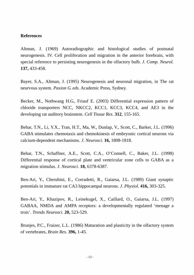
morphology and location (Fig. 2A). At P1 and P6, granule cells were depolarized by GABA (50 μM) application. P1 granule cells were rather less-excitable, in which spontaneous and GABA-evoked action potentials were not frequently observed (Fig. 2B). At P6, GABA could evoke an action potential firing (Fig. 2C). At P16, granule cells were hyperpolarized and their firings were inhibited by GABA (Fig. 2D). In contrast to granule cells, mitral cells were hyperpolarized and their firings were inhibited by GABA at any age (Fig. 2E-G). Thus, there were no developmental changes in GABA responses in postnatal mitral cells. The results indicate that granule cells are depolarized and/or excited by GABA in the early postnatal period, whereas mitral cells are already hyperpolarized and inhibited.
Second, we studied changes in Ca2+ transients evoked by GABA in olfactory bulb neurons during development. The olfactory bulb slice preparations were loaded with fura-2 (n=3 for each experiment), the ratio of the fluorescence intensities excited at 340 nm and 380 nm (RF340/F380) was obtained to evaluate [Ca2+]i changes (Fig. 3). We identified mitral cells and granule cells by their morphology (Fig. 3A). In P1 granule neurons, GABA evoked marked [Ca2+]i increases (Fig. 3B). However, [Ca2+]i increases were not apparently evoked by GABA in P1 mitral cells (Fig. 3E). The GABA-evoked [Ca2+]i increases in granule cells gradually declined with age (Fig. 3C), then, it was hardly observed at P14 (Fig. 3D). No developmental changes in [Ca2+]i transients in response to GABA application were observed in mitral cells. These data are consistent with the electrophysiological aspects of GABA responses, indicating that GABA-induced depolarizations in granule cells during the early postnatal period would exceed the threshold of voltage-dependent Ca2+ channels.

- 10 -
Discussion The rat olfactory bulb is anatomically immature at birth and considerable neurogenesis and synaptogenesis are known to take place postnatally (Bayer and Altman 1995; Brunjes and Fraizer, 1986; Hinds and Kinds, 1976). Interestingly, significant physiological changes have also been reported in the early postnatal period. For example, spontaneous activity rates of mitral cells at P5 are much lower than those in adult (Shafa et al., 1981). In addition, granule cell-mediated inhibition following electrical stimulations to the lateral olfactory tract is robust during the first postnatal week, then subsequently and abruptly decreases after the second week (Wilson and Leon, 1986, 1987). These findings indicate that in the early postnatal period mitral cells are strongly inhibited by granule cells. However, the mechanism(s) underlying this enhanced inhibition remains to be elucidated. In the present study, we revealed that granule cells do not express KCC2 mRNA in the early postnatal period, resulting in depolarizing responses to GABA. In contrast, mitral cells already express KCC2 mRNA, showing hyperpolarizing responses to GABA. What happens in the olfactory bulb under such conditions? An important local circuit in the bulb occurs at synaptic contacts formed between the lateral dendrites of mitral cells and the dendrites of GABAergic granule cells. Mitral cell dendrites release glutamate onto the dendritic spines of granule cells, which in turn release GABA back onto mitral cell dendrites. This reciprocal synaptic circuit underlies self and lateral dendrodendritic inhibition (Mori and Takagi, 1978). Namely, granule cells control the activity of mitral cells through these dendrodendritic inhibitory synapses (Shepherd, 1972). What controls the activity of granule cells? Interestingly, GABAergic granule cells also express GABAA receptors and receive GABAergic inputs (Laurie et al., 1992, Nusser et al., 1999). Where are the sources for these GABAergic inputs? One is the input from GABAergic short axon cells present in the granule cell layer (Schneider and Macrides, 1978). The second source may be interconnection of granule cells through dendritic synapses. Finally, the basal forebrain (diagonal band nuclei) and, to a lesser extent, the ventral pallidum, anterior amygdala, and the nucleus of the lateral olfactory tract could also provide a GABAergic innervation of the granule

- 11 -
cells (Zaborsky et al., 1986). In adult, these GABAergic innervations inhibit granule cell activities. Indeed, Nusser et al. have reported that selective disruption of GABAA receptors on granule cells leads to the augmentation of IPSCs in mitral cells (Nusser et al., 2001). However, in the early postnatal period, granule cells are supposed to be depolarized and/or excited by GABA as we have shown in this study, indicating that all GABAergic innervations robustly activate granule cells. Thus, we speculate that this GABA-induced activation of granule cells plays an important role in the genesis of the enhanced inhibition in mitral cells. Of course, there remains a possibility that these GABA-mediated depolarizations may act inhibitory due to shunting of membrane currents. What is the physiological significance of this phenomenon? Many mammals are born with their ears and eyes closed. As a result, the olfactory system must be the primary mechanism for receiving information about the environment (Teicher and Blass, 1977). Indeed, the early olfactory experience and learning has repeatedly been shown to be essential for locating sustenance, identifying nest mates and conspecifics, finding home, and learning what is safe to eat (Brunjes and Greer, 2003). However, while mitral cell neurogenesis is almost completed at birth, relatively few granule cells are present at this time (Altman, 1969; Hinds, 1968; Mair et al., 1982). Thus, in the early postnatal olfactory bulb each granule cell must show stronger inhibition on mitral cells than in adult to compensate their paucity, so that they could keep normal olfactory functions. To achieve this, it is strategically advantageous if granule cells are excited by each other with their own neurotransmitter GABA. Besides above, this phenomenon may be indispensable for the normal development of the olfactory bulb. Since GABA-mediated depolarization plays functional roles in activity-dependent neuronal maturation and synapse formation during early development (Ben-Ari et al., 1997) and in promoting the migration of newly generated neurons (Behar et al., 1996, 1998). Indeed, the early developmental period is exactly the period when granule cells increase in number and make dendritic branches, dendritic spines and synapses (Bayer and Altman 1995; Brunjes and Fraizer, 1986; Hinds and Kinds, 1976), indicating granule cells also need GABA-mediated depolarization to develop normally. Recently, Ganguly et al. reported that GABA itself promotes the

- 12 -
developmental switch of neuronal GABAergic responses from excitation to inhibition (Gangly et al., 2001). Thus, granule cells might need excitatory GABAergic inputs both for their normal development and for keeping the adequate function of the early postnatal olfactory bulb. Recently, Becker et al. (2003) have examined the expression of six secondary active chloride transporter genes in the developing rat auditory brainstem and found that a HCO3
-/Cl- exchanger AE3 plays an important role in chloride homeostasis in the CNS, suggesting that at least two different mechanisms exist to control chloride homeostasis. Namely, in the hippocampus and cortex, an age-related upregulation of KCC2 expression plays a role (Rivera et al., 1999; Yamada et al., 2004), while in the brainstem AE3 does (Becker et al., 2003). In the present study, we found a dramatic upregulation of KCC2 expression in the olfactory bulb, indicating that at least in the olfactory bulb KCC2 might be the main player. Further studies might reveal how chloride homeostasis is regulated in other brain regions
Acknowledgements Part of this work was supported by the Ministry of Education, Science and Culture of Japan and the Ministry of Health and Welfare of Japan. Abbreviations KCC1, 2, K-Cl cotransporter 1, 2; NKCC1, Na-K-2Cl cotransporter 1, 2.
- 13 -
References Altman, J. (1969) Autoradiographic and histological studies of postnatal neurogenesis. IV. Cell proliferation and migration in the anterior forebrain, with special reference to persisting neurogenesis in the olfactory bulb. J. Comp. Neurol. 137, 433-458. Bayer, S.A., Altman, J. (1995) Neurogenesis and neuronal migration, in The rat neurvous system. Paxion G. eds. Academic Press, Sydney. Becker, M., Nothwang H.G., Friauf E. (2003) Differential expression pattern of chloride transporters NCC, NKCC2, KCC1, KCC3, KCC4, and AE3 in the developing rat auditory brainstem. Cell Tissue Res. 312, 155-165. Behar, T.N., Li, Y.X., Tran, H.T., Ma, W., Dunlap, V., Scott, C., Barker, J.L. (1996) GABA stimulates chemotaxis and chemokinesis of embryonic cortical neurons via calcium-dependent mechanisms. J. Neurosci. 16, 1808-1818. Behar, T.N., Schaffner, A.E., Scott, C.A., O’Connell, C., Baker, J.L. (1998) Differential response of cortical plate and ventricular zone cells to GABA as a migration stimulus. J. Neurosci. 18, 6378-6387. Ben-Ari, Y., Cherubini, E., Corradetti, R., Gaiarsa, J.L. (1989) Giant synaptic potentials in immature rat CA3 hippocampal neurons. J. Physiol. 416, 303-325. Ben-Ari, Y., Khazipov, R., Leinekugel, X., Caillard, O., Gaiarsa, J.L. (1997) GABAA, NMDA and AMPA receptors: a developmentally regulated ‘menage a trois’. Trends Neurosci. 20, 523-529. Brunjes, P.C., Fraizer, L.L. (1986) Maturation and plasticity in the olfactory system of vertebrates, Brain Res. 396, 1-45.

- 14 -
Brunjes, P.C., Greer, C.A. (2003) Progress and directions in olfactory development. Neuron 38, 371-374. Fukuda, A., Muramatsu, K., Okabe, A., Shimano, Y., Hida, H., Fujimoto, I., Nishino, H. (1998) Changes in intracellular Ca2+ induced by GABAA receptor activation and reduction in Cl- gradient in neonatal rat neocortex. J. Neurophysiol. 79, 439-446. Ganguly, K., Schinder, A.F., Wong, S.T., Poo, M. (2001) GABA itself promotes the developmental switch of neuronal GABAergic responses from excitation to inhibition. Cell 105, 521-532. Hinds, J.W. (1968) Autographic study of histogenesis in the mouse olfactory bulb. I. time of origin of neurons and neuroglia J. Comp. Neurol. 134, 287-304. Hinds, J.W., Kinds, P.L. (1976) Synapse formation in the mouse olfactory bulb. 1. Quantitative studies J. Comp. Neurol. 169, 15-40. Kaila, K., Voipio, J., Paalasmaa, P.J., Pasternack, M., Deisz, R.A. (1993) The role of bicarbonate in GABAA receptor-mediated IPSPs of rat neocortical neurons. J. Physiol. 464, 273-289. Kanaka, C., Ohno, K., Okabe, A., Kuriyama, K., Itoh, T., Fukuda, A., Sato, K. (2001) The differential expression patterns of messenger RNAs encoding K-Cl cotransporters (KCC1,2) and Na-K-2Cl cotransporter (NKCC1) in the rat nervous system. Neuroscience 104, 933-946. Kaplan, M.S. and Hinds, J.W. (1977) Neurogenesis in the adult rat: electron microscopic analysis of light radioautographs, Science 197, 1092-1094. Laurie, D.J., Seeburg, P.H., Wisden, W. (1992) The distribution of 13 GABAA receptor subunit messenger RNAs in the rat brain: 2. olfactory bulb and cerebellum
- 15 -
J. Nerosci. 12, 1063-1076. Luhmann, H.J., Prince, D.A. (1991) Postnatal maturation of the GABAergic system in rat neocortex. J. Neurophysiol. 65, 247-263. Mair, R.G., Gellman, R.L., Gesteland, R.C. (1982) Postnatal proliferation and maturation of olfactory bulb neurons in the rat. Neuroscience 7, 3105-3116. Mori, K., Takagi, S.F. (1978) An intracellular study of dendrodendritic inhibitory synapses on mitral cells in the rabbit olfactory bulb. J. Physiol. (London) 279, 569-588. Nusser, Z., Sieghart, W., Mody, I. (1999) Differential regulation of synaptic GABAA receptors by cAMP-dependent protein kinase in mouse cerebellar and olfactory bulb neurons. J. Pysiol. (Lond) 521, 421-435. Nusser, Z., Kay, L.M., Laurent, G., Homanics, G.E., Mody, I. (2001) Disruption of GABAA receptors on GABAergic interneurons leads to increased oscillatory power in the olfactory bulb network. J. Neurophysiol. 86, 2823-2833. Payne, J.A. (1997) Functional characterization of the neuronal-specific K-Cl cotransporter: implications for [K+]o regulation. Am. J. Physiol. 273, C1516-1525. Rivera, C., Voipio, J., Payne, J.A., Ruusuvuori, E., Lahtinen, H., Lamsa, K., Pivola, U., Saarma, M., Kaila, K. (1999) The K+/Cl- co-transporter KCC2 renders GABA hyperpolarizing during neuronal maturation. Nature 397, 251-255. Sato, K., Kiyama, H., Tohyama, M. (1993) The differential expression patterns of messenger RNAs enconding non-N-methyl-D-aspartate glutamate receptor subsunits (GluR1-4) in the rat brain. Neuroscience 52, 515-539.

- 16 -
Schneider, S.P., Macrides, F. (1978) Laminar distributions of interneurons in the main olfactory bulb o fthe adult hamster. Brain Res. Bull. 3, 73-82. Shafa, F., Shineh, S.N., Bidanjiri, A. (1981) Development of spontaneous activity in the olfactory bulb neurons of postnatal rat. Brain Res. 223, 409-412. Shepherd, G.M. (1972) Synaptic organization of the mammalian olfactory bulb. Physiol. Rev. 52, 864-917. Sun, D., Murali, S.G. (1999) Na-K-2Cl cotransporter in immature cortical neurons: a role in intracellular Cl regulation. J. Neurophysiol. 81, 1939-1948. Teicher, M.H., Blass, E.M. (1977) First suckling response of the newborn albino rat: the roles of olfaction and amniotic fluid. Science 198, 635-636. Toyoda, H., Ohno, K., Okabe, A., Ikeda, M., Yamada, J., Sato, K., Hashimoto, K., Fukuda, A. (2003) Induction of NMDA and GABAA receptors-mediated Ca2+
oscillations with KCC2 mRNA downregulation in injured facial motoneurons. J. Neurophysiol. 89, 1353-1362. Wilson, D.A., Leon, M. (1986) Early appearance of inhibition in the neonatal rat olfactory bulb. Dev. Brain Res. 26, 289-292. Wilson, D.A., Leon, M. (1987) Abrupt decrease in synaptic inhibition in the postnatal rat olfactory bulb. Dev. Brain Res. 33, 134-138. Yamada, J., Okabe, A., Toyoda, H., Kilb, W., Luhmann, H.J., Fukuda, A. Cl- uptake promoting depolarizing GABA actions in immature rat neocortical neurones is mediated by NKCC1. J. Physiol. in press. Zaborszky, L., Carlsen, J., Brashear, H.R., Heimer, L. (1986) Cholingergic and GABAergic afferents to the olfactory bulb in the rat with special emphasis on the

- 17 -
projection neurons in the nucleus of the horizontal limb of the diagonal band. J. Comp. Neurol. 243, 488-509. Table 1. Relative abundance of KCC1, KCC2, NKCC1 mRNAs in the developing rat olfactory bulb
Cell type mRNA P1 P3 P7 P14 Adult KCC1 + + + + + + + + + + + + + KCC2 + + + + + + + + + + + + + + + + + Mitral NKCC1 + + + + + + + + + + + + + + + KCC1 + + + + + + + + + + + + KCC2 n.d. n.d. + + + + + + + Granule NKCC1 + + + + + + + + + + + + + +
Relative expression levels were estimated by visual comparison of exposed emulsion-coated slides. n.d., not detected; +, weak; + +, moderate; + + +, strong; + + + +, very strong.

- 18 -
Figure legends
Fig. 1. Expression of KCC1 mRNA (A, D, G, J), KCC2 mRNA (B, F, H, K), and NKCC1 mRNA (C, F, I, L) in the
developing rat olfactory bulb. Gl, glomerular layer; Gr, granule cell layer; Mi, mitral cell layer.
- 19 -
Fig. 2. Effects of GABA in the developing rat olfactory bulb neurons. (A) IR-DIC images of granule cells (Gr:
arrow head) and mitral cells (Mi: arrow) at P16. Thus, olfactory bulb neurons were distinguished by their location
and morphology. Bar=50 μm. By gramicidin-perforated patch-clamp recordings in the current clamp mode, GABA
(50 μM) depolarized P1 and P6 granule cells (B,C), and hyperpolarized P16 granule cell (D). Note that GABA
application evoked an action potential firing in a granule cell (* in C). In contrast, mitral cells were hyperpolarized
by the application of GABA at any age (E-G). Resting potentials are indicated at left.

- 20 -
Fig. 3. Effects of GABA on [Ca2+]i in the developing rat olfactory bulb neurons. (A). Fura-2 fluorescence image
(excited at 380 nm) of granule cells (Gr) and mitral cells (Mi) at P7. Bar = 50 μm. In P1 granule cells, application
of GABA (100 μM) evoked marked increases in [Ca2+]i (B), which gradually declined with age (C), and almost
disappeared by P14 (D). In contrast, application of GABA (100 μM) to mitral cells evoked no apparent [Ca2+]i
responses at any age (E-G).