Constitutive overexpression of the Drosophila period protein inhibits

JOURNAL OF VIROLOGY, Sept. 2011, p. 9614–9622 Vol. 85, No. 180022-538X/11/$12.00 doi:10.1128/JVI.00480-11Copyright © 2011, American Society for Microbiology. All Rights Reserved.
The Leader Protein of Cardioviruses InhibitsStress Granule Assembly�
Fabian Borghese and Thomas Michiels*Universite catholique de Louvain, de Duve Institute, MIPA-VIRO 74-49, 74, avenue Hippocrate, B-1200 Brussels, Belgium
Received 9 March 2011/Accepted 1 July 2011
Stress granules (SG) are cytoplasmic aggregates of stalled translation preinitiation complexes that form incells exposed to various environmental stresses. Here, we show that stress granules assemble in cells infectedwith Theiler’s murine encephalomyelitis virus (TMEV) mutants carrying alterations in the leader (L) protein,but not in cells infected with wild-type TMEV. Stress granules also formed in STAT1-deficient cells, suggestingthat SG formation was not a consequence of increased type I interferon (IFN) production when cells wereinfected with the mutant virus. Ectopic expression of the wild-type L protein was sufficient to inhibit stressgranule formation induced by sodium arsenite or thapsigargin treatment. In conclusion, TMEV infectioninduces stress granule assembly, but this process is inhibited by the L protein. Unlike poliovirus-induced stressgranules, TMEV-induced stress granules did not contain the nuclear protein Sam68 but contained poly-pyrimidine tract binding protein (PTB), an internal ribosome entry site (IRES)-interacting protein. Moreover,G3BP was not degraded and was found in SG after TMEV infection, suggesting that SG content could be virusspecific. Despite the colocalization of PTB with SG and the known interaction of PTB with viral RNA, in situhybridization and immunofluorescence assays failed to detect viral RNA trapped in infection-induced SG.Recombinant Theiler’s viruses expressing the L protein of Saffold virus 2 (SAFV-2), a closely related humantheilovirus, or the L protein of mengovirus, an encephalomyocarditis virus (EMCV) strain, also inhibitedinfection-induced stress granule assembly, suggesting that stress granule antagonism is a common feature ofcardiovirus L proteins.
Theiler’s murine encephalomyelitis virus (TMEV) belongsto the Cardiovirus genus, within the picornavirus family. Othercardioviruses are Saffold virus (SAFV), a recently describedhuman virus closely related to TMEV, and encephalomyocar-ditis virus (EMCV). The genomes of these viruses are com-posed of nonsegmented positive-stranded RNA molecules ofapproximately 8 kb. During infection, these viruses produce ashort protein cleaved from the amino-terminal end of the viralpolyprotein and therefore called leader (L) protein (21).
The DA strain of TMEV is responsible for persistent infec-tion of the central nervous system of the mouse, leading tochronic demyelinating lesions reminiscent of those found inmultiple sclerosis (5, 22). Disruption of the host immune re-sponse is critical for the establishment of viral persistence, andthe L protein of the virus plays a crucial role in this process.Cardiovirus L proteins are closely related multifunctional pro-teins shown to interfere with critical cellular processes such asinterferon (IFN) and chemokine production (16, 32), nucleo-cytoplasmic trafficking (4, 8, 17, 27, 30), apoptosis (12, 31), andmitogen-activated protein (MAP) kinase activity (28). Cardio-virus L proteins are very small proteins (about 70 amino acids)in which several domains have been described (Fig. 1): (i) anamino-terminal zinc finger, (ii) a glutamate/aspartate-rich do-main, which confers a very acidic character to the protein (pIabout 3.8), (iii) a serine/threonine-rich domain, and (iv) acarboxy-terminal domain, present in the L proteins of TMEV
and SAFV (theilovirus species) but lacking in EMCV L andtherefore called the Theilo domain. Mutations introduced ei-ther in the zinc finger or in the Theilo domain strongly inhibitall the known activities of the TMEV L protein (29).
Polypyrimidine tract binding protein (PTB) is a RNA-bind-ing protein involved in splicing regulation and 3� end process-ing of RNA molecules. In TMEV-infected cells, PTB wasfound to relocalize from the nucleus to the cytoplasm (8),where it is believed to interact with the internal ribosome entrysite (IRES) of viral RNA (24). Binding of PTB to the IRES isthought to stabilize the RNA structure, thereby promotingtranslation of viral proteins. Such translation-modulating ac-tivity of PTB has been confirmed for the GDVII TMEV strain(25).
Stress granules (SG) are cytoplasmic foci appearing in cellsexposed to various environmental stresses, such as UV expo-sure, oxidative stress, hypoxia, and heat shock. They are mainlycomposed of stalled translation preinitiation complexes thataggregate in response to stress (3). The best-known event trig-gering SG assembly is the phosphorylation of the alpha subunitof eukaryotic translation initiation factor 2 (eIF2�). eIF2�phosphorylation can be mediated by four kinases activated inresponse to different stress signals: PKR, activated by cytosolicdouble-stranded RNA; PERK, activated by endoplasmic retic-ulum stress; GCN2, activated by amino acid starvation; andHRI, activated by oxidative stress or heme depletion (9). Phos-phorylation of eIF2� leads to translational arrest and to theassembly of incomplete translation preinitiation complexes(lacking eIF2) on the caps of mRNA molecules. In this pro-cess, TIA-1, a nuclear prion-like protein, delocalizes from thenucleus to the cytoplasm and binds the eIF2-deficient preini-tiation complexes, inducing their aggregation and thereby as-
* Corresponding author. Mailing address: Universite catholique deLouvain, de Duve Institute, MIPA-VIRO 74-49, 74, avenue Hippro-crate, B-1200 Brussels, Belgium. Phone: 32 2 764 74 29. Fax: 32 2 76474 95. E-mail: [email protected].
� Published ahead of print on 13 July 2011.
9614
Dow
nloa
ded
from
http
s://j
ourn
als.
asm
.org
/jour
nal/j
vi o
n 25
Feb
ruar
y 20
22 b
y 22
0.12
2.15
4.18
.

sembly of SG (13). SG are thought to play a role in translationinhibition during stress, through sequestration of cellularmRNA. When cells recover from stress, SG disperse andmRNA can be sent back to effective translation or targeted toprocessing bodies (P-bodies). P-bodies are cytosolic complexescontaining enzymes for decapping and degradation of mRNA,as well as proteins involved in small interfering RNA (siRNA)-mediated repression of mRNA translation. Therefore, P-bod-ies are thought to be a place where mRNA is degraded (2).Factors regulating the fate of sequestered mRNA during re-covery are incompletely known.
In recent years, several viruses have been shown to induce astress granule response in host cells. Depending on the virus,the response can be pro- or antiviral. For example, respiratorysyncytial virus (RSV) preferentially replicates in host cellsforming stress granules (18). Conversely, poliovirus inhibitsboth P-body and SG formation (11, 33). Poliovirus 3C proteasehas been shown to block infection-induced SG formation bycleaving G3BP, a host cell protein involved in the SG assemblyprocess. Expression of a noncleavable form of G3BP in hostcells significantly lowered poliovirus replication (33).
Some cellular proteins, such as eIF3, TIA-1, and G3BP, areconcentrated in SG and are thus used as ubiquitous SG mark-ers. However, certain proteins are associated only with a par-ticular type of SG. For example, Sam68, a nuclear factor in-volved in splicing regulation, was reported to be associatedwith poliovirus-induced SG, but not with oxidative stress orheat shock-induced SG (26).
We previously observed a stress granule-like cytoplasmicdistribution of PTB in cells infected with TMEV expressing amutated L protein, but not in cells infected with a wild-type
(wt) virus. This observation led us to investigate the relation-ship between TMEV infection and the stress granule response.
MATERIALS AND METHODS
Cells, viruses, and plasmids. HeLa, U3A 2FTGH (19) (kindly provided by IanKerr), and L929 cells were maintained in Dulbecco’s modified Eagle medium(Lonza) supplemented with 10% fetal calf serum (MP Biomedicals), 100 IUpenicillin/ml, and 100 �g streptomycin/ml.
Viruses used in this study (Table 1) were derivatives of the DA1 molecularclone of the persistent Daniels (DA) TMEV strain (7, 20). L-mutant virusesderived from DA1 were TM564 carrying a deletion encompassing codons 6 to 67of the L region (L�6–67), TM598 carrying mutations disrupting the L zinc finger(LZn) (32), and FB05 carrying a M60V substitution in the L Theilo domain(LM60V) (29).
For infection of L929 and U3A cells, we used virus KJ6 and L mutants thereof.KJ6 is a DA1 derivative carrying capsid mutations enabling the virus to infectL929 cells with high efficiency (15). This virus is further referred to as wild typein this work as it expresses a wild-type L protein (Lwt). L mutant KJ6 derivativeswere SB3 (L�6–67), TM659 (LZn) (32), and FB09 (LM60V) (29). KJ6 derivativesexpressing the wild-type L protein of mengovirus (SPA24) and its correspondingzinc finger mutant (SPA28) were described previously (23). FB26 and FB27 areKJ6 derivatives expressing the wild-type L protein and an M55V mutant of theL protein from Saffold virus 2 (SAFV-2; GenBank accession no. AM922293),respectively. These viruses were constructed as follows. The LSAFV-2 codingsequence was subcloned as a BamHI-XbaI restriction fragment from a syntheticplasmid (ordered to MrGene) in pBS-KS�, giving pFB16. This fragment wasalso cloned in pTM624, a pcDNA3 derivative containing the TMEV IRESfollowed by the enhanced green fluorescent protein (eGFP) coding sequence,giving pFB14. Plasmid pFB14 thus allows expression of a bicistonic LSAFV-2-IRES-eGFP construct from the cytomegalovirus (CMV) immediate-early pro-moter. The M55V mutation was introduced in LSAFV-2 by PCR amplification ofthe L region of pFB16 with primers TM952 (5�AAA GGA TCC GCC ACC ATGGCG TGC) and TM956 (5� GGT AGA TCT GTC CAT TCC ACA TGG AGGTCA TCA GGA TAA). TM956 encodes the M55V mutation (A163TG3GTG).The resulting PCR product was used to replace the corresponding NcoI-BglIIrestriction fragment in pFB16 to yield pFB18. To construct TMEV cDNA clones
FIG. 1. Alignment of L protein sequences used in this work. Shown are the sequences of L proteins from TMEV (strain DA1), SAFV-2, andmengovirus. Protein domains are indicated. Cysteine and histidine residues forming the Zn finger, and the M60/M55 residue of the Theilo domain,are framed. L mutations are indicated under the alignment.
TABLE 1. Plasmids carrying full-length viral genomes
Plasmid Virus L proteina Virus background, characteristics
pTMDA1 DA1 Lwt Molecular clone of TMEV DA strainpTM564 TM564 L�6–67 DA1pTM598 TM598 LZn DA1pFB05 FB05 LM60V DA1pKJ6 KJ6 Lwt DA1, capsid adapted to infect L929 cellspSB3 SB3 L�6–67 KJ6, capsid adapted to infect L929 cellspTM659 TM659 LZn KJ6, capsid adapted to infect L929 cellspFB09 FB09 LM60V KJ6, capsid adapted to infect L929 cellspFB26 FB26 SAFV-2 Lwt KJ6, capsid adapted to infect L929 cellspFB27 FB27 SAFV-2 LM55V KJ6, capsid adapted to infect L929 cellspSPA24 SPA24 Mengovirus Lwt KJ6, capsid adapted to infect L929 cellspSPA28 SPA28 Mengovirus LZn KJ6, capsid adapted to infect L929 cells
a Unless specified, the leader protein was that of TMEV DA1.
VOL. 85, 2011 CARDIOVIRUSES AND STRESS GRANULES 9615
Dow
nloa
ded
from
http
s://j
ourn
als.
asm
.org
/jour
nal/j
vi o
n 25
Feb
ruar
y 20
22 b
y 22
0.12
2.15
4.18
.

expressing the SAFV-2 L protein, the mutated coding region of the LSAFV-2 genewas then transferred back from pFB18 to pFB14 as a BamHI-XbaI restrictionfragment. The resulting plasmid was called pFB19. The wild-type and mutatedLSAFV-2 gene coding sequences were then PCR amplified from pFB14 andpFB19 with primers TM952 and TM954 (5� AAA CCT GAG GAC TGG GAGTTA CTC TTG TCA GAT GAA GAG GCG TTT CCT TGT GGT TCC ATTTCA ATG TC). The resulting PCR products were cloned into pTM565 (asubclone of pTMDA1) as NcoI-Bsu36I restriction fragments to yield pFB20 andpFB21. Finally, the XbaI-MscI fragment carrying the L gene coding region wasextracted from these plasmids and used to replace the corresponding fragment inpKJ6. The resulting plasmids, pFB26 and pFB27, carry the full-length cDNA ofKJ6 derivatives coding for the wild-type and M55V mutant SAFV-2 L proteins,respectively. Synthetic and PCR-amplified regions of all constructs were se-quenced to check that no unexpected mutation occurred during the cloningsteps. Plasmid constructs carrying full-length virus cDNA and the correspondingviruses are presented in Table 1.
All wild-type and mutant viruses were produced from the correspondingcDNA clones, as described previously (20). Viruses were collected 48 to 72 hafter electroporation of BHK-21 cells with in vitro-transcribed viral RNA. Vi-ruses were titrated in parallel by standard plaque assay in BHK-21 cells.
Bicistronic constructs expressing IRES-eGFP alone (pTM624), LTMEVwt-
IRES-eGFP (pTM625), or LTMEVM60V-IRES-eGFP (pCER48) were described
previously (30).Immunostaining and in situ hybridization. Immunostainings and in situ hy-
bridizations were performed on cells cultivated on glass coverslips treated withpoly-L-lysine and placed in 24-well plates. Prior to immunostaining, cells werefixed for 4 to 10 min in 300 �l of phosphate-buffered saline (PBS)–4% paraform-aldehyde (PFA). Cells were then washed in 500 �l of PBS and permeabilized for5 min at room temperature in 500 �l of PBS–0.1% Triton X-100. Blockingoccurred for 1 h at room temperature in 300 �l of TNB blocking reagent (PerkinElmer). Cells were next incubated with the primary antibody diluted in TNB atthe following dilutions: PTB (mouse; Zymed 32-4800), 1/50; VP1 (mouse; F12B3clone; kind gift from M. Brahic), 1/10; eIF3 (goat; Santa Cruz sc-16377), 1/200;TIA-1 (goat; Santa Cruz sc-1751 C20), 1/100; K1 or J2 (mouse, anti-double-stranded RNA [anti-dsRNA], English & Scientific Consulting Bt.), 1/200; Sam68(rabbit; Santa Cruz sc-333; C20), 1/100; G3BP (mouse; BD Transduction Lab-oratories 611126), 1/500. After 1 h of incubation at room temperature, cells werewashed 3 times for 5 min in 500 �l PBS–0.1% Tween 20. Secondary antibodies(Alexa Fluor 488- or 594-conjugated antibodies; Invitrogen) were incubated for1 h at a 1/800 dilution in TNB. Finally, cells were washed 3 times in 500 �lPBS–0.1% Tween 20 and mounted with Mowiol for fluorescence microscopy.
Experiments involving in situ hybridization coupled to eIF3 immunostainingwere performed according to a protocol adapted from that of Chakraborty et al.(6) using a 3� biotinylated DNA probe (Eurogentec) complementary to thepositive strand of viral RNA (5� AGG GGT GCC TTT TCT TTC CAG GTGAGC CAT ATT CGG GAG AAA ATT). All reagents were prepared in 0.5%diethyl pyrocarbonate (DEPC)-treated water or PBS. Cells were fixed for 8 minin 500 �l of PBS–4% PFA prior to in situ hybridization. After 3 washes in 500 �lPBS, cells were permeabilized for 5 min at 4°C in 500 �l PBS–0.1% Triton X-100.Fifty microliters of prehybridization solution (2� SSC [1� SSC is 0.15 M NaClplus 0.015 M sodium citrate] containing 1 mg/ml of Escherichia coli tRNA[Roche 109 541], 10% dextran sulfate [Sigma D-6001], and 25% formamide) wasthen carefully pipetted over the cells on the coverslips, which were in a humid-ifying chamber. After incubations of 15 min at room temperature and 15 min at42°C, the coverslips were drained on a piece of Tork paper and flipped over a50-�l drop of hybridization solution (prehybridization solution containing 100�g/ml of the biotinylated DNA probe) in the humidifying chamber at 42°C. Afterone night, the cells were washed two times in 2� SSC and one time in 0.5� SSCat 42°C for 15 min. Cells were then fixed for 8 min in PBS–4% PFA and washed3 times in PBS. Coverslips were then turned on a drop of PBS containingCy3-conjugated streptavidin (Sigma) diluted at 1/100 and anti-eIF3 antibodydiluted at 1/200. After 1 h, the cells were washed two times in PBS–0.2% TritonX-100 and incubated with the secondary antibody (chicken anti-goat IgG–Alexa488) diluted at 1/400 in PBS. Cells were washed twice for 15 min in PBS–0.2%Triton X-100 and twice for 15 min in PBS before being mounted with Mowiol formicroscopy.
Fluorescence microscopy was performed with a DMIRB inverted microscope(Leica) equipped with a DC200 digital camera (Leica), a microscope (Zeiss)equipped for confocal microscopy (Bio-Rad; MRC-1024), or a spinning diskconfocal microscope (Zeiss). Intensity, contrast, and color balance of imageswere equilibrated using ImageJ or Adobe Photoshop.
Plasmid transfection. Transfection of plasmid DNA was performed on cellsgrown on poly-L-lysine-treated coverslips placed in 24-well plates. Cells were
plated the day before transfection at a density of 6 � 104 cells per well. TransIT-LT1 (Mirus) was used as the transfection reagent, with a DNA/transfectionreagent ratio of 1 �g/3 �l, according to the manufacturer’s recommendations.
Immunoblotting. Protein extracts were run on Tris-glycine-sodium dodecylsulfate–8 or 10% polyacrylamide gels and transferred on polyvinylidene difluo-ride (PVDF) membranes (Immobilon P; Millipore). Primary antibodies wereanti-VP1 (F12B3 monoclonal antibody; kindly provided by M. Brahic) and anti-G3BP (reference no. 611126; BD Transduction Laboratories).
RESULTS
TMEV infection induces SG assembly, but the process isinhibited by the leader protein. We analyzed whether stressgranules (SG) are formed during infection of cells with TMEV.Therefore, HeLa cells were infected with the wild-type DA1strain of TMEV or with L-mutant viruses carrying a deletionencompassing residues 6 to 67 of the L region (L�6–67), muta-tions in the Zn finger domain of L (LZn), or an M60V substi-tution in the Theilo domain of L (LM60V). Twelve and 16 hpostinfection, cells were immunostained for viral capsid anti-gen VP1 and for eIF3, used as an SG marker. As shown in Fig.2A and C, eIF3 was distributed homogeneously in the cyto-plasm of mock-infected cells or of cells infected with the wild-type virus. In contrast, granular cytoplasmic aggregates ofeIF3, evoking SG, were clearly visible in cells infected with thethree L-mutant viruses. Identical results were obtained afterimmunostaining of TIA-1, another SG marker (Fig. 2B). Intime course experiments, SG appeared from 8 h postinfectionin HeLa cells infected with the L-mutant viruses and did notdisappear before the development of cytopathic effect (CPE),around 24 h postinfection (Fig. 2D). In the case of the wild-type virus, SG were not detected in infected cells at any timepoint. In contrast to what was observed for poliovirus (26), wedid not detect transient SG assembly early after infection witheither the wild-type virus or mutant viruses (data not shown).The lack of SG in cells infected with the wild-type virus was nota consequence of faster CPE occurrence for this virus since SGwere detected from 8 h postinfection, long before CPE devel-opment with either virus (Fig. 2D).
Thus, our results suggest that TMEV infection triggers SGformation but that this process is inhibited by the L protein.
L protein blocks SG formation, independently of its inhibi-tion of interferon production. Due to L’s antagonism of type Iinterferon (IFN) production, viruses carrying mutations in theZn finger or in the Theilo domain of L were shown to triggerenhanced IFN production in infected cells, compared to wild-type virus. Since type I IFN can sensitize cells to translationblockade and apoptosis through PKR-mediated eIF2� phos-phorylation, we tested whether SG formation in cells infectedwith L-mutant viruses was a consequence of the type I IFNresponse. To this end, SG formation was monitored in STAT-1-deficient U3A cells, which are unresponsive to type I IFN. Asin other cell types, SG were detected in U3A cells after infec-tion with the L-mutant viruses but not with the wt virus (Fig.3). Kinetics of SG appearance did not differ from that observedin STAT-1�/� parental 2ftgh cells (data not shown). In con-clusion, type I IFN is not required for SG formation in TMEV-infected cells and L-mediated inhibition of SG formation is nota consequence of IFN antagonism by L.
L can block the assembly of SG induced by nonviralstresses. The absence of SG in cells infected with the wild-type
9616 BORGHESE AND MICHIELS J. VIROL.
Dow
nloa
ded
from
http
s://j
ourn
als.
asm
.org
/jour
nal/j
vi o
n 25
Feb
ruar
y 20
22 b
y 22
0.12
2.15
4.18
.

virus can result either from a lack of SG induction, possibly dueto a lack of virus detection by cell sensors, or from an “active”blockade of the SG formation process. To discriminate be-tween these possibilities, we checked if the virus was able toinhibit SG assembly induced by stresses other than infection.To this end, HeLa cells infected for 12 h with wild-type andL-mutant viruses were treated with sodium arsenite, a strong
SG inducer. eIF3 labeling was performed 45 min after sodiumarsenite treatment. As shown in Fig. 4A, oxidative stress-in-duced SG formation was strongly inhibited in cells infected bythe wild-type virus. This was due to the L protein because SGformation was not inhibited in cells infected with L-mutantviruses (not shown). These results show that inhibition of in-fection-induced SG results from an active process of TMEV L.
FIG. 2. TMEV infection induces stress granule assembly. (A) HeLa cells were infected with 10 PFU/cell of wild-type TMEV (Lwt) or withL�6–67, LZn, or LM60V mutant virus. Twelve hours postinfection cells were fixed, coimmunostained for the VP1 capsid viral antigen (red) and eIF3(green), and examined by confocal microscopy. Arrowheads point to infected cells (i.e., VP1 positive). The right column (zoom) shows ahigher-magnification view of the region framed in the middle column. Note that some VP1-negative cells also display SG in the wells infected withthe L-mutant viruses. This is probably due to the fact that VP1 had not reached a detectable level at the time when the cells were fixed. (B) Sameexperiment as in panel A but with immunostaining of VP1 (red) and TIA-1 (green). (C) Histogram showing the percentage (mean of twoindependent experiments) of VP1-positive cells displaying SG according to the virus used. Note that cells presenting CPE were excluded from thecounts. (D) Percentages (means of two independent experiments) of cytopathic effect observed in HeLa cells infected with the wild-type andmutant viruses at 16 and 24 h postinfection.
VOL. 85, 2011 CARDIOVIRUSES AND STRESS GRANULES 9617
Dow
nloa
ded
from
http
s://j
ourn
als.
asm
.org
/jour
nal/j
vi o
n 25
Feb
ruar
y 20
22 b
y 22
0.12
2.15
4.18
.

We next asked whether the L protein inhibited SG assemblywhen expressed ectopically, in the absence of other virus com-ponents. HeLa cells were transfected with bicistronic con-structs coexpressing eGFP and either Lwt or LM60V or with acontrol plasmid expressing eGFP alone. Sixteen hours post-transfection, cells were treated with sodium arsenite for 45min, and the formation of SG was monitored by immunofluo-rescent staining of eIF3 (red) in transfected cells expressingeGFP (green) (Fig. 4B). Transfection of pTM624, expressingeGFP alone, did not induce SG formation in untreated cellsand did not affect SG formation in cells treated with sodiumarsenite (not shown). In contrast, expression of Lwt clearlyinhibited sodium arsenite-induced SG assembly (Fig. 4B andC). This inhibition was not observed in cells expressing themutated LM60V protein (Fig. 4B and C).
Thapsigargin treatment of the cells induced SG that wereless conspicuous than those induced by sodium arsenite treat-ment. Again, assembly of these SG was inhibited after ectopicexpression of the wild-type protein but not of the mutantLM60V protein (Fig. 4B and C). When SG assembly was in-duced by heat shock (50 min at 44°C), ectopic expression of Lwt
triggered rapid apoptosis of most cells, preventing assessmentof SG formation (not shown).
Taken together, these results show that TMEV infectiontriggers SG assembly but that the L protein produced by thevirus inhibits this process, in a way that is independent of itsantagonism of IFN production.
PTB but not Sam68 partially colocalizes with TMEV-in-duced SG. To further characterize the composition of SG in-duced by mutant TMEVs, we performed different combina-tions of double immunofluorescent labeling in infected and, asa control, in sodium arsenite-treated HeLa cells. Granulesinduced by TMEV infection were much more heterogenousthan sodium arsenite-induced SG (not shown). However, inboth infected and arsenite-treated cells, we observed a perfectmatch between eIF3-G3BP and G3BP–TIA-1 localizations,suggesting that infection, like arsenite treatment, triggered theassembly of bona fide SG (Fig. 5A).
We previously observed PTB aggregates in the cytoplasm of
L929 and HeLa cells infected with L-mutant viruses. We thusexplored whether these PTB aggregates colocalized with SGusing confocal fluorescence microscopy. As shown in Fig. 5B,all infection-induced SG contained PTB. In contrast, somePTB aggregates did not colocalize with SG, as detected by eIF3labeling. These additional granules are either not SG or SGwith prominent PTB and low eIF3 content. Similar resultswere obtained after PTB/TIA-1 coimmunostaining. Interest-ingly, small amounts of PTB were also systematically detectedin sodium arsenite-induced stress granules (not shown).
Sam68 is a nuclear protein that was shown to be incorpo-rated into poliovirus-induced SG but not into oxidative stress-induced SG (26). We tested whether Sam68 incorporation intoSG was a hallmark of virus-induced SG or whether the com-position of SG varied according to the virus involved. As pre-viously observed in the case of PTB, the virus expressing Lwt
induced some diffusion of Sam68 out of the nucleus. However,unlike PTB, Sam68 failed to form visible spots in the cytoplasmof cells infected by L-mutant viruses and was therefore notvisible in SG (Fig. 5C). Thus, stress granule composition ap-pears to vary according to the inducing stimulus. Figure 5Dsums up the detected SG markers, according to the stressstimulus used.
Viral RNA is not detected in stress granules. Stress granuleshave been shown to trap cellular mRNA during stress (2). Wehypothesized that infection-induced SG could sequester viralRNA and thereby negatively impact the viral cycle. The as-sumption that SG could sequester viral RNA was reinforced bythe fact that PTB, which is reported to interact with TMEVRNA, was detected in TMEV-induced SG. To test whetherviral RNA was trapped in SG, we used combined in situ hy-bridization for detection of positive-stranded viral RNA andimmunofluorescence for detection of eIF3. In cells infectedwith the wild-type virus, positive-stranded viral RNA was de-tected in dense and large, sometimes focal perinuclear areas(Fig. 6A). Intriguingly, in cells infected with L-mutant viruses,positive-stranded viral RNA was detected in a punctated pat-tern. Yet the spots of viral RNA did not colocalize with SG.
Viral double-stranded RNA, considered to be characteristic
FIG. 3. Inhibition of SG assembly is independent of L inhibition of interferon production. U3A cells were infected with 5 PFU/cell of wild-typeTMEV (Lwt) or with LZn and LM60V mutant viruses. At 8 hours postinfection, cells were processed for VP1 and eIF3 coimmunolabeling andexamined by fluorescence microscopy. SG appear as bright spots of eIF3 staining in the cytoplasm.
9618 BORGHESE AND MICHIELS J. VIROL.
Dow
nloa
ded
from
http
s://j
ourn
als.
asm
.org
/jour
nal/j
vi o
n 25
Feb
ruar
y 20
22 b
y 22
0.12
2.15
4.18
.

of replication complexes, was detected in infected cells usingthe J2 or K1 anti-dsRNA monoclonal antibodies. Here, dou-ble-stranded RNA was detected as a spotted pattern for thewild-type and mutant viruses. Again, this form of viral RNAwas not detected in stress granules (Fig. 6B).
Inhibition of SG assembly is a common activity of cardio-virus L proteins. The L proteins of TMEV and EMCV share35% identity. This percentage increases to 60% for identitybetween TMEV and Saffold virus L proteins (Fig. 1). We usedrecombinant TMEV derivatives to test whether EMCV L(mengovirus strain) or Saffold virus L (SAFV-2 strain) alsoantagonized SG formation. Therefore, U3A cells were infectedin parallel with wild-type TMEV or with TMEV derivativesexpressing TMEV LZn, TMEV LM60V, SAFV-2 Lwt, SAFV-2LM55V, mengovirus Lwt, or mengovirus LZn. Coimmunolabel-ing of eIF3 and VP1 was performed 8 h postinfection. Asshown in Fig. 7A, SG appeared in cells infected with the re-combinants expressing mutated L proteins but not in cellsinfected with the recombinant viruses expressing the wt L pro-tein of TMEV, SAFV-2, or EMCV. Thus, the L proteins of thedifferent cardioviruses share the ability to inhibit infection-induced SG assembly. Western blot analysis of infected cell
extracts failed to show G3BP degradation, suggesting that Lproteins act in a different fashion than poliovirus 3C (Fig. 7B).
DISCUSSION
The leader proteins of cardioviruses are very small proteinsendowed with pleiotropic functions (1). They interfere withIFN and chemokine production, thereby slowing down innateimmune responses against the virus; they promote hyperphos-phorylation of nucleoporins that are critical components of thenuclear pore complex and perturb nucleocytoplasmic traffick-ing of mRNA and proteins; they also modulate the apoptoticresponse of the cell, either positively or negatively, accordingto the experimental conditions. Recently, EMCV L proteinwas found to trigger activation of extracellular signal-regulatedkinase 1/2 (ERK1/2) and p38 kinase (28). Here, we report anew activity of cardiovirus L proteins: the inhibition of stressgranule formation in infected cells. It is not clear how this newL activity relates to the previously identified L functions. Wecould exclude the possibility that SG formation inhibition wasa mere consequence of IFN production antagonism by L. In-deed, SG formation was also promoted by L-mutant viruses in
FIG. 4. Ectopic expression of L is sufficient to inhibit arsenite- or thapsigargin-induced SG assembly. (A) HeLa cells were infected with awild-type TMEV (Lwt) or with L-mutant viruses (not shown). Twelve hours postinfection, cells were treated with 0.5 mM sodium arsenite for 45min, fixed, and processed for coimmunolabeling of VP1 and eIF3 (confocal microscopy images). (B) HeLa cells were transfected with bicistronicconstructs expressing Lwt and eGFP or LM60V and eGFP. Sixteen hours posttransfection cells were mock treated or treated with 0.5 mM sodiumarsenite for 45 min or with 15 �M thapsigargin for 50 min, fixed, and processed for eIF3 immunolabeling. White arrowheads indicate transfectedcells. Note that cells transfected with the Lwt-IRES-eGFP construct had a much lower eGFP fluorescence level because Lwt expression represseseGFP expression (30). Moreover, eIF3 exhibited a partially nuclear localization in many cells expressing Lwt, in agreement with the reported effectof this protein on nucleocytoplasmic transport. (C) Histogram showing the percentages of transfected (eGFP-positive) cells displaying total orpartial inhibition or no inhibition of arsenite- and thapsigargin-induced SG. In view of ectopically expressed L protein toxicity, only cells withunaltered morphology were taken into account.
VOL. 85, 2011 CARDIOVIRUSES AND STRESS GRANULES 9619
Dow
nloa
ded
from
http
s://j
ourn
als.
asm
.org
/jour
nal/j
vi o
n 25
Feb
ruar
y 20
22 b
y 22
0.12
2.15
4.18
.

STAT-1-deficient cells, which are not responsive to IFN. Theeffect of L on SG formation might relate to nucleocytoplasmictrafficking alteration since factors involved in SG assembly, likeTIA-1, are known to shuttle between nucleus and cytoplasm. Infavor of a link between the different activities of L is theobservation that mutations in either the Zn finger or in theTheilo domain abrogate all known activities of TMEV L (29),including SG formation inhibition (this work).
Poliovirus, which also belongs to the picornavirus family, wassimilarly reported to inhibit SG formation. However, in thecase of poliovirus, SG formation inhibition involves the pro-
teolytic activity of protein 3C, which was shown to cleaveG3BP, a factor involved in the formation of SG (11, 33). Incontrast, G3BP clearly accumulated in TMEV-induced SG.Moreover, in cells infected with TMEV derivatives expressingthe various cardiovirus L proteins, there was no evidence ofG3BP cleavage. This is in line with the fact that cardiovirus Ldoes not possess protease activity. It is interesting to note theconvergent evolution of cardiovirus L proteins and of poliovi-rus and rhinovirus proteases. Although these proteins appearto act by totally different mechanisms, they play very similarroles (10). Among the functions exerted by L proteins, IFN
FIG. 5. PTB, but not Sam68, partially colocalizes with TMEV infection-induced SG. (A) Confocal microscopy images showing coimmunos-taining of eIF3 and G3BP or TIA-1 and G3BP in HeLa cells infected for 16 h (10 PFU/cell) with the LM60V mutant virus. (B) Confocal microscopyimages showing coimmunostaining of eIF3 and PTB in HeLa cells infected for 12 h (10 PFU/cell) with wild-type TMEV or with LZn and LM60V
mutant viruses. Yellow arrowheads indicate colocalization between PTB and stress granules. Red arrowheads indicate cytoplasmic PTB foci inwhich no eIF3 was detected. (C) Double fluorescence microscopy images showing representative U3A cells coimmunostained for Sam68 and eIF3,8 h after infection with 5 PFU/cell of wild-type or L-mutant (LM60V) TMEV. (D) Detected stress granule markers associated with arsenite-inducedSG or TMEV infection-induced SG.
9620 BORGHESE AND MICHIELS J. VIROL.
Dow
nloa
ded
from
http
s://j
ourn
als.
asm
.org
/jour
nal/j
vi o
n 25
Feb
ruar
y 20
22 b
y 22
0.12
2.15
4.18
.
antagonism and nucleocytoplasmic trafficking perturbation areexerted by the poliovirus and rhinovirus 2A proteases while SGformation inhibition is exerted by poliovirus protease 3C.
A negative impact of SG assembly on viral replication haspreviously been demonstrated in the case of poliovirus (33). Amechanism by which SG could affect viral production would bethe sequestration of viral RNA and the consequent inhibitionof viral RNA translation. However, in situ hybridization exper-
iments and immunolabeling experiments failed to detect anyviral RNA in infection-induced SG. A similar observation wasmade in the case of poliovirus (26). One cannot, however,exclude the possibility that the sensitivity of the method used todetect viral RNA is not high enough to detect a minor pool ofviral RNA trapped in SG. Another hypothesis to explain thenegative impact of SG on viral replication would be that trans-lation factors and other proteins required for the expression ofthe picornavirus genome (eIF3, PTB, etc.) would be made un-available for virus genome translation when sequestered in SG.
An intriguing observation is the fact that positive-strandedTMEV RNA from wild-type and L-mutant strains yieldedquite different patterns in infected cells. Although wild-typepositive-stranded genomes were detected in large focal areas,generally perinuclear, genomes from L-mutant viruses weredetected as small patches scattered into the cell cytoplasm. Thereason for this is unknown.
Finally, it appears that SG formed in different experimentalconditions might differ in their content: PTB was associatedwith TMEV-induced SG, while Sam68 was detected in polio-virus-induced but not in TMEV-induced SG. However, such
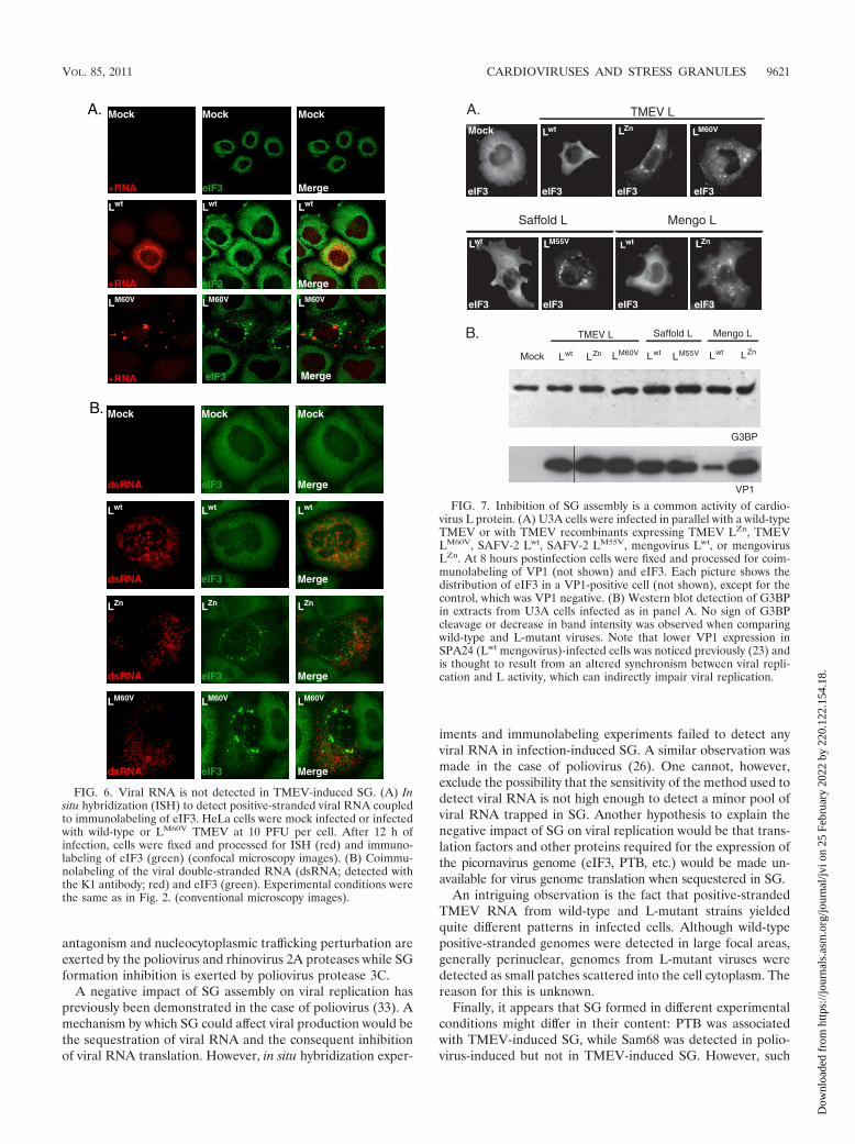
FIG. 6. Viral RNA is not detected in TMEV-induced SG. (A) Insitu hybridization (ISH) to detect positive-stranded viral RNA coupledto immunolabeling of eIF3. HeLa cells were mock infected or infectedwith wild-type or LM60V TMEV at 10 PFU per cell. After 12 h ofinfection, cells were fixed and processed for ISH (red) and immuno-labeling of eIF3 (green) (confocal microscopy images). (B) Coimmu-nolabeling of the viral double-stranded RNA (dsRNA; detected withthe K1 antibody; red) and eIF3 (green). Experimental conditions werethe same as in Fig. 2. (conventional microscopy images).
FIG. 7. Inhibition of SG assembly is a common activity of cardio-virus L protein. (A) U3A cells were infected in parallel with a wild-typeTMEV or with TMEV recombinants expressing TMEV LZn, TMEVLM60V, SAFV-2 Lwt, SAFV-2 LM55V, mengovirus Lwt, or mengovirusLZn. At 8 hours postinfection cells were fixed and processed for coim-munolabeling of VP1 (not shown) and eIF3. Each picture shows thedistribution of eIF3 in a VP1-positive cell (not shown), except for thecontrol, which was VP1 negative. (B) Western blot detection of G3BPin extracts from U3A cells infected as in panel A. No sign of G3BPcleavage or decrease in band intensity was observed when comparingwild-type and L-mutant viruses. Note that lower VP1 expression inSPA24 (Lwt mengovirus)-infected cells was noticed previously (23) andis thought to result from an altered synchronism between viral repli-cation and L activity, which can indirectly impair viral replication.
VOL. 85, 2011 CARDIOVIRUSES AND STRESS GRANULES 9621
Dow
nloa
ded
from
http
s://j
ourn
als.
asm
.org
/jour
nal/j
vi o
n 25
Feb
ruar
y 20
22 b
y 22
0.12
2.15
4.18
.

differences might be more subtle than they appear since im-munofluorescent labeling does not allow comparisons of therelative abundances of different proteins and since detectionthresholds might vary according to the experimental set-up.For instance, conflicting data regarding Sam68 detection inoxidative stress-induced SG have been reported (14, 26).
In conclusion, it comes to light that more and more viruseshave developed strategies to affect the stress granule responsein host cells. However, the impact of SG formation on virusreplication and spread and on cell resistance to viral infectionis still not fully understood and warrants further studies.
ACKNOWLEDGMENTS
F.B. is the recipient of a fellowship from the Belgian FRIA. Thiswork was supported by a DIANE convention of the Walloon region, byARC (communaute francaise de Belgique), and by FRSM (Fondsnational de la recherche Medicale convention 3.4576.08 and credit auxchercheurs).
REFERENCES
1. Agol, V. I., and A. P. Gmyl. 2010. Viral security proteins: counteracting hostdefences. Nat. Rev. Microbiol. 8:867–878.
2. Anderson, P., and N. Kedersha. 2009. RNA granules: post-transcriptionaland epigenetic modulators of gene expression. Nat. Rev. Mol. Cell Biol.10:430–436.
3. Anderson, P., and N. Kedersha. 2009. Stress granules. Curr. Biol. 19:R397–R398.
4. Bardina, M. V., et al. 2009. Mengovirus-induced rearrangement of the nu-clear pore complex: hijacking cellular phosphorylation machinery. J. Virol.83:3150–3161.
5. Brahic, M., J. F. Bureau, and T. Michiels. 2005. The genetics of the persis-tent infection and demyelinating disease caused by Theiler’s virus. Annu.Rev. Microbiol. 59:279–298.
6. Chakraborty, P., N. Satterly, and B. M. Fontoura. 2006. Nuclear exportassays for poly(A) RNAs. Methods 39:363–369.
7. Daniels, J. B., A. M. Pappenheimer, and S. Richardson. 1952. Observationson encephalomyelitis of mice (DA strain). J. Exp. Med. 96:517–530.
8. Delhaye, S., V. van Pesch, and T. Michiels. 2004. The leader protein ofTheiler’s virus interferes with nucleocytoplasmic trafficking of cellular pro-teins. J. Virol. 78:4357–4362.
9. Dever, T. E. 2002. Gene-specific regulation by general translation factors.Cell 108:545–556.
10. Dougherty, J. D., N. Park, K. E. Gustin, and R. E. Lloyd. 2010. Interferencewith cellular gene expression, p. 165–180. In E. Ehrenfeld, E. Domingo, andR. P. Roos (ed.), The picornaviruses. ASM Press, Washington, DC.
11. Dougherty, J. D., J. P. White, and R. E. Lloyd. 2011. Poliovirus-mediateddisruption of cytoplasmic processing bodies. J. Virol. 85:64–75.
12. Fan, J., K. N. Son, S. Y. Arslan, Z. Liang, and H. L. Lipton. 2009. Theiler’smurine encephalomyelitis virus leader protein is the only nonstructural pro-tein tested that induces apoptosis when transfected into mammalian cells.J. Virol. 83:6546–6553.
13. Gilks, N., et al. 2004. Stress granule assembly is mediated by prion-likeaggregation of TIA-1. Mol. Biol. Cell 15:5383–5398.
14. Henao-Mejia, J., and J. J. He. 2009. Sam68 relocalization into stress granulesin response to oxidative stress through complexing with TIA-1. Exp. CellRes. 315:3381–3395.
15. Jnaoui, K., and T. Michiels. 1998. Adaptation of Theiler’s virus to L929 cells:mutations in the putative receptor binding site on the capsid map to neu-tralization sites and modulate viral persistence. Virology 244:397–404.
16. Kong, W. P., G. D. Ghadge, and R. P. Roos. 1994. Involvement of cardiovirusleader in host cell-restricted virus expression. Proc. Natl. Acad. Sci. U. S. A.91:1796–1800.
17. Lidsky, P. V., et al. 2006. Nucleocytoplasmic traffic disorder induced bycardioviruses. J. Virol. 80:2705–2717.
18. Lindquist, M. E., A. W. Lifland, T. J. Utley, P. J. Santangelo, and J. E.Crowe, Jr. 2010. Respiratory syncytial virus induces host RNA stress gran-ules to facilitate viral replication. J. Virol. 84:12274–12284.
19. McKendry, R., et al. 1991. High-frequency mutagenesis of human cells andcharacterization of a mutant unresponsive to both alpha and gamma inter-ferons. Proc. Natl. Acad. Sci. U. S. A. 88:11455–11459.
20. Michiels, T., V. Dejong, R. Rodrigus, and C. Shaw-Jackson. 1997. Protein 2Ais not required for Theiler’s virus replication. J. Virol. 71:9549–9556.
21. Michiels, T., and R. P. Roos. 2010. Theiler’s virus central nervous systeminfection, p. 411–428. In E. Ehrenfeld, E. Domingo, and R. P. Roos (ed.),The picornaviruses. ASM Press, Washington, DC.
22. Oleszak, E. L., J. R. Chang, H. Friedman, C. D. Katsetos, and C. D. Plat-soucas. 2004. Theiler’s virus infection: a model for multiple sclerosis. Clin.Microbiol. Rev. 17:174–207.
23. Paul, S., and T. Michiels. 2006. Cardiovirus leader proteins are functionallyinterchangeable and have evolved to adapt to virus replication fitness.J. Gen. Virol. 87:1237–1246.
24. Pilipenko, E. V., et al. 2000. A cell cycle-dependent protein serves as atemplate-specific translation initiation factor. Genes Dev. 14:2028–2045.
25. Pilipenko, E. V., E. G. Viktorova, S. T. Guest, V. I. Agol, and R. P. Roos.2001. Cell-specific proteins regulate viral RNA translation and virus-induceddisease. EMBO J. 20:6899–6908.
26. Piotrowska, J., et al. 2010. Stable formation of compositionally unique stressgranules in virus-infected cells. J. Virol. 84:3654–3665.
27. Porter, F. W., Y. A. Bochkov, A. J. Albee, C. Wiese, and A. C. Palmenberg.2006. A picornavirus protein interacts with Ran-GTPase and disrupts nucle-ocytoplasmic transport. Proc. Natl. Acad. Sci. U. S. A. 103:12417–12422.
28. Porter, F. W., B. Brown, and A. C. Palmenberg. 2010. Nucleoporin phos-phorylation triggered by the encephalomyocarditis virus leader protein ismediated by mitogen-activated protein kinases. J. Virol. 84:12538–12548.
29. Ricour, C., et al. 2009. Random mutagenesis defines a domain of Theiler’svirus leader protein which is essential for antagonism of nucleocytoplasmictrafficking and of cytokine gene expression. J. Virol. 83:11223–11232.
30. Ricour, C., et al. 2009. Inhibition of mRNA export and dimerization ofinterferon regulatory factor 3 by Theiler’s virus leader protein. J. Gen. Virol.90:177–186.
31. Romanova, L. I., et al. 2009. Antiapoptotic activity of the cardiovirus leaderprotein, a viral “security” protein. J. Virol. 83:7273–7284.
32. van Pesch, V., O. van Eyll, and T. Michiels. 2001. The leader protein ofTheiler’s virus inhibits immediate-early alpha/beta interferon production.J. Virol. 75:7811–7817.
33. White, J. P., A. M. Cardenas, W. E. Marissen, and R. E. Lloyd. 2007.Inhibition of cytoplasmic mRNA stress granule formation by a viral pro-teinase. Cell Host Microbe 2:295–305.
9622 BORGHESE AND MICHIELS J. VIROL.
Dow
nloa
ded
from
http
s://j
ourn
als.
asm
.org
/jour
nal/j
vi o
n 25
Feb
ruar
y 20
22 b
y 22
0.12
2.15
4.18
.