1. Smell Taste Hearing, equilibrium (balance) Vision (separate lecture) 2.
73
1
-
Upload
louisa-wilkins -
Category
Documents
-
view
265 -
download
3
Transcript of 1. Smell Taste Hearing, equilibrium (balance) Vision (separate lecture) 2.

1

Smell Taste Hearing, equilibrium (balance) Vision (separate lecture)
2

Olfactory receptors are CHEMORECEPTORS; a special type of neuron which senses particular chemicals and triggers an action potential.
Chemoreceptors are at the roof of the nasal cavity. There are hundreds of thousands of types, and they can smell a wide variety of substances.
They are extremely sensitive, and can detect parts per billion, as in the scent of natural gas…just a few molecules!
The olfactory nerve goes through the cribiform plate to the OLFACTORY BULB (one of the shortest nerves in the body) and into the limbic system.
3

Scientists who are trying to find a way to make neurons divide to heal nerve injuries often study the body’s only mitotic neurons (undergo mitosis).
These neurons are the olfactory receptors.
4

The Bulgarian man, who was paralyzed after a knife attack in 2010, can walk after doctors in Poland transplanted nerve cells from his nose into his severed spinal cord.
5
The successful operation was the first of its kind for regenerative medicine, and Fidyka is believed to be the first man to walk again after having a completely severed spinal cord.

People who experience imaginary odors have what are called “unicate fits”.
6
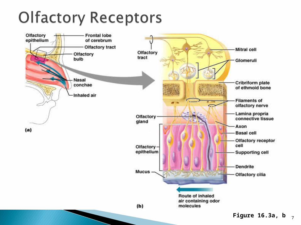
Figure 16.3a, b7

8

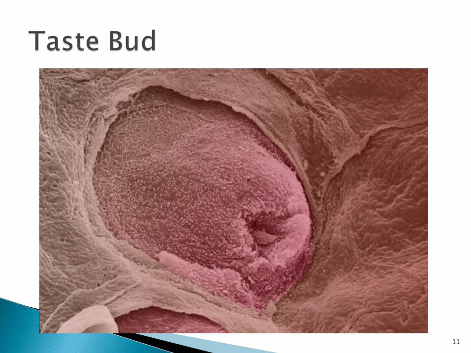
Sensed on taste buds, which are located mostly on the tongue surface, but are also on the palate, pharynx, and a few on the lips.
Taste buds have specialized cells, which increase surface area and have chemoreceptors.
They are surrounded by support cells (like glia). They synapse on sensory neurons, which go to the facial nerve.
Someone with a damaged facial nerve can not easily taste sweet, sour, or salty substances. Taste buds are the only parts of the nervous system that can regenerate completely.
The taste information is sent to the primary gustatory (taste) cortex, located in the parietal lobe of the brain.
9

Figure 16.1a, b10
11

12

How many different tastes are there? Dozens. Salt, sweet, bitter, and sour are only a few.
Where are they located on the tongue? All tastes are located all over the tongue.
The picture in the book was drawn 120 years ago by an anatomist that knew his drawing was not right; he just wanted to use it as a starting point for further experimentation.
13

Taste appreciation is also involved in texture (a mealy apple is not as good), temperature (cold pizza tastes different than warm), and smell (perfume or cigarette smoke clog the senses and decrease taste).
There are dozens of taste receptors, hundreds of thousands of smell receptors, so the subtly of taste is from smell.
14

Foods people like are in opposite proportion to the numbers of taste receptors for that.
People that love sweets have FEWER taste receptors for sweets, so they crave more taste of sweet things.
If you dislike something, it’s because you have lots of receptors for it.
Also, as you get older, you become less tolerant of sweets and more tolerant of bitter tastes (like beer and coffee).
15

The catfish has over 27,000 taste buds. (What could be so tasty on the bottom of a pond?)
Flies taste with their feet.
16

17
Outer Ear Middle Ear Inner Ear
18

1. OUTER EAR consists of the PINNA and the EXTERNAL AUDITORY CANAL.
The pinna is the cartilage of the ear; it acts as a funnel to capture the sound.
If you cup your hands to your ears (do it now), you’ll notice the sound of my voice is louder.
If you rolled up a piece of paper like a funnel and put it to your ear, it functions like the pinna.
The transmission of sound vibrations through the outer ear occurs chiefly through AIR.
19

The Outer (External) Ear
Figure 16.17a20

Ear Crease and Heart Disease• A diagonal earlobe crease is a potential indicator of coronary artery disease.
This crease is called “Frank’s sign”
• Whereas a “normal” earlobe is smooth, an earlobe with a crease has a fold, straight line, or wrinkle that appears to cut the earlobe in half.
• Having an ear crease predicts an 80% chance of coronary artery disease in individuals younger than 40 years. The poor vascularity of this area allows it to show signs of clogged arteries.
21

2. MIDDLE EAR is an AIR filled space with structures.
The TYMPANIC MEMBRANE (ear drum) vibrates in response to sound.
Attached to it are 3 bones: The MALLEUS (hammer), INCUS (anvil), and the STAPES (stirrup) are the smallest bones in the body. Together, they are only one inch long.
Their function is to amplify sound vibrations. The malleus vibrates the incus, which vibrates the stapes.
22

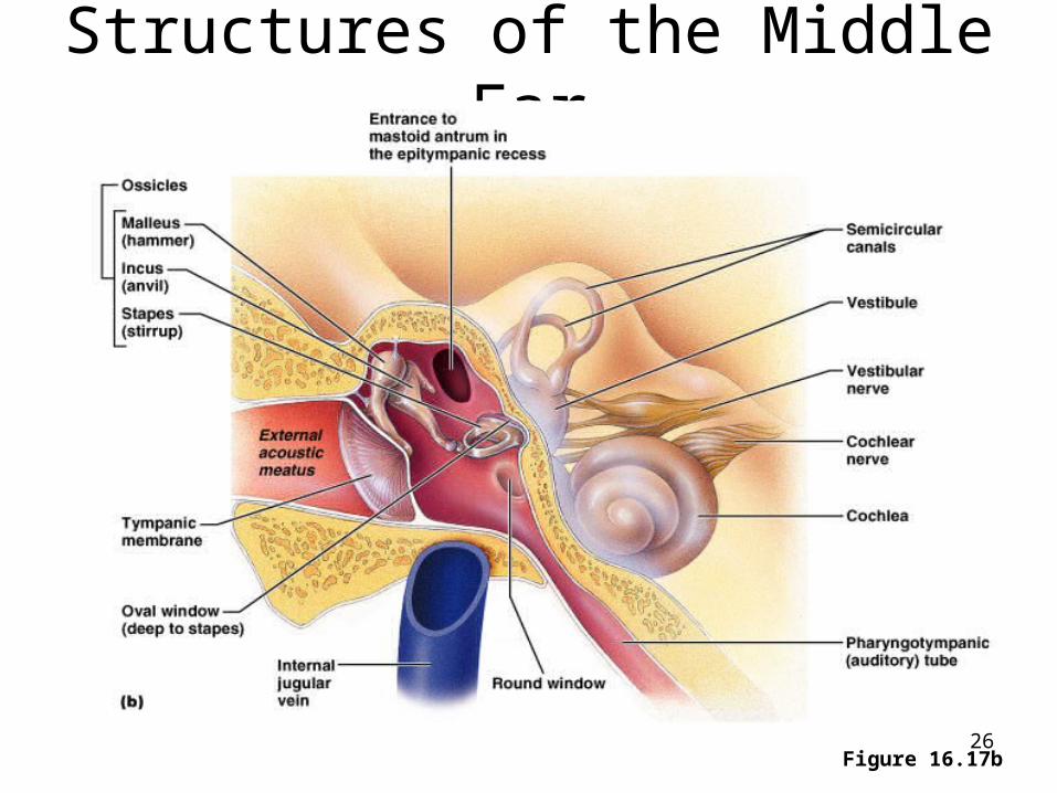
Structures of the Middle Ear
Figure 16.17b23
The middle ear is open to the nasopharynx by way of the AUDITORY TUBE (also called eustachian tube or nasopharyngeal tube), which is only the thickness of a pencil lead.
If this tube is closed, the ears feel plugged up. The function of the auditory tube is to equalize the
pressure of the middle ear and the outside air so the ear bones can vibrate.
Tubes are put in the tympanic membrane to drain fluids in kids with frequent ear infections.
24

25
Doctors issue new guidelines for treating kids' ear infections, http://fxn.ws/YRnUv3
Structures of the Middle Ear
Figure 16.17b26

27
3. INNER EAR exists within the temporal bone (petrous portion).
It is a complex structure. It is located in a bony cavity called the BONY LABYRINTH (“maze”).
The bony labyrinth is filled with a fluid called PERILYMPH, which is similar to CSF. The bony labyrinth is the only place where perilymph is found.
28
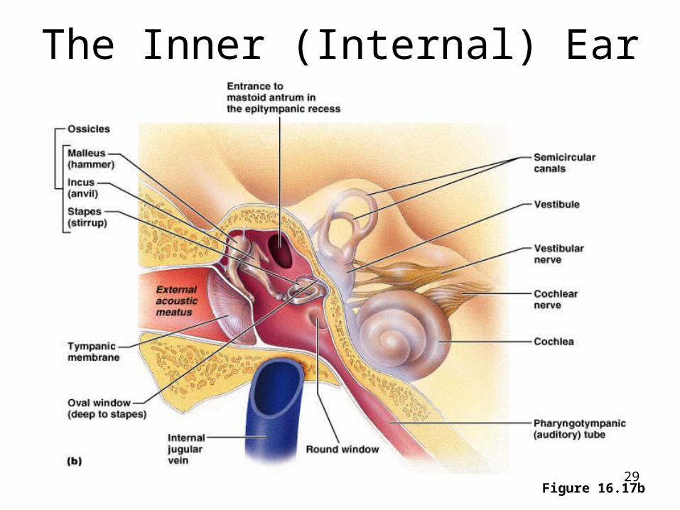
The Inner (Internal) Ear
Figure 16.17b29

Inner Ear• Within the bony labyrinth is a snail-shaped
structure, called the MEMBRANOUS LABYRINTH, which is filled with ENDOLYMPH.
• The snail-shaped structure is divided into two main components. One is the COCHLEA (“snail shell”). This is responsible for hearing.
• The other structure is called the vestibular system. It is responsible for balance and consists of three parts:– Semicircular Canals– Utricle– Saccule
30
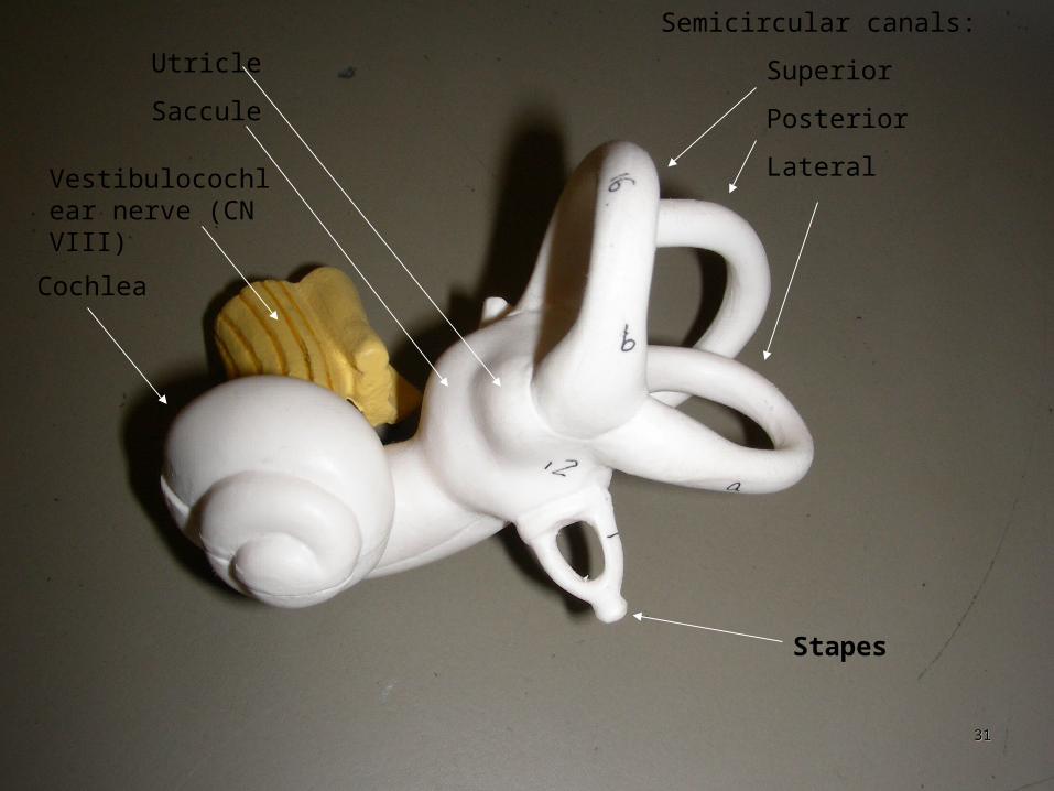
Semicircular canals:
Superior
Posterior
Lateral
Utricle
Saccule
Cochlea
Stapes
Vestibulocochlear nerve (CN VIII)
3131

Inside the cochlea are special neurons called HAIR CELLS; their axons form CN VIII.
The stapes is attached to the OVAL WINDOW, and vibrations cause the endolymph to vibrate; the hair cells here transmit this vibration.
Therefore the HAIR CELLS in this region are receptors for HEARING.
32

COCHLEA
33
34
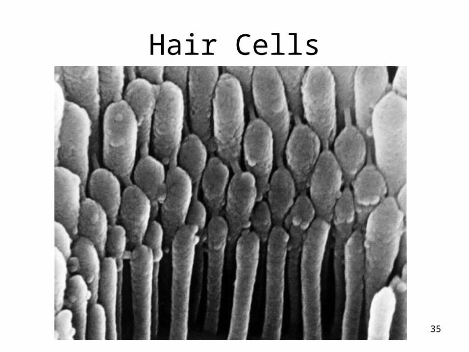
Hair Cells
35
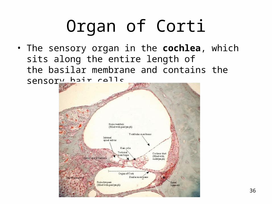
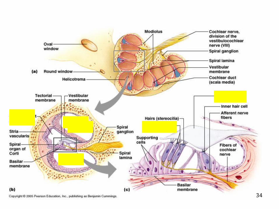
Organ of Corti• The sensory organ in the cochlea, which sits along the
entire length of the basilar membrane and contains the sensory hair cells.
36

• Within the organ of Corti, there is a difference in electrical charge (electrical potential) between inside and outside the hair cells.
37

Organ of Corti
• When the basilar membrane moves in response to sound waves entering the cochlea, the organ of Corti moves with it, and this movement causes inner hair cells in a small area to open tiny transport gates (similar to trapdoors), which allows electrically charged ions to flow through. This causes an action potential to be sent along the auditory nerve to the brain.
• The area from which the action potential arises provides information to the brain as to the pitch of the sound (rather as if the organ of Corti was a piano), and the rate of nerve firing provides information about the loudness of the sound. ORGAN OF CORTI VIDEO
• HOW HEARING WORKS VIDEO
38

Low frequencies (like the longer strings of a piano) cause a response in the tip of the cochlea.
High frequencies cause a response at the larger end of the cochlea.
39

The axons of the hair cells form CN VIII, the VESTIBULOCOCHLEAR NERVE, which takes the signals to the brain.
Therefore, the cochlea is where the hearing receptors are located, so the cochlea is responsible for all of the hearing of sounds.
However, the ear does more than just hear; it is also responsible for balance and equilibrium.
40

This system regulates balance. It is also within the inner ear. SEMI-CIRCULAR CANALS (Three of them,
all in different planes) determine your head’s movement in three planes.
Within each semi-circular canal is endolymph and hair cells, whose axons go to the cerebellum.
41

When you move in one direction, like sliding across the room, the fluid sloshes like a cup of coffee, and it triggers the hair cells.
42

Attached to the semi-circular canals are two joined structures called the UTRICLE and the SACCULE.
These also contain HAIR CELLS and ENDOLYMPH. Within the endolymph here are OTOLITHS (“ear rocks”)
which are calcium deposits. When you stand perfectly upright, these otoliths float
above the hair cells, but if you tip your head to the side, they will land on and stimulate the hairs on that side.
That tells you what position your head is in and gives you a sense of equilibrium.
Therefore, the HAIR CELLS in this region are receptors for equilibrium and the OTOLITHS are an essential component of this process.
43

Anatomy and Function of the Otoliths
Figure 16.21b44

Inflammation of the semi-circular canals give you a sense of motion when you’re not moving. The symptom is called VERTIGO (dizziness). The diagnosis is LABYRINTHITIS.
This can be debilitating. Sometimes only one canal is affected, so you
only get dizzy if you turn your head one way. It can be caused by sinus infections, excess
salt consumption, viral infections, stress. It is more common in smokers than non-smoking.
45

Benign paroxysmal positional vertigo (BPPV) The Epley maneuver helps relieve the
dizziness and spinning symptoms by moving the calcium particles out of the sensitive semicircular canals and into another inner chamber of the ear, where they don’t cause symptoms.
The exercise simply involves lying down and rolling your head in such way so that the particles fall out.
Epley Maneuver 1 http://www.youtube.com/watch?v=ZqokxZRbJfw
Epley Maneuver 2 http://www.youtube.com/watch?v=pa6t-Bpg494
46

House Ear Clinic◦ Dr. Luxford ◦ Chapman Ave, Orange
◦ARTIFICIAL EARS VIDEO
47

When your eyes get one set of information that conflicts with the vestibular structures, such as when you are high up in the air or strobe lights flashing, or reading in a car.
Whether the vertigo is from visual or vestibular disturbances, your body interprets the signals as a poison invasion, so it initiates a vomit reflex.
48

“Cauliflower Ear”Hematoma auris or Traumatic auricular hematoma
Common in boxers and wrestlers
A blood clot or other fluid collects under the perichondrium. This separates the cartilage from the overlying perichondrium that is its source of nutrients, causing the cartilage to die.
This leads to a formation of fibrous tissue in the overlying skin.
49

Conductive hearing loss happens when there is a problem conducting sound waves through the outer ear, tympanic membrane (eardrum) or middle ear (ossicles). It may be caused from excess wax, damaged eardrum, or arthritis of the ossicles.
Hearing loss from nerve damage (sensorineural) is a problem in the vestibulocochlear nerve (Cranial nerve VIII), the inner ear, or central processing centers of the brain.
50

Weber Test: only tests unilateral problems. A tuning fork is touched to the middle of the forehead:◦ Conductive hearing loss: sound is heard louder
in the problem ear (earwax, etc) because reflected soundwaves cannot escape the ear canal, so they penetrate deeper into the inner ear.
◦ Nerve damage: sound is heard louder in the normal ear because the damage is to the nerve, so bone conduction of the sound is ineffective.
51

Performed by placing a vibrating tuning fork on the mastoid process until sound is no longer heard, the fork is then immediately placed just outside the ear. Normally, the sound is audible at the ear, indicating a positive Rinne test.
If they cannot hear the sound at the ear, it is a negative Rinne test, and indicates Sensorineural hearing loss
VIDEO
52

Do NOT hold your nose and blow. That can rupture the tympanic membrane!
Try chewing gum or swallowing. You could hold your nose and swallow…
that is gentler than blowing.
53

Hearing Loss
• Hearing aids can help with partial hearing loss such as that caused by chronic noise exposure, but they do so by amplifying sound waves rather than restoring lost hearing. Most amplify all sound waves equally, which is why it may be difficult for someone wearing a hearing aid to carry on a conversation in a noisy restaurant.
• Some of the newer and more expensive hearing aids are programmable, so the level of amplification can be adjusted depending on the noise level of one's surroundings. Through the use of cochlear implants, there is hope today for people who have little or no hearing due to cochlear damage.
• A person with a cochlear implant wears a microphone picks up sounds and sends them to a processor, which relays signals to a small receiver implanted beneath the scalp. The receiver transmits signals through a number of electrodes to the origin of the cochlear nerve. Cochlear implants are not yet capable of producing all of these sounds of normal hearing, but they do allow people who have them to understand speech and perceive sounds such as alarms and telephones.
54

A cochlear implant is a small, complex electronic device that can help to provide a sense of sound to a person who is profoundly deaf or severely hard-of-hearing. The implant consists of an external portion that sits behind the ear and a second portion that is surgically placed under the skin. An implant has the following parts:
A microphone, which picks up sound from the environment. A speech processor, which selects and arranges sounds picked up
by the microphone. A transmitter and receiver/stimulator, which receive signals from
the speech processor and convert them into electric impulses. An electrode array, which is a group of electrodes that collects the
impulses from the stimulator and sends them to different regions of the auditory nerve.
An implant does not restore normal hearing. Instead, it can give a deaf person a useful representation of sounds in the environment and help him or her to understand speech.
Cochlear Implant http://www.youtube.com/watch?v=SmNpP2fr57A
55

56

Hearing Loss • Otitis media is an infection of the middle ear that can lead to
conduction deafness. It typically occurs when an upper respiratory infection, such as a cold, leads to swelling of the auditory tibe, creating a vacuum that pulls fluid into the middle ear. The fluid provides an ideal environment for bacteria or viruses, and the pressure it exerts causes symptoms such as pain, ringing in the ears, and hearing loss. The loss is usually incomplete, and rarely permanent. However, when such hearing loss occurs in very young children, it can delay their speech development. If the tympanic membrane ruptures under pressure, it usually heals on its own in a matter of weeks. If it does not rupture, fluid may linger in the middle ear, and in some cases a tube must be surgically inserted for drainage.
57

Hearing Loss • Noise exposure is a common and usually preventable cause of
nerve deafness. Noise volume is measured in units called decibels. Any noise above a level of 80 dB could result in damage to the hair cells of the ear. Eventually, the hair cells disappear completely. If listening to city traffic for extended periods can damage hearing, it stands to reason that frequent attendance at rock concerts, constantly playing a stereo loudly, or using earphones at high volume is also damaging to hearing. The first sign of danger could be temporary hearing loss, a feeling of fullness in the ears, muffled hearing, or ringing in the ears. If you have any of these symptoms, modify your listening habits immediately to prevent further damage. If exposure to noise is unavoidable, specially designed noise reduction earmuffs are available, and it is also possible to purchase earplugs made from a compressible, spongelike material at the drugstore or sporting goods store. These earplugs are not the same as those worn for swimming, and they should not be used interchangeably.
58

Hearing Loss
• Aside from loud music, noisy indoor or outdoor equipment, such as a rug cleaning machine or a chainsaw, is also troublesome. Even motorcycles and recreational vehicles such as snowmobiles can contribute to gradual loss of hearing.
• Exposure to intense sounds of short duration, such as a burst of gunfire, can result in an immediate hearing loss. Hunters may have a significant hearing reduction in the ear opposite of the shoulder where the gun is carried. The butt of the rifle offers some protection to the ear nearest the gun when it is shot.
59
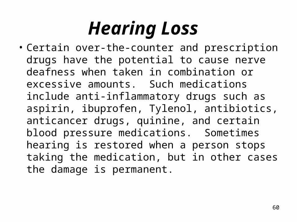
Hearing Loss • Certain over-the-counter and prescription
drugs have the potential to cause nerve deafness when taken in combination or excessive amounts. Such medications include anti-inflammatory drugs such as aspirin, ibuprofen, Tylenol, antibiotics, anticancer drugs, quinine, and certain blood pressure medications. Sometimes hearing is restored when a person stops taking the medication, but in other cases the damage is permanent.
60

Hearing Loss • One uncommon cause of deafness is
autoimmune inner ear disease. This occurs when a person's immune system turns against normal, healthy tissue.
• The person experiences symptoms such as dizziness and ringing in the ears in addition to hearing loss. Although it can be treated with drugs that suppress the immune system, the drugs themselves can have serious side effects, and may only slow the disease.
61

Excess noise Frequent sinus infections (or allergies) Medicines
◦ Excess Tylenol or aspirin, antibiotics, sedatives, antidepressants
Lack of blood flow (anemia, hypertension, diabetes, age) Drugs (marijuana, caffeine) Foods (soy, wheat, chocolate, red wine)
Symptoms of nerve damage:◦ Tinnitus: ringing in the ears◦ If damage is not severe, axons can regenerate and tinnitus will
go away◦ http://health.learninginfo.org/tinnitus.htm
62

When a person dies, hearing is the last sense to go. The first sense lost is sight.
The roar that we hear when we place a seashell next to our ear is not the ocean, but rather the sound of blood surging through the veins in the ear.
63

Deaf Baby with Hearing Aid: https://www.youtube.com/watch?v=UUP02yTKWWo
64
The rest of this lecture is not test material
65

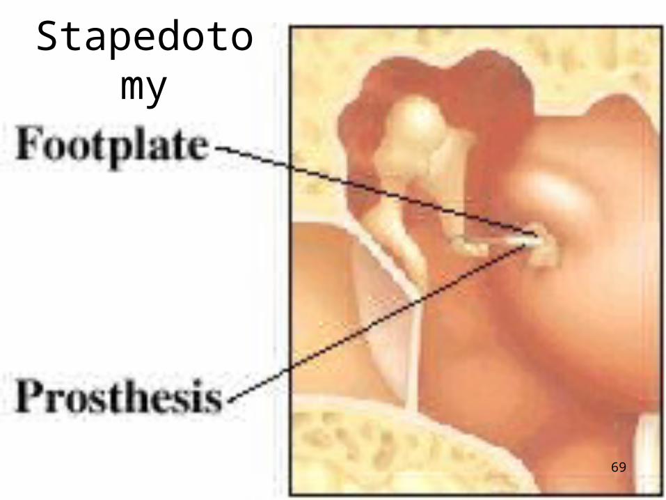
The stapes becomes fixed, cannot move, and dampens sound conduction.
Stapedotomy: A portion of the stapes is removed and replaced with a titanium-nickel prosthesis.
66

Stapedotomy
67

Stapedotomy
Prosthesis
68
Stapedotomy
69

Hearing damage from headphones is more common than from loudspeakers, because people listen at higher volumes.
Even at comparable volumes, hearing damage from headphones is higher than with loudspeakers, due to the close coupling of the transducers to the ears.
70

How long can you listen at certain volumes without damage?
90 dbA 8 hrs
92 dbA 6 hrs
95 dbA 4 hrs
97 dbA 3 hrs
100 dbA 2 hrs
102 dbA 1.5 hrs
105 dbA 1 hr
110 dbA 0.5 hr
115 dbA 0.25 hr or less
71

60 dB Everyday conversation, ringing telephone.
70 dB Restaurant.
80 dBHeavy city traffic, alarm clock at 2 feet, factory noise, vacuum cleaner, garbage disposal.
90 dBSubway trains, motorcycle, workshop tools, lawn mower. 8 hrs
100 dB Chain saw, pneumatic drill. 2 hrs
110 dB Dance club. 30 minutes
120 dBRock concert speaker sound, sandblasting, thunderclap. 15 minutes or less
130 dB Jet take off.
140 dB gunfire Nerve damage occurs immediately
150 dB rock music peak
72

73