The process by which our sensory systems receive stimuli from our environment.
Upload
jonas-leonardCategory
view
239download
1
1
Senses• Sense:
ability to perceive stimuli
• Sensation:conscious awareness of stimuli received by sensory neurons
• Sensory receptors:sensory nerve endings that respond to stimuli by developing action potentials

Types of Senses• General senses:
- receptors over large part of body
- somatic provide info. about body and env’t
- visceral provide info. about internal organs,
pain, pressure
- touch, pressure, pain, temp., and itch
• Special senses:
smell, taste, sight, hearing, and balance

3
Types of Receptors• Mechanoreceptors:
- detect movement - Ex. touch, pressure, vibration
• Chemoreceptors:- detect chemicals - Ex. Odors
• Photoreceptors:detect light

4
• Thermoreceptors:
detect temp. changes
• Nociceptors:
detect pain
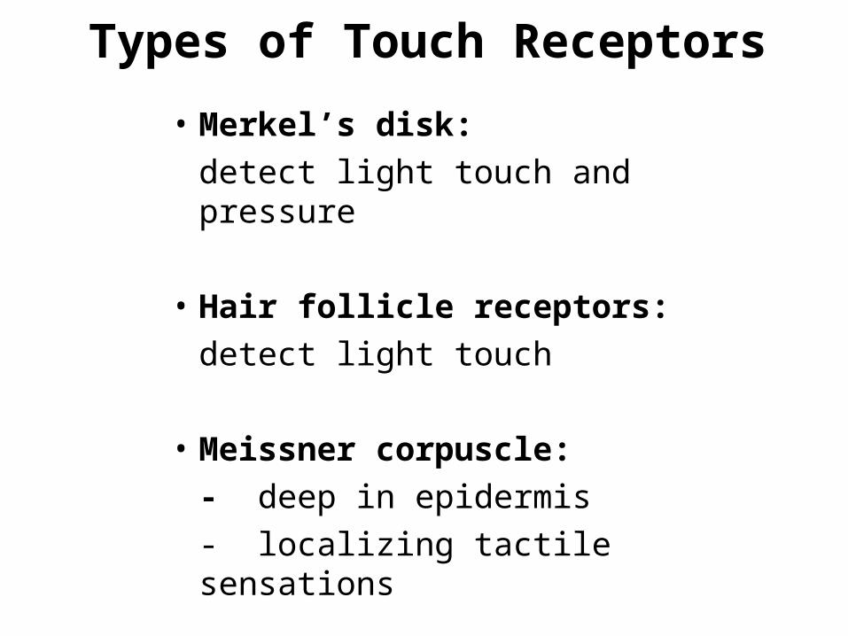
Types of Touch Receptors
• Merkel’s disk:
detect light touch and pressure
• Hair follicle receptors:
detect light touch
• Meissner corpuscle:
- deep in epidermis
- localizing tactile sensations

• Ruffini corpuscle:
- deep tactile receptors
- detects continuous pressure in skin
• Pacinian corpuscle:
- deepest receptors
- associated with tendons and joints
- detect deep pressure, vibration, position
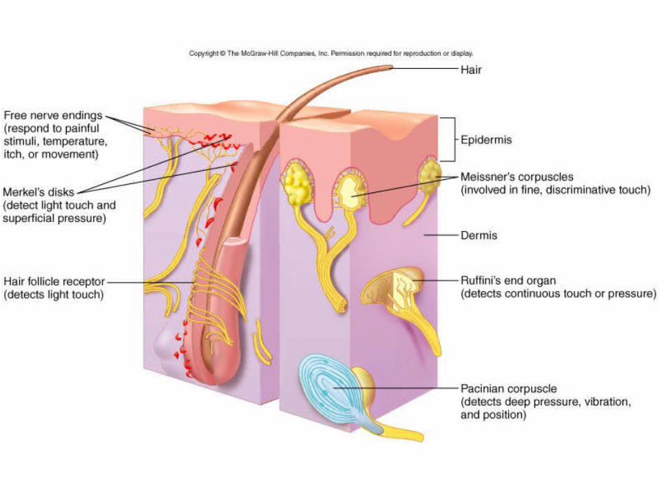
Figure 9.1

Pain
• What is it?
unpleasant perceptual and emotional
experience
8
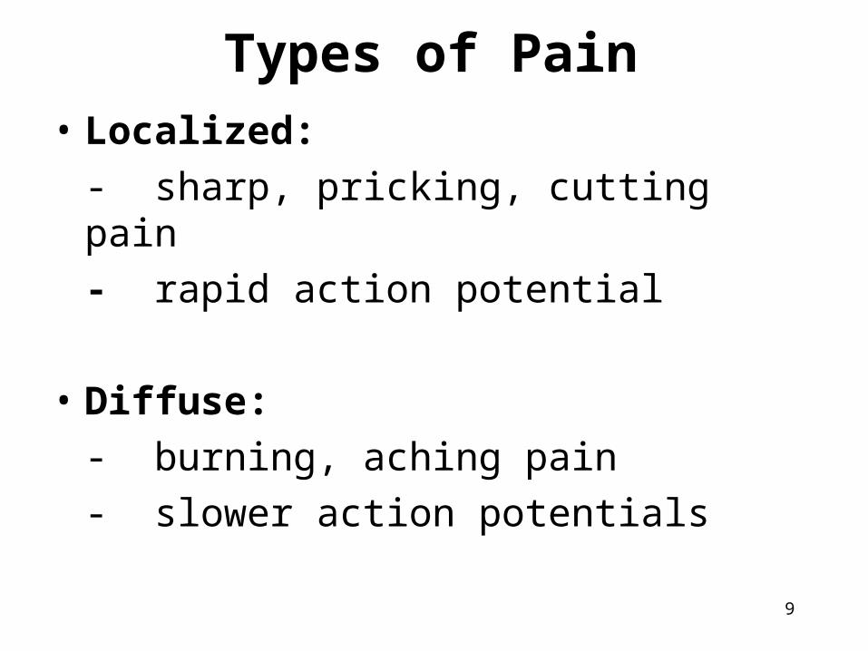
Types of Pain• Localized:
- sharp, pricking, cutting pain
- rapid action potential
• Diffuse:
- burning, aching pain
- slower action potentials
9

Pain Control• Local anesthesia:
- action potentials suppressed from pain
receptors in local areas
- chemicals are injected near sensory nerve
• General anesthesia:
- loss of consciousness
- chemicals affect reticular formation
10
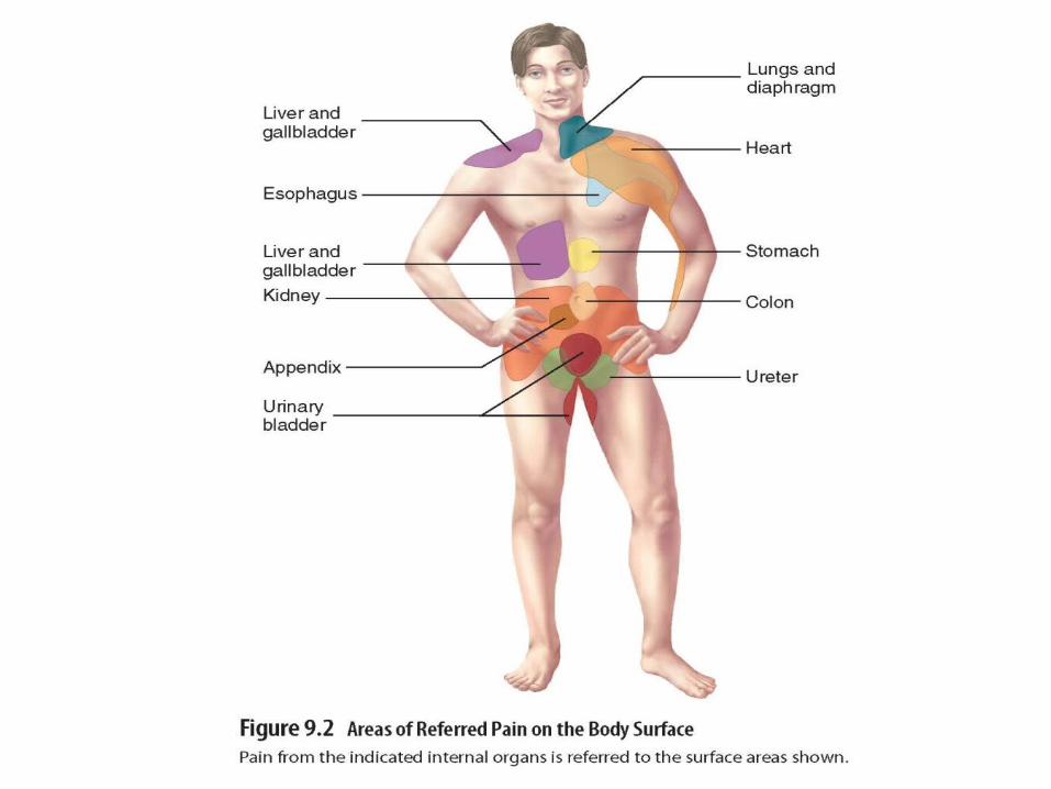
Referred Pain• What is it?
- originates in a region that is not source of pain
stimulus
- felt when internal organs are damaged or
inflamed
- sensory neurons from superficial area and
neurons of source pain converge onto same
ascending neurons of spinal cord

13
Olfaction• What is it?
- sense of smell
- occurs in response to
odorants
- receptors are located
in nasal cavity and
hard palate
- we can detected
10,000 different smells
14
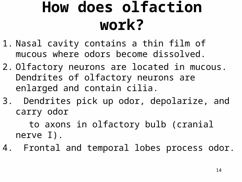
How does olfaction work?
1. Nasal cavity contains a thin film of mucous where odors become dissolved.
2. Olfactory neurons are located in mucous. Dendrites of olfactory neurons are enlarged and contain cilia.
3. Dendrites pick up odor, depolarize, and carry odor
to axons in olfactory bulb (cranial nerve I).
4. Frontal and temporal lobes process odor.

Figure 9.3b

16
Taste• Taste buds:
- sensory structures that detect taste
- located on papillae on tongue, hard palate,
throat
• Inside each taste bud are 40 taste cells
• Each taste cell has taste hairs that extend into taste pores

Figure 9.4

18
How does taste work?
1. Taste buds pick up taste and send it to taste cells.
2. Taste cells send taste to taste hairs.
3. Taste hairs contain receptors that initiate an action potential which is carried to parietal lobe.
4. Brain processes taste.

19
Types of Tastes• Sweet• Sour• Salty• Bitter• Umami
• Certain taste buds are more sensitive to certain tastes.
• Taste is also linked to smell.
20
VisionAccessory Structures
• Eyebrow:
- protects from sweat
- shade from sun
• Eyelid/Eyelashes:
- protects from foreign objects
- lubricates by blinking


22
• Conjunctiva:
thin membrane that covers inner surface of
eyelid
• Lacrimal apparatus:
produces tears
• Extrinsic eye muscles:
help move eyeball

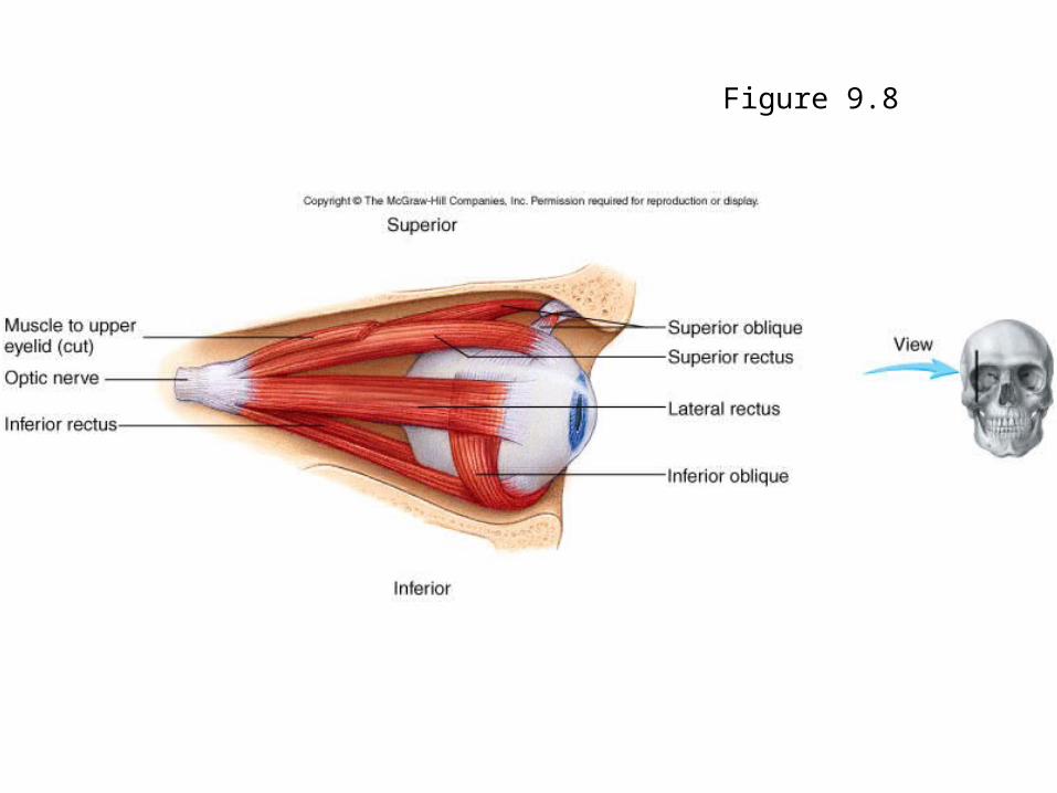
Figure 9.8

26
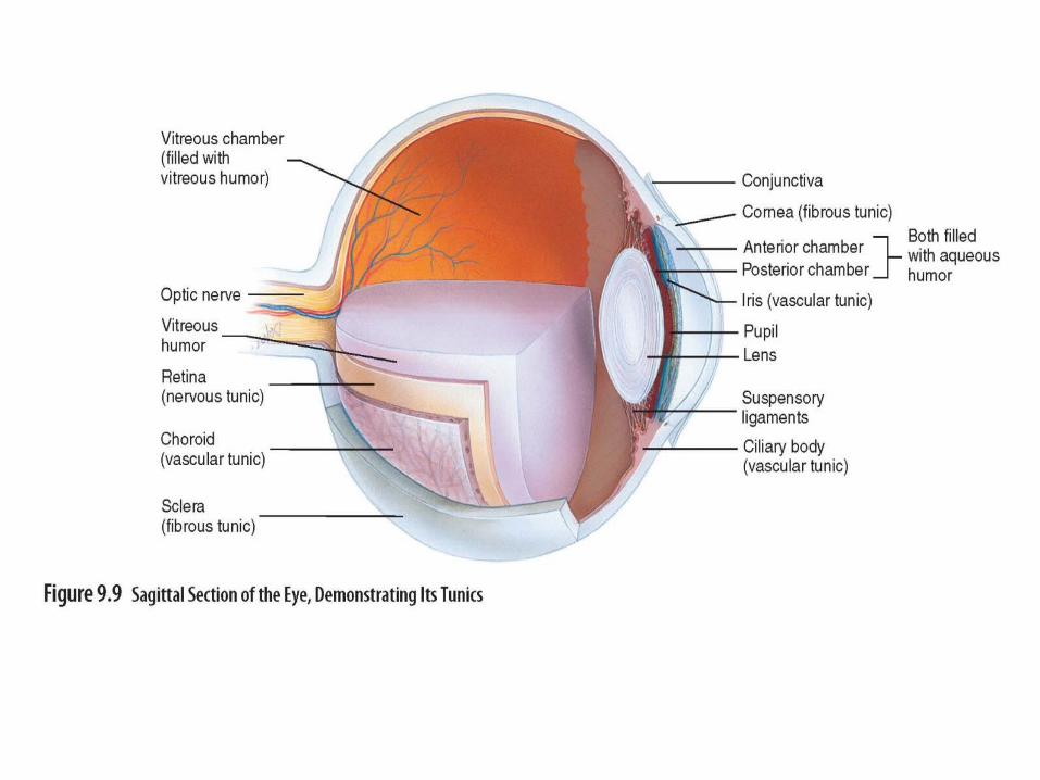
Anatomy of Eye
• Hollow, fluid filled sphere
• Composed of 3 layers (tunics)
• Divided into chambers

Fibrous Tunic
• Outermost layer
• Sclera:
- firm, white outer part
- helps maintain eye shape, provides attachment
sites, protects internal structures
• Cornea:- transparent structure that covers iris and pupil
- allows light to enter and focuses light

28
Vascular Tunic• Middle layer
• Contains blood supply
• Choroid:
- black part (melanin)
- delivers O2 and nutrients to retina
• Ciliary body:
helps hold lens in place
• Suspensory ligaments:
help hold lens in place

29
• Lens:
- flexible disk
- focuses light onto retina
• Iris:
- colored part
- surrounds and regulates pupil
• Pupil:
- regulates amount of light entering
- lots of light = constricted
- little light = dilated


Figure 9.10
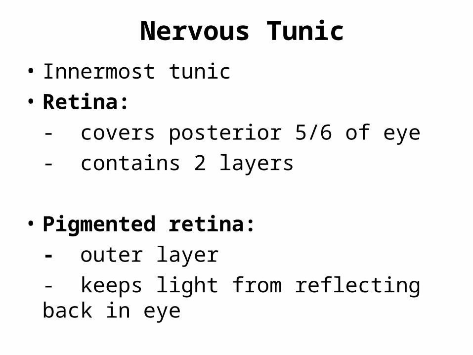
Nervous Tunic
• Innermost tunic
• Retina:
- covers posterior 5/6 of eye
- contains 2 layers
• Pigmented retina:
- outer layer
- keeps light from reflecting back in eye
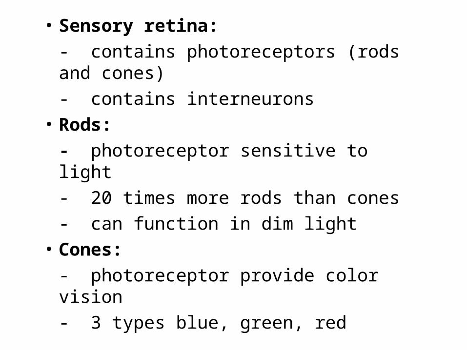
• Sensory retina:
- contains photoreceptors (rods and cones)
- contains interneurons
• Rods:
- photoreceptor sensitive to light
- 20 times more rods than cones
- can function in dim light
• Cones:
- photoreceptor provide color vision
- 3 types blue, green, red

Figure 9.12b

Figure 9.12c
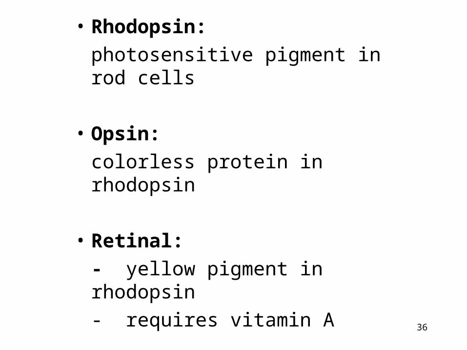
• Rhodopsin:
photosensitive pigment in rod cells
• Opsin:
colorless protein in rhodopsin
• Retinal:
- yellow pigment in rhodopsin
- requires vitamin A
36

Effects of Light on Rhodopsin1. Light strikes rod cell
2. Retinal changes shape
3. Opsin changes shape
4. Retinal dissociates from opsin
5. Change rhodopsin shape stimulates response in rod cell which results in vision
6. Retinal detaches from opsin
7. ATP required to reattach retinal to opsin and return rhodopsin to original shape

Figure 9.13

Retina Structures Continued• Rods and cones synapse with bipolar cells of
sensory retina
• Horizontal cells of retina modify output of rods and cones
• Bipolar and horizontal cells synapse with ganglion cells
• Ganglion cells axons’ converge to form optic nerve

40
Nervous Tunic (Retina)
• Innermost layer
• 2 parts of retina: sensory and pigmented
• Keeps light from reflecting back into eye
• Rods:
photoreceptors that detect amount light
• Cones:
- photoreceptors that detect colors
- 3 types: red, blue, green
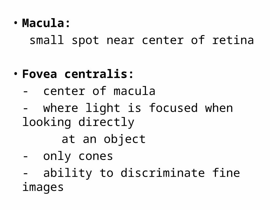
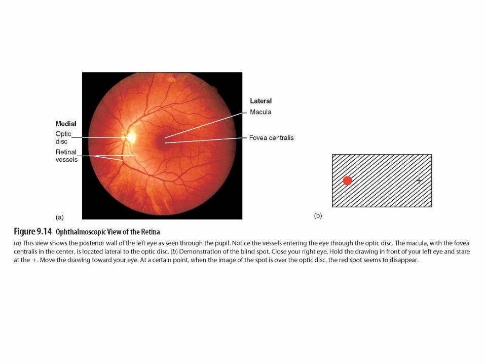
• Macula:
small spot near center of retina
• Fovea centralis:
- center of macula
- where light is focused when looking directly
at an object
- only cones
- ability to discriminate fine images

• Optic disk:
- white spot medial to macula
- blood vessels enter eye and spread over retina
- axons exit as optic nerve
- no photoreceptors
- called blindspot
44
Chambers of Eye• Anterior chamber:
- located between cornea and lens
- filled with aqueous humor (watery)
- aqueous humor helps maintain pressure, refracts
light, and provide nutrients to inner surface of
eye
• Posterior chamber:
- located behind anterior chamber
- contains aqueous humor

45
• Vitreous chamber:
- located in retina region
- filled with vitreous humor: jelly-like substance
- vitreous humor helps maintain pressure, holds
lens and retina in place, refracts light

Functions of Eye
Light Refraction
Bending of light
• Focal point:
- point where light rays converge
- occurs anterior to retina
- object is inverted

Focusing Images on Retina
• Accommodation:
- lens becomes less rounded and image can be
focused on retina
- enables eye to focus on images closer than
20 feet


Neuronal Pathway for Vision
• Optic nerve:
leaves eye and exits orbit through optic foramen to enter cranial cavity
• Optic chiasm:
where 2 optic nerves connect
• Optic tracts:
route of ganglion axons

Figure 9.16b

51
Eye Defects• Myopia:
- nearsightedness
- image is in front of retina
• Hyperopia:
- farsightedness
- image is behind retina
• Presbyopia:
- lens becomes less elastic
- reading glasses required

Clinical Focus 9A

53
• Astigmatism:
- irregular curvature of lens
- glasses or contacts required to correct
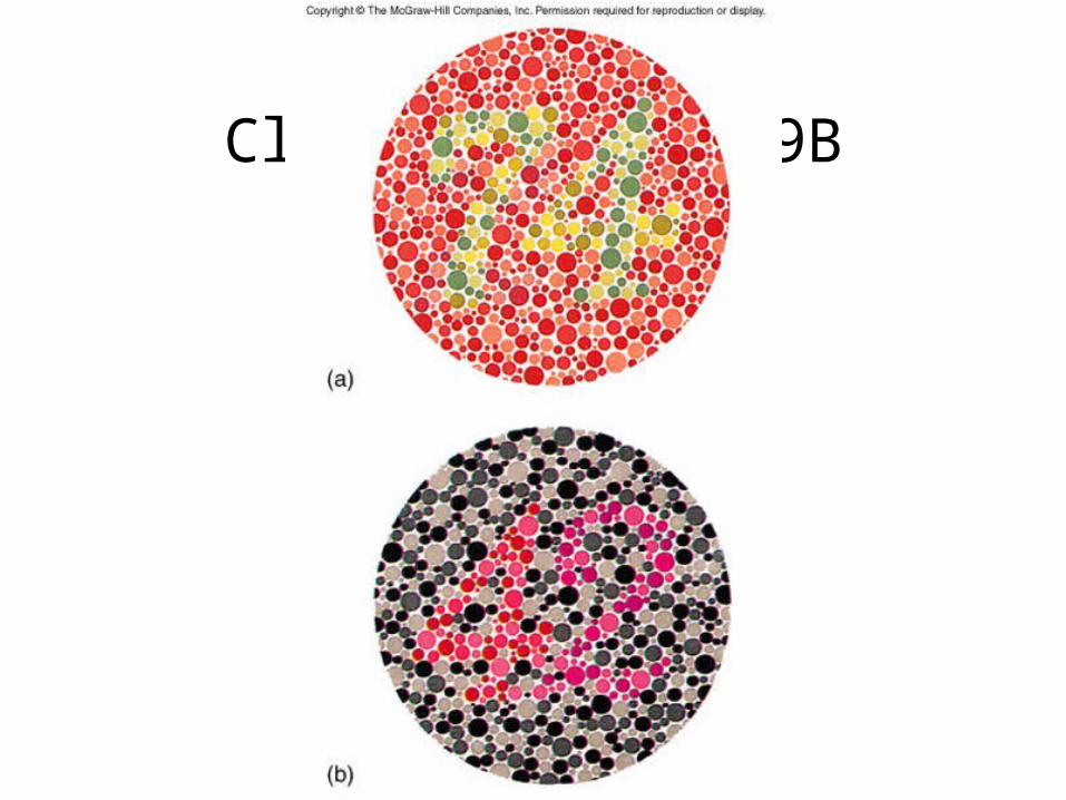
• Colorblindness:
- absence or deficient cones
- primarily in males
• Glaucoma:
- decreased pressure in eye
- can lead to blindness
Clinical Focus 9B
55
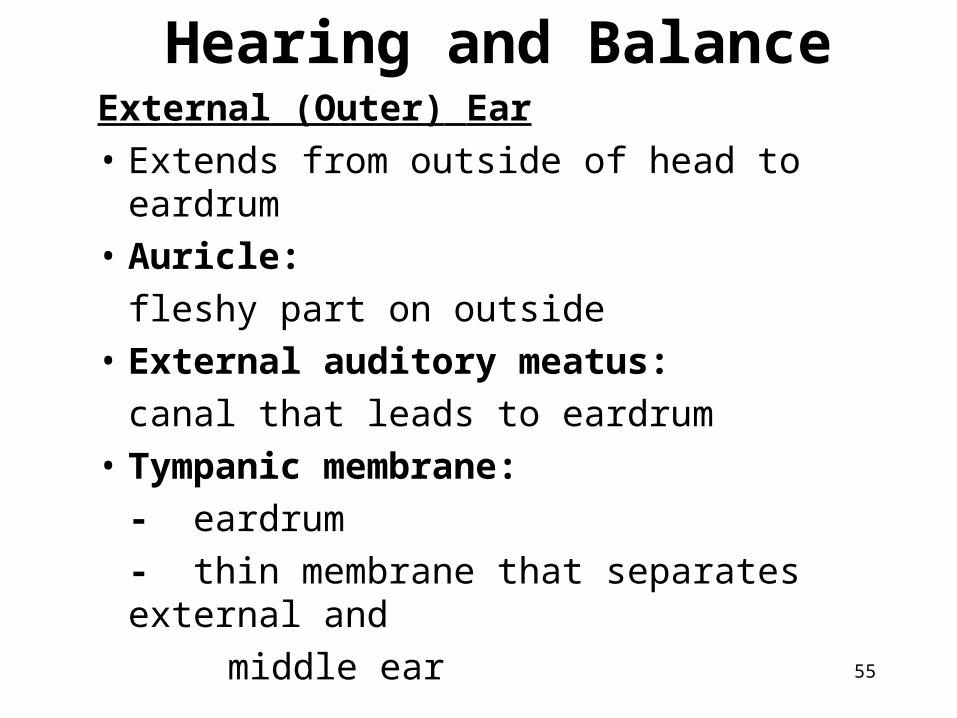
Hearing and BalanceExternal (Outer) Ear
• Extends from outside of head to eardrum
• Auricle:
fleshy part on outside
• External auditory meatus:
canal that leads to eardrum
• Tympanic membrane:
- eardrum
- thin membrane that separates external and
middle ear

56
Middle Ear
• Air filled chamber
• Malleus (hammer):
bone attached to tympanic membrane
• Incus (anvil):
bone that connects malleus to stapes
• Stapes (stirrup):
bone located at base of oval window
57
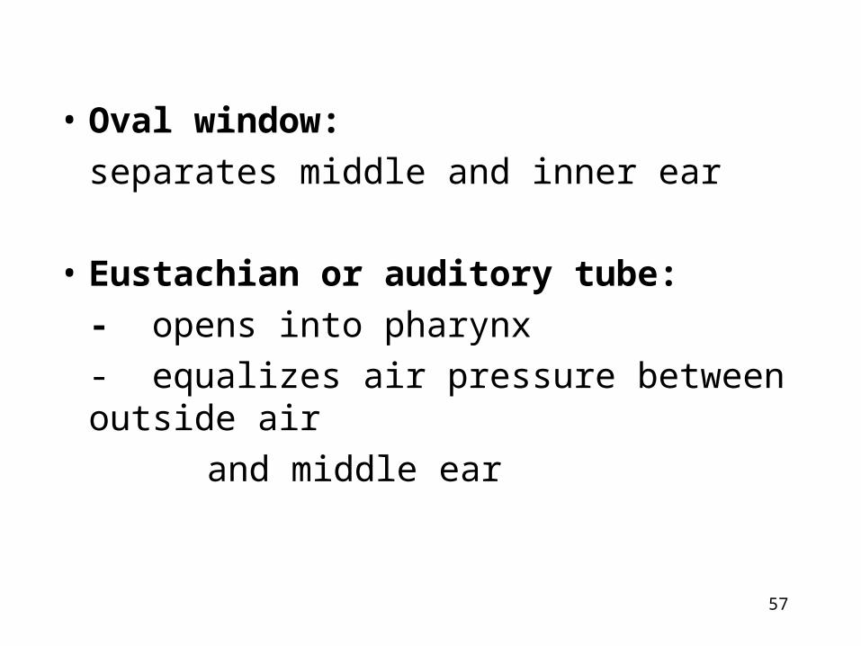
• Oval window:
separates middle and inner ear
• Eustachian or auditory tube:
- opens into pharynx
- equalizes air pressure between outside air
and middle ear

58
59
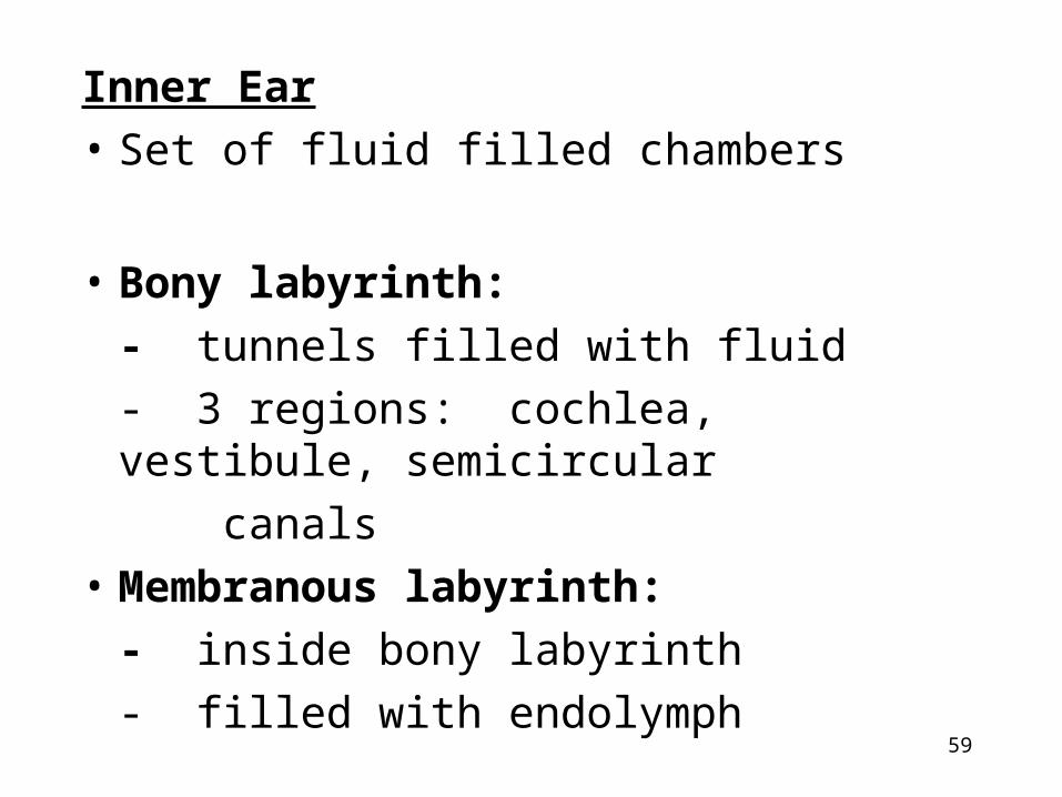
Inner Ear
• Set of fluid filled chambers
• Bony labyrinth:
- tunnels filled with fluid
- 3 regions: cochlea, vestibule, semicircular
canals
• Membranous labyrinth:
- inside bony labyrinth
- filled with endolymph

• Endolymph:
clear fluid in membranous labyrinth
• Perilymph:
fluid between membranous and bony labyrinth
• Cochlea:
- snail-shell shaped structure
- where hearing takes place
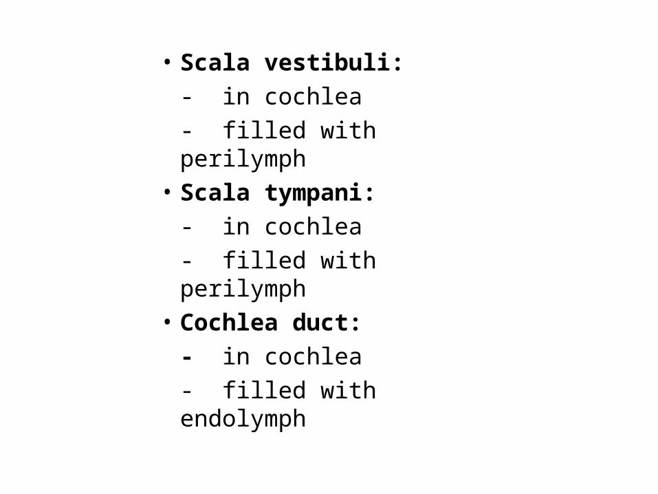
• Scala vestibuli:
- in cochlea
- filled with perilymph
• Scala tympani:
- in cochlea
- filled with perilymph
• Cochlea duct:
- in cochlea
- filled with endolymph
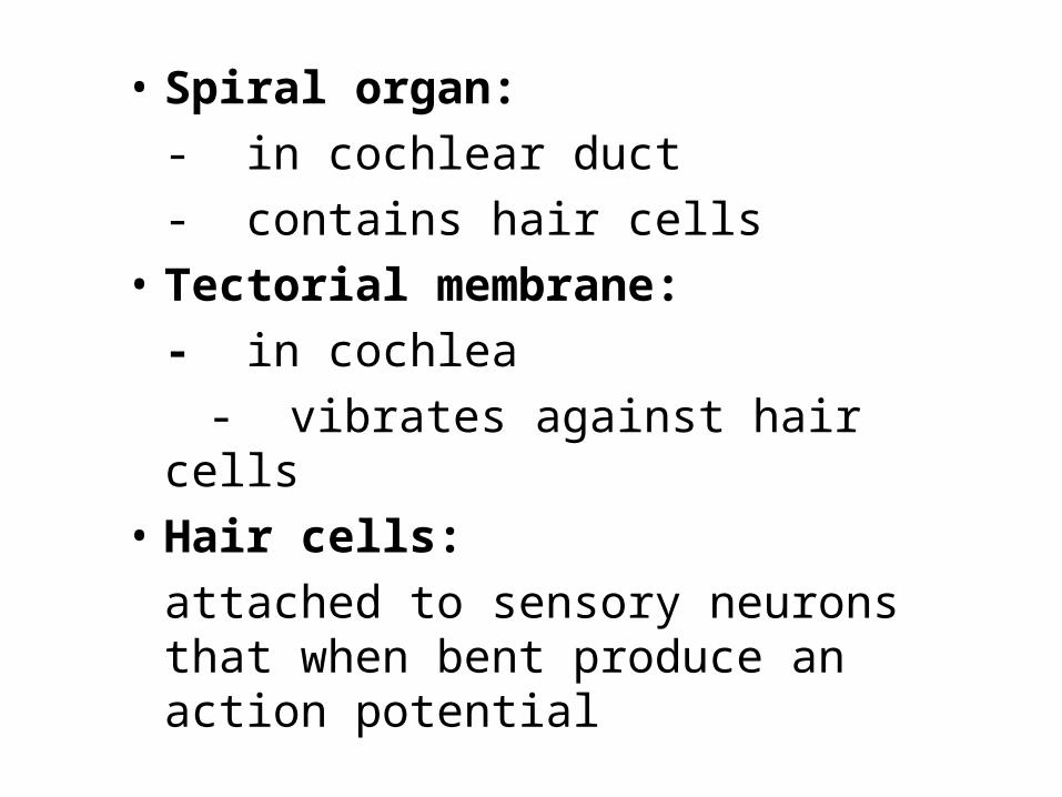
• Spiral organ:
- in cochlear duct
- contains hair cells
• Tectorial membrane:
- in cochlea
- vibrates against hair cells
• Hair cells:
attached to sensory neurons that when bent produce an action potential

• Vestibular membrane:
wall of membranous labyrinth that lines scala vestibuli
• Basilar membrane:
wall of membranous labyrinth that lines scala tympani

65
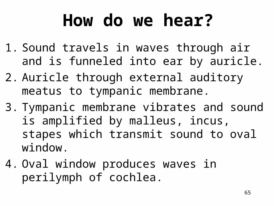
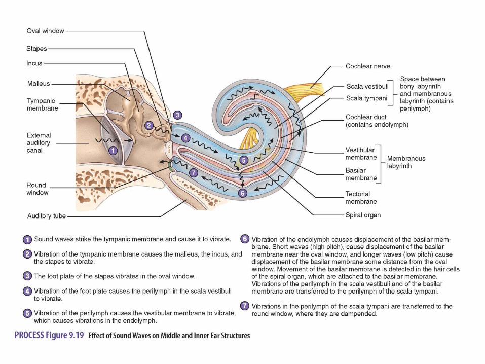
How do we hear?
1. Sound travels in waves through air and is funneled into ear by auricle.
2. Auricle through external auditory meatus to tympanic membrane.
3. Tympanic membrane vibrates and sound is amplified by malleus, incus, stapes which transmit sound to oval window.
4. Oval window produces waves in perilymph of cochlea.

66
5. Vibrations of perilymph cause vestibular membrane and endolymph to vibrate.
6. Endolymph cause displacement of basilar membrane.
7. Movement of basilar membrane is detected by hair hairs in spiral organ.
8. Hair cells become bent and cause action potential is created.

Balance (Equilibrium)
• Static equilibrium:
- associated with vestibule
- evaluates position of head relative to gravity
• Dynamic equilibrium:
- associated with semicircular canals
- evaluates changes in direction and rate of head
movement

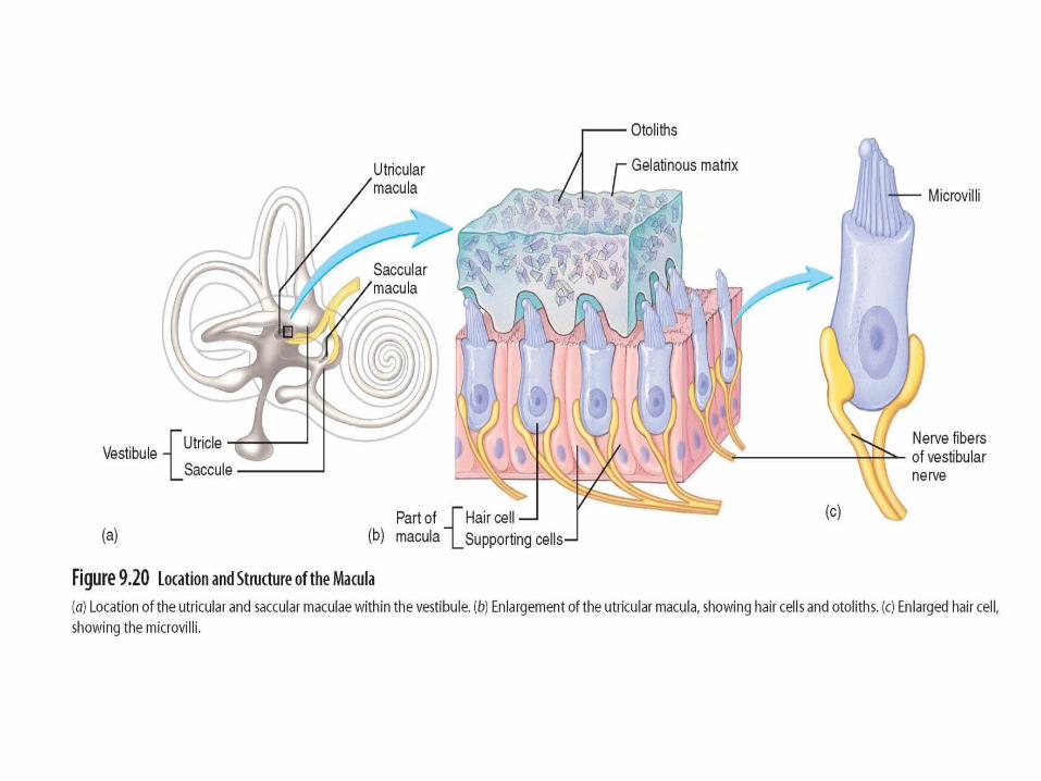
• Vestibule:
- inner ear
- contains utricle and saccule• Maculae:
- specialized patches of epithelium in utricle
and saccule surround by endolymph
- contain hair cells• Otoliths:
- gelatinous substance that moves in response
to gravity
- attached to hair cell microvilli which initiate action
potentials


72
• Semicircular canals:- dynamic equil.- sense movement if any direction
• Ampulla:base of semicircular canal
• Crista ampullaris:in ampulla
• Cupula:- gelatinous mass - contains microvilli- float that is displaced by endolymph movement