1-s2.0-S154996341100520X-main
8
Research Article Photothermal release of small molecules from gold nanoparticles in live cells Wesley F. Zandberg, PhD a , Amir Bahman Samsam Bakhtiari, MSc a , Zach Erno, BSc a , Dennis Hsiao, BSc a , Byron D. Gates, PhD a , Thomas Claydon, PhD b , Neil R. Branda, PhD a, ⁎ a 4D LABS, Department of Chemistry, Simon Fraser University, Burnaby, British Columbia, Canada b Department of Biomedical Physiology and Kinesiology, Simon Fraser University, Burnaby, British Columbia, Canada Received 10 January 2011; accepted 23 October 2011 Abstract The ability of gold (Au) nanoparticles (NPs) to generate heat efficiently by absorbing visible and near-infrared (NIR) light holds great promise as a means to trigger chemical and biochemical events near the NPs. Previous demonstrations show that pulsed laser irradiation can selectively elicit the release of a fluorescent dye covalently anchored to the NP surface through a heat-labile linker without measurably changing the temperature of the surroundings. This article reports that the authors demonstrate the biological efficacy of this approach to photodelivery by showing that the decorated Au NPs are rapidly internalized by cells, are stable under physiological conditions, are nontoxic, and exhibit nonlethal photorelease following exposure to pulsed laser radiation. These observations, further supported by the versatility of our delivery motif, reaffirm the potential for further development of nonlethal photothermal therapeutics and their future relevance to such fields as gene therapy and stem-cell differentiation. From the Clinical Editor: The authors further refine previous observations suggesting that Au NP-s may be useful in targeted drug or gene delivery systems. Due to a strong photothermal release effect and their generally low toxicity, Au NP-s may become an important subject of choice in targeted delivery systems. © 2012 Elsevier Inc. All rights reserved. Key words: Gold nanoparticles; Photothermal effect; Drug delivery; Live cells The challenge of developing pharmaceuticals capable of distinguishing between healthy and diseased cells remains a major impediment to the treatment of many illnesses and has inspired the development of various strategies designed to facilitate the delivery of therapeutic agents in a spatially and temporally controlled manner. 1 These strategies benefit from the use of delivery vehicles that are nontoxic, compatible with targeting modalities, and able to circumvent biological barriers that would otherwise render their therapeutic payloads ineffective. 2 Gold nanoparticles (Au NPs) excel in many of these aspects. 3 They exhibit low cytotoxicities, 4-6 are readily internalized by cells, 7-9 and are amenable to functionalization with biomolecules and passivating agents that have been shown to confer enhanced cell- targeting 10,11 and immunoevasive 12,13 properties, respectively. Au NPs also possess intense absorbance bands that can be tuned to specific regions throughout the visible and near-infrared (NIR) spectrum by judicious control over their sizes and shapes. These bands correspond to excitation of localized surface plasmons and have extinction coefficients approaching 10 11 M –1 cm –1 , which are several orders of magnitude greater than archetypal organic dyes. 14 Subsequent plasmonic relaxation is accompanied by localized heating, a phenomenon that has been successfully exploited to target and destroy cancer cells. 11,15 However, the gradual dissipation of the heat with increasing distance from the NP suggests an additional possibility: the use of the photothermal effect as a means to selectively drive temperature-sensitive chemical reactions at the particle surface without affecting the temperature of the surroundings. The nearly ubiquitous BASIC SCIENCE Nanomedicine: Nanotechnology, Biology, and Medicine 8 (2012) 908 – 915 nanomedjournal.com This research was supported by the Natural Sciences and Engineering Research Council (NSERC) of Canada, the Canada Research Chairs Program (Branda and Gates) and Simon Fraser University (SFU) through the Community Trust Endowment Fund. This work made use of 4D LABS shared facilities supported by the Canada Foundation for Innovation (CFI), British Columbia Knowledge Development Fund (BCKDF), Western Economic Diversification Canada, and Simon Fraser University. The authors report that there are no commercial associations, current and within the past five years, that might pose a potential, perceived, or real conflict of interest. ⁎ Corresponding author: 8888 University Drive, Burnaby, B.C. V5A 1S6, Canada. E-mail address: [email protected] (N.R. Branda). 1549-9634/$ – see front matter © 2012 Elsevier Inc. All rights reserved. doi:10.1016/j.nano.2011.10.012 Please cite this article as: W.F., Zandberg, et al, Photothermal release of small molecules from gold nanoparticles in live cells. Nanomedicine: NBM 2012;8:908-915, doi:10.1016/j.nano.2011.10.012
-
Upload
roxana-cristina-popescu -
Category
Documents
-
view
2 -
download
1
description
sfjkjfkdjf
Transcript of 1-s2.0-S154996341100520X-main
BASIC SCIENCE
Nanomedicine: Nanotechnology, Biology, and Medicine8 (2012) 908–915
Research Article
Photothermal release of small molecules from gold nanoparticles inlive cells
Wesley F. Zandberg, PhDa, Amir Bahman Samsam Bakhtiari, MSca, Zach Erno, BSca,Dennis Hsiao, BSca, Byron D. Gates, PhDa, Thomas Claydon, PhDb, Neil R. Branda, PhDa,⁎
a4D LABS, Department of Chemistry, Simon Fraser University, Burnaby, British Columbia, CanadabDepartment of Biomedical Physiology and Kinesiology, Simon Fraser University, Burnaby, British Columbia, Canada
Received 10 January 2011; accepted 23 October 2011
nanomedjournal.com
Abstract
The ability of gold (Au) nanoparticles (NPs) to generate heat efficiently by absorbing visible and near-infrared (NIR) light holds greatpromise as a means to trigger chemical and biochemical events near the NPs. Previous demonstrations show that pulsed laser irradiation canselectively elicit the release of a fluorescent dye covalently anchored to the NP surface through a heat-labile linker without measurablychanging the temperature of the surroundings. This article reports that the authors demonstrate the biological efficacy of this approach tophotodelivery by showing that the decorated Au NPs are rapidly internalized by cells, are stable under physiological conditions, are nontoxic,and exhibit nonlethal photorelease following exposure to pulsed laser radiation. These observations, further supported by the versatility ofour delivery motif, reaffirm the potential for further development of nonlethal photothermal therapeutics and their future relevance to suchfields as gene therapy and stem-cell differentiation.
From the Clinical Editor: The authors further refine previous observations suggesting that Au NP-s may be useful in targeted drug or genedelivery systems. Due to a strong photothermal release effect and their generally low toxicity, Au NP-s may become an important subject ofchoice in targeted delivery systems.© 2012 Elsevier Inc. All rights reserved.
Key words: Gold nanoparticles; Photothermal effect; Drug delivery; Live cells
The challenge of developing pharmaceuticals capable ofdistinguishing between healthy and diseased cells remains amajor impediment to the treatment of many illnesses and hasinspired the development of various strategies designed to facilitatethe delivery of therapeutic agents in a spatially and temporallycontrolled manner.1 These strategies benefit from the use ofdelivery vehicles that are nontoxic, compatible with targeting
This research was supported by the Natural Sciences and EngineeringResearch Council (NSERC) of Canada, the Canada Research Chairs Program(Branda and Gates) and Simon Fraser University (SFU) through theCommunity Trust Endowment Fund. This work made use of 4D LABSshared facilities supported by the Canada Foundation for Innovation (CFI),British Columbia Knowledge Development Fund (BCKDF), WesternEconomic Diversification Canada, and Simon Fraser University.
The authors report that there are no commercial associations, current andwithin the past five years, that might pose a potential, perceived, or realconflict of interest.
⁎Corresponding author: 8888 University Drive, Burnaby, B.C. V5A 1S6,Canada.
E-mail address: [email protected] (N.R. Branda).
1549-9634/$ – see front matter © 2012 Elsevier Inc. All rights reserved.doi:10.1016/j.nano.2011.10.012
Please cite this article as: W.F., Zandberg, et al, Photothermal release of sm2012;8:908-915, doi:10.1016/j.nano.2011.10.012
modalities, and able to circumvent biological barriers that wouldotherwise render their therapeutic payloads ineffective.2 Goldnanoparticles (Au NPs) excel in many of these aspects.3 Theyexhibit low cytotoxicities,4-6 are readily internalized by cells,7-9
and are amenable to functionalization with biomolecules andpassivating agents that have been shown to confer enhanced cell-targeting10,11 and immunoevasive12,13 properties, respectively.
AuNPs also possess intense absorbance bands that can be tunedto specific regions throughout the visible and near-infrared (NIR)spectrum by judicious control over their sizes and shapes. Thesebands correspond to excitation of localized surface plasmons andhave extinction coefficients approaching 1011 M–1 cm–1, whichare several orders of magnitude greater than archetypal organicdyes.14 Subsequent plasmonic relaxation is accompanied bylocalized heating, a phenomenon that has been successfullyexploited to target and destroy cancer cells.11,15 However, thegradual dissipation of the heat with increasing distance from theNPsuggests an additional possibility: the use of the photothermaleffect as a means to selectively drive temperature-sensitivechemical reactions at the particle surface without affecting thetemperature of the surroundings. The nearly ubiquitous
all molecules from gold nanoparticles in live cells. Nanomedicine: NBM

Figure 1. Photothermolysis of a tailor-made tether anchoring a fluorescentdye to the surface of a Au NP, resulting in a decrease in quenching efficiencyas the lumophore diffuses away from the particle surface. The two weakbonds programmed into the linker are highlighted.
909W.F. Zandberg et al / Nanomedicine: Nanotechnology, Biology, and Medicine 8 (2012) 908–915
dependence of chemical reaction rates on local temperature impliesthat such a universal approach could be used in conjunctionwith analmost limitless variety of drug-delivery motifs.
A scant collection of reports describe photothermal chemical-release methodologies, either by eliciting direct dissociation ofsurface ligands16,17 or by enhancing the permeability of polymericvesicles encapsulating therapeutic agents.18 Of these systems, thestudies reported by Lee17 and Yavuz20 are the only ones thatevaluate cellular uptake of the delivery vehicle and cell viabilityfollowing laser irradiation. However, these elegant examples havesome limitations. The former system, though potentially compatiblewith a wide range of oligonucleic acid therapeutics, is not as wellsuited to release small molecules. The composite delivery vehiclesdeveloped by Yavuz are ultimately derived from silver nanocubeprecursors whose preparation requires stringent optimization.19
A more versatile photorelease system that overcomes theselimitations would be one capable of delivering almost anymolecular or macromolecular payload in response to nonlethalphotothermal stimuli using simple metal NPs. We recentlydescribed two successful systems consisting of 16 nm Au or200 nm silica-Au core-shell NPs decorated with a modifiedfluorescein dye.16 In these examples, the fluorescent dye wastethered to the NP surface using a linker containing alkanethioland oxabicycloheptene moieties (Figure 1). The former acts toanchor the dye to the Au surface to allow fluorescent quenchingby the NP whereas the latter (a versatile motif used in numerousstudies requiring the reversible assembly of covalent bonds)20-24
provides the programming of the photothermal release due to itsreadily undergoing predictable bond breaking when thetemperature is raised to above 60°–70°C. Pulsed laser irradiationof the delivery vehicle with 532 nm light (Au NPs) or 800 nmlight (core-shell NPs) led to the efficient photothermolysis of theoxabicycloheptene motif via a retro-Diels-Alder reaction andsubsequent diffusion of the fluorescent dye away from the NP,resulting in visible luminescence. In contrast to the lethal
temperatures generated during photothermal tumor ablation,these conditions confine the heating to the surfaces of the NPswithout measurably increasing the temperature of the surround-ings, offering the possibility of photorelease without killing cells.However, several issues critical to the efficacy of our deliveryapproach in biological settings were not addressed in the originalmanuscript,16 which include cellular uptake and cytoxicity of thedelivery vehicle, stability of the decorated Au NPs under cellularphysiological conditions, cell viability following laser irradia-tion, and the in vivo photothermal release of the NP's payload.These outstanding issues are addressed within this article.
Methods
General preparations
DecoratedAuNPswere prepared as previously described.16 Allother chemicalswere purchased fromSigma-Aldrich (Mississauga,Ontario) unless otherwise stated. Oocytes were surgically isolatedfromXenopus frogs in accordancewith the policies and proceduresof the Simon Fraser University Animal Care Committee (License862K-08) and the Canadian Council of Animal Care.
Oocyte preparation and injection
Oocytes were acutely isolated from gravid Xenopus laevisfrogs following terminal anesthesia by immersion in tricainemethanesulphonate (2 mg/mL). Stage V-VI oocytes were isolatedand defolliculated using a combination of collagenase treatment (1hour in 1mg/mL collagenase type 1A) andmanual defolliculation.Oocytes were injected with 50 nL of decorated GNPs using aDrummond digital microdispenser (Fisher Scientific, Nepean,Canada) and then incubated in Barth's medium (in mM: 88 NaCl,1 KCl, 2.4NaHCO3, 0.82MgSO4, 0.33 Ca(NO3)2, 0.41CaCl2, 20HEPES, pH 7.4). Following microinjection, oocytes wereincubated at 19°C for 1 hour prior to laser exposure.
Sample irradiation
A subset of the injected oocytes was irradiated with 532 nmlight (10 Hz, 90–100 mW cm–2, 4 ns) emitted from a nanosecondNd:YAG (neodymium-doped yttrium aluminium garnet, Nd:Y3Al5O12) PL8000 laser (Continuum). All samples were irradiatedover four 60-second intervals set 30 seconds apart, and all theexperiments subsequently described were carried out using thesameNPs andmethod of irradiation. A lens of 500mm focal lengthwas used to ensure that oocytes were evenly irradiated; this wasomitted from the experiments described for tissue-cultured cellsand for capillary electrophoresis samples. After irradiation, someoocytes were transferred onto glass slides and visualized bywidefield fluorescence microscopy at 5 × magnification on anAxio Imager uprightmicroscope (Carl Zeiss CanadaLtd., Toronto,Canada) equipped with a mercury lamp as a light source. Imageswere acquired with an AxioCam MRc5 color camera (Zeiss) at450–490 nm and 515–565 nm excitation and emission wave-lengths, respectively, and 400-millisecond exposure times. Lightbelow 510 nm was excluded with a band pass filter.

910 W.F. Zandberg et al / Nanomedicine: Nanotechnology, Biology, and Medicine 8 (2012) 908–915
Electrophysiology
Following laser exposure, oocyte health and qualitywas assessedelectrophysiologically using the two-electrode voltage clamptechnique. Microelectrodes with a resistance of 0.5–2.0 MΩ (tipdiameter, 1–5 μm) filled with 3 M KCl were used. Restingmembrane potential and membrane currents were recorded undervoltage clamp using anAxoclamp 900 amplifier (Axon Instruments,Foster City, California) with computer-driven voltage protocols(Clampex software and Digidata 1440A interface, Axon Instru-ments). Currents were recorded during 200-millisecond voltagepulses from a holding potential of –80 mV to a test potential of +60mV. During experiments, oocytes were perfused with ND96solution (mM: 96 NaCl, 3 KCl, 1 MgCl2, 2 CaCl2 and 5 HEPES,pH 7.4). Experiments were performed at room temperature (20°–22°C). Data are expressed as means ± standard deviation (SD).
Cell culture
Chinese hamster ovary (CHO)-K1 cells were routinely grown ina 1:1 (v/v)mixture ofDulbeco'smodifiedEagle'smedium (DMEM)and Ham's F12 medium (Invitrogen, Carlsbad, California) supple-mentedwith 5% fetal bovine serum (FBS;Gibco,Grand Island,NewYork). Cells weremaintained at 37°C and 5%CO2 unless indicated.Cells were passed at a dilution of 1/20 every 5 or 6 days.
Au NP uptake, sample irradiation, and cell viability assay
CHO K1 cells were added to 3.5-cm tissue culture dishes(Sarstedt, Numbrecht, Germany), each containing one flame-sterilized 12-mm diameter glass coverslip (VWR, Radnor,Pennsylvania), at a seeding density of 3 × 10⁴ cells / plate. Then,24 hours post seeding, plates were washed with 1 × 1 mLphosphate buffered saline (PBS), pH 7.4, and 1 mL mediacontaining 5% FBS, decorated Au NPs (20 μL Au NP stock/mLmedia) and 100 ug/mL AlexaFluor 561 transferin-conjugate(Invitrogen) was added. Cells were incubated in the presence ofAuNPs for 20minutes, afterwhich themediawas removed and thecellswerewashedwith 1 × 1mL19-22 °CPBS. Room temperatureDMEM:F12 without serum or the phenol red pH indicator (Gibco)was then added to each sample. It was necessary to add themedia atroom temperature to prevent the build-up of condensation dropletsunder the lid of the tissue culture dish, which could scatter the laserbeam. Sampleswere irradiatedwith 532-nm laser light (see above).Radiant flux through the samples was confirmed to be between90–100mWcm–2 by placing a radiometer below the tissue culturedish. Care was taken to ensure that each coverslip was directlycentered under the laser beam.
Cell viability, n = 8 for all conditions, was assessed withAlamarBlue (Invitrogen) according to the manufacturer's in-structions. Cells were seeded into 96-well plates (Corning, Inc.,Corning, New York) 24 hours prior to Au NP or laser exposure.All washes, Au NP/media concentrations, incubation times, andirradiation protocols were identical to those described aboveexcept that the volumes used were scaled down from 1 mL to 200μL. After irradiation, the media were replaced with fresh mediacontaining 5% FBS and the AlamarBlue reagent was added tocells immediately or after 20 hours. For both groups, cells wereincubated for 3 hours before fluorescence was measured on a
fmax Microplate reader (Molecular Devices, Sunnyvale, Cali-fornia) at 544/590 nm (Ex/Em). Lactacystin (Toronto ResearchChemicals, Inc., North York, Canada) was used as a positivecontrol and included in the culture media (50 μM) before andduring the viability assays.
Microscopy
Immediately after irradiation cells were washed with 1 × 1 mLPBS and fixed in a solution of 4% paraformaldehyde in PBSat 37°C for 10 minutes. After washing with 4 × 1 mL PBS(5 minutes/wash) coverslips were mounted in VectaShield(Vector Laboratories) containing fluorescent 4′,6-diamidino-2-phenylindole (DAPI). Images were acquired with a WaveFXspinning disc confocal microscope (QuorumTechnologies, Inc.,Guelph, Canada), equipped with 491 and 561 nm solid statelasers, using a 63 ×, 1.4 numerical aperture oil immersionobjective lens (Zeiss). Fluorescein (491/520 nm, Ex/Em),AlexaFluor 561 (561/590 nm, Ex/Em) and DAPI (acquiredusing an X-Cite series 120 UV lamp (Exfo, Quebec, Canada) inwide-field mode) fluorescent signals were sequentially collected,in the order listed, using an EMCCD camera (HamamatsuPhotonics, Hamamatsu, Japan). All images are composed of aseries of XY planes acquired in increments of 0.25 μm in the z-axis. The fluorescein and AlexaFluor 561 images were deconvo-luted, and the fluorescence intensity was quantified usingVelocity 5.0 software (Improvision/Perkin-Elmer, Waltham,Massachusetts). Parameters for quantification were selected sothat whole cells were defined as regions of interest. All imageswere acquired and displayed using the same optical settings.
Capillary electrophoresis
Capillary electrophoresis was performed on a Beckman-CoulterPA800 equippedwith a laser-induced fluorescence detector (Ar laserat 488 nm) using a boric acid buffer (10 mM, pH 9.2) containingsodium dodecylsulfate (50 mM). The buffer was prepared bydissolving analytical grade boric acid in 18MΩwater and adjustingthe pH with aqueous NaOH (1.0 M). Sodium dodecylsulfate wasthen added, and the resulting solution was filtered before use. Thefused silica capillary was purchased fromBeckman-Coulter and hadan internal diameter of 75 μMand a total length of 60 cm; the lengthfromdetection to injectionwas 50 cm.The capillarywas conditionedprior to sample injection by rinsing for 120 seconds with aqueousNaOH (0.1 M), water (120 seconds), and then with buffer (120seconds), each under an applied pressure of 20 psi. Samples wereinjected into the cathode end of the capillary using hydrodynamicpressure (0.5 psi) for 3 seconds, and electropheragrams wereacquired at a constant voltage of –30 kV at 22°C.
Results
Wide-field fluorescence microscopy revealed little to noobservable difference in luminescence intensity between thecontrol, untreated oocytes (Figure 2, A) and those microinjectedwith 50 nL of an aqueous dispersion containing 16 nm decoratedAu NPs 1 hour before imaging (Figure 2, B). Only a smallamount of background autofluorescence was observed in both

Figure 2. Wide-field fluorescence micrographs of Xenopus laevis oocytes at 5× magnification (A) before and (B) after injection with the delivery vehicle, and(C) after injection with the NP vehicle and exposure to pulsed laser irradiation. (D) Comparison of average resting membrane potentials and average holdingcurrents (required to maintain a –80 mV membrane potential) among samples of oocytes injected with the delivery vehicle and noninjected oocytes, measuredbefore and after pulsed laser irradiation (n = 19 for all samples except untreated cells, for which n = 20). The inset shows the number of cells (which werenot subjected to patch-clamp analysis) visually inspected to be alive after 48 hours. In addition, n = 20 for both irradiated (open circles) and control (closedcircles) oocytes.
911W.F. Zandberg et al / Nanomedicine: Nanotechnology, Biology, and Medicine 8 (2012) 908–915
cases. Bright fluorescence was visually observed only when thecells were exposed to pulsed laser irradiation with 532 nm light(10 Hz, 90–100 mW/cm2, 4 nanoseconds; Figure 2, C).
Oocyte viability was assessed based on resting membranepotentials (RMPs) and leak current measurements in ND96(Figure 2, D). The values measured for untreated oocytes prior toirradiation (–48 ± 0.09 mV, –0.09 ± 0.01 μA, n = 20) weresimilar to those acquired after exposure to the laser light (–49 ±0.06 mV, –0.06 ± 0.01 μA, n = 19). Analogous results wereobtained from oocytes injected with 16 nm decorated Au NPsbefore (–44 ± 1 mV, –0.15 ± 0.03 μA, n = 19) and afterirradiation (–45 ± 2 mV, –0.08 ± 0.01 μA, n = 19).
Internalization of the NP vehicle and subsequent photothermalrelease were also studied using CHO cells. Confluent monolayersof CHO cells grown on glass coverslips were incubated withmedia containing 2% (v/v) of a decorated NP solution, 5% FBS,and a fluorescent transferin (Tfn) derivative. Cells were exposedto the NPs, under sterile conditions, for 20 minutes at 37°C, afterwhich they were washed once and the media were replaced withfresh, NP-free media. The cells were then kept in the dark orirradiated over four 1-minute intervals separated by 30 seconds,with 532 nm laser light (10 Hz, 90–100 mW/cm2, 4
nanoseconds). Irradiation was performed directly through thelid of the tissue culture dish as with the oocytes to maintain sterileconditions. The cells were then fixed with paraformaldehyde andvisualized in three dimensions using a spinning-disc confocalmicroscope (Figure 3). Cell nuclei were stained with DAPI andappear in blue, and early endosomes stained with the luminescentTfn derivative are shown in red. Luminescence from the modifiedfluorescein ligand, shown in green, is visible only in the group ofcells exposed to pulsed laser irradiation (Figure 3, bottom panel).Quantification of the fluorescence microscope images based onvoxel intensities reveals similar amounts of emission from the Tfnderivative in both irradiated and nonirradiated cells (Table 1). Theaverage voxel intensity per cell attributable to emission fromfluorescein, however, was 1.8 times greater among irradiatedCHO cells than values obtained from cells kept in the dark.
Before fixation with paraformaldehyde, CHO cells wereexamined using an AlamarBlue viability assay in whichmetabolic activity, used as a proxy for cell viability, wasquantified by measuring the reduction of the nonfluorescent dye,resazurin, to the highly fluorescent dye, resorufin. Lactacystin (aspecific inhibitor of the 20S proteasome21 that induces apoptosisin some cell lines22 by inhibition of cell cycle progression)23,24

Figure 3. Cells were incubated with the functionalized NP vehicle for 20 min before pulsed laser irradiation (bottom panel); control cells (top panel) were notirradiated. From left to right: DAPI-stained nuclei visualized in wide-field mode appear in blue; Tfn-AlexaFluor 561-stained early endosomes appear in red andthe released fluorescein derivative appears in green, both of which are visualized as stacks of multiple confocal planes; the final image is an overlay of the sameconfocal plane acquired using each of the three separate filters.
Table 1Quantification of fluorescence data acquired from CHO cells
Laser Voxel count Volume (μm3) # of cells AlexFluor 561 voxel intensity Fluorescein voxel intensity
min max avg avg/cell min max avg avg/cell
– 2.15 × 10⁷ 129570 22.5 0 789119 7686 342 664 127048 3370 150+ 1.99 × 10⁷ 120089 21 0 846329 8755 417 490 643952 5616 267
TFn and fluorescein fluorescence was quantified after image deconvolution. The average number of cells was visually estimated from the DAPI signal.
912 W.F. Zandberg et al / Nanomedicine: Nanotechnology, Biology, and Medicine 8 (2012) 908–915
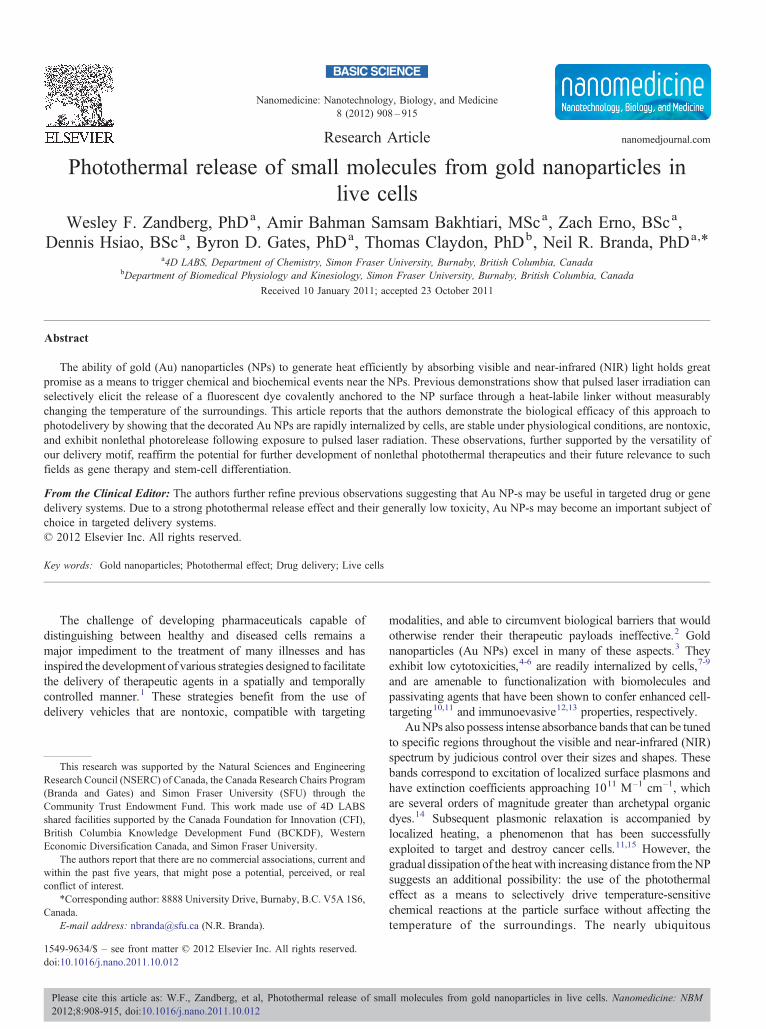
was utilized as a positive control for the viability assays. Fourhours after treatment with resazurin, cells exposed to the NPvector and pulsed laser irradiation, independently or incombination with one another, exhibited luminescence intensi-ties similar to those observed in control populations (Figure 4).When cells were exposed to resazurin and examined 24 hoursafter laser irradiation, an increase in fluorescence was observedin all populations except the ones treated with Lactacystin. Toeliminate the possibility that the fluorescence observed fromcells treated with the NPs and exposed to laser light was due tofree fluorescein, the fluorescence of a solution of the original NPmixture added to the cells was measured before and afterirradiation using the AlamarBlue filter set (Figure 4, inset).Although there was a slight increase in fluorescence uponirradiation of the NP solution (0.92 ± 0.13 increased to 1.62 ±0.04 RFU), the AlamarBlue signal in a sample treated with theNPs and irradiated cells was much greater (5094 ± 948 RFU).
Capillary electrophoretic analysis of the mixture generated bypulsed laser irradiation of the decorated NPs revealed two peaks
(Figure 5, top). Comparison with authentic samples of furan-functionalized fluorescein and free fluorescein (Figure 5,bottom) enabled identification of the major product as theformer and the minor product as the latter (see SupplementaryMaterials [available online at http://www.nanomedjournal.com]for peak assignments).
Discussion
The ability of our delivery vehicle to function in live cells wasdemonstrated in two systems. We selected Xenopus laevisoocytes as a model cellular system because their large sizespermit direct microinjection of the decorated Au NPs, allowingus to measure the efficacy of the photothermal release undercellular physiological conditions without the added complicationof addressing whether the delivery vehicles would undergo facileendocytotic uptake. Furthermore, previous electrophysiologicalcharacterization of the oocytes enables analysis of RMPs and
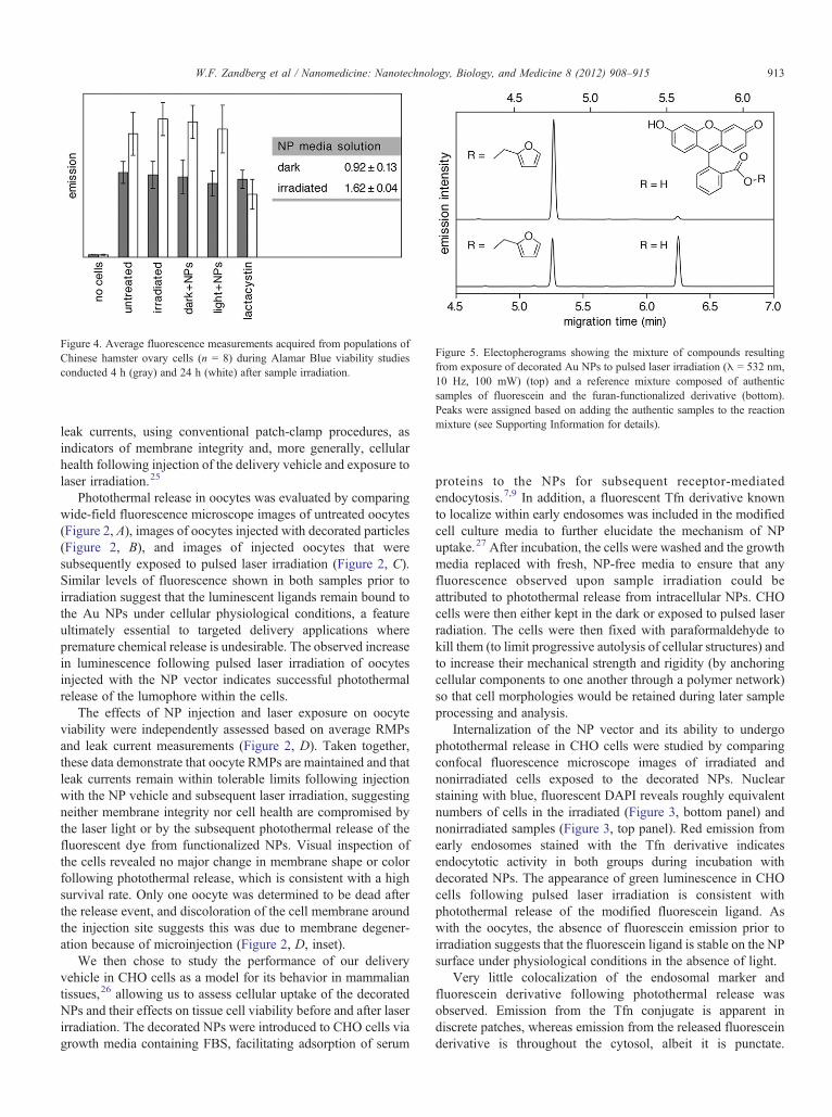
Figure 4. Average fluorescence measurements acquired from populations ofChinese hamster ovary cells (n = 8) during Alamar Blue viability studiesconducted 4 h (gray) and 24 h (white) after sample irradiation.
Figure 5. Electopherograms showing the mixture of compounds resultingfrom exposure of decorated Au NPs to pulsed laser irradiation (λ = 532 nm,10 Hz, 100 mW) (top) and a reference mixture composed of authenticsamples of fluorescein and the furan-functionalized derivative (bottom).Peaks were assigned based on adding the authentic samples to the reactionmixture (see Supporting Information for details).
913W.F. Zandberg et al / Nanomedicine: Nanotechnology, Biology, and Medicine 8 (2012) 908–915
leak currents, using conventional patch-clamp procedures, asindicators of membrane integrity and, more generally, cellularhealth following injection of the delivery vehicle and exposure tolaser irradiation.25
Photothermal release in oocytes was evaluated by comparingwide-field fluorescence microscope images of untreated oocytes(Figure 2, A), images of oocytes injected with decorated particles(Figure 2, B), and images of injected oocytes that weresubsequently exposed to pulsed laser irradiation (Figure 2, C).Similar levels of fluorescence shown in both samples prior toirradiation suggest that the luminescent ligands remain bound tothe Au NPs under cellular physiological conditions, a featureultimately essential to targeted delivery applications wherepremature chemical release is undesirable. The observed increasein luminescence following pulsed laser irradiation of oocytesinjected with the NP vector indicates successful photothermalrelease of the lumophore within the cells.
The effects of NP injection and laser exposure on oocyteviability were independently assessed based on average RMPsand leak current measurements (Figure 2, D). Taken together,these data demonstrate that oocyte RMPs are maintained and thatleak currents remain within tolerable limits following injectionwith the NP vehicle and subsequent laser irradiation, suggestingneither membrane integrity nor cell health are compromised bythe laser light or by the subsequent photothermal release of thefluorescent dye from functionalized NPs. Visual inspection ofthe cells revealed no major change in membrane shape or colorfollowing photothermal release, which is consistent with a highsurvival rate. Only one oocyte was determined to be dead afterthe release event, and discoloration of the cell membrane aroundthe injection site suggests this was due to membrane degener-ation because of microinjection (Figure 2, D, inset).
We then chose to study the performance of our deliveryvehicle in CHO cells as a model for its behavior in mammaliantissues,26 allowing us to assess cellular uptake of the decoratedNPs and their effects on tissue cell viability before and after laserirradiation. The decorated NPs were introduced to CHO cells viagrowth media containing FBS, facilitating adsorption of serum
proteins to the NPs for subsequent receptor-mediatedendocytosis.7,9 In addition, a fluorescent Tfn derivative knownto localize within early endosomes was included in the modifiedcell culture media to further elucidate the mechanism of NPuptake.27 After incubation, the cells were washed and the growthmedia replaced with fresh, NP-free media to ensure that anyfluorescence observed upon sample irradiation could beattributed to photothermal release from intracellular NPs. CHOcells were then either kept in the dark or exposed to pulsed laserradiation. The cells were then fixed with paraformaldehyde tokill them (to limit progressive autolysis of cellular structures) andto increase their mechanical strength and rigidity (by anchoringcellular components to one another through a polymer network)so that cell morphologies would be retained during later sampleprocessing and analysis.
Internalization of the NP vector and its ability to undergophotothermal release in CHO cells were studied by comparingconfocal fluorescence microscope images of irradiated andnonirradiated cells exposed to the decorated NPs. Nuclearstaining with blue, fluorescent DAPI reveals roughly equivalentnumbers of cells in the irradiated (Figure 3, bottom panel) andnonirradiated samples (Figure 3, top panel). Red emission fromearly endosomes stained with the Tfn derivative indicatesendocytotic activity in both groups during incubation withdecorated NPs. The appearance of green luminescence in CHOcells following pulsed laser irradiation is consistent withphotothermal release of the modified fluorescein ligand. Aswith the oocytes, the absence of fluorescein emission prior toirradiation suggests that the fluorescein ligand is stable on the NPsurface under physiological conditions in the absence of light.
Very little colocalization of the endosomal marker andfluorescein derivative following photothermal release wasobserved. Emission from the Tfn conjugate is apparent indiscrete patches, whereas emission from the released fluoresceinderivative is throughout the cytosol, albeit it is punctate.

914 W.F. Zandberg et al / Nanomedicine: Nanotechnology, Biology, and Medicine 8 (2012) 908–915
Interpretation of these results as evidence of nonendocytoticinternalization of the NP vehicle would conflict with a previouslyreported example of receptor-mediated endocytosis facilitated byadsorption of serum proteins to Au NPs, although it cannot beruled out that a different entry mechanism was utilized by ourdecorated NPs.8 Alternatively, the possibility of rapid diffusionof the released fluorescein derivative through the endosomalmembrane prior to fixation with paraformaldehyde wouldreconcile our results with those reported in the literature.
The fluorescence microscopy data acquired from both groupsof CHO cells were quantified and compared in terms of voxelintensities (Table 1). These results suggest that similar quantitiesof the fluorescent Tfn derivative were endocytosed by theirradiated cells and by the control group, supporting thereasonable assumption that both populations internalizedcomparable amounts of the NP vehicle. Additionally, theaverage voxel intensity per cell attributable to emission fromthe dissociated fluorescein derivative was much greater inirradiated cells than in control groups. These data indicatesuccessful photothermal release of the fluorescent dye in tissue-cultured cells irradiated under sterile conditions. Given theamount of background fluorescein emission observed innonirradiated samples, it can be concluded that the functiona-lized NP vehicle is also reasonably stable in the cellularenvironment and that very small quantities of the fluoresceinderivative are released in the absence of laser irradiation. Similarlevels of background fluorescence were observed from cells thathad been incubated with functionalized NPs for 4 hours prior tofixation and confocal microscopic analyses.
The effects of NP uptake and pulsed laser irradiation onCHO cell viability and proliferation were examined using anassay that quantifies cellular physiological activity based on theamount of a luminescent dye, resorufin, generated metabolicallyfrom an administered precursor, the nonfluorescent dyeresazurin. As a positive control, CHO cells were separatelytreated with the proteasome inhibitor Lactacystin. Results fromexperiments performed 4 hours after laser exposure suggestthat uptake of the decorated Au NPs, laser irradiation or acombination of both had little to no immediate effect on cellviability. An increase in luminescence intensity observed amongall groups except those treated with lactacystin was observed24 hours after laser irradiation, which indicates that neither thepresence of the Au NPs nor laser exposure had reduced the ratesof cell proliferation. In contrast, cells exposed to lactacystindid not exhibit a corresponding increase in luminescence after24 hours, which is consistent with cell-cycle inhibition andaffirms the validity of our AlamarBlue viability data.
The possibility that luminescence from the modifiedfluorescein ligand might interfere with the results of theAlamarBlue assay was addressed by emission measurementsperformed on a solution of the decorated NPs using theAlamarBlue filter set, before and after exposure to pulsed laserirradiation. The slight increase in luminescence intensity thatwas observed indicates a small amount of bleed-through.However, the amount of interference is negligible in compa-rison with the increase in luminescence intensity observed incells treated with the NP vector following irradiation. Thisfinding confirms that the observed increase in fluorescence is not
due to free fluorescein adducts but to metabolized resazurin. Italso confirms that these procedures were not cytotoxic, nor didthey affect cell proliferation.
To identify what fluorescent compound or compounds arereleased as a result of the photothermal reaction, samples of AuNPs were irradiated (532 nm) and analyzed by capillaryelectrophoresis, whereby the electrophoretic mobility of eachcomponent in the mixture was characterized based on the timeit took to migrate past an excitation light source (Ar laser, 488nm) and fluorescence detector positioned alongside thecapillary. The mixture was shown to consist of a major and aminor product. The major product was identified as the furan-functionalized fluorescein, which confirms that the increase influorescence intensity accompanying pulsed laser irradiation ofthe decorated Au NPs is due primarily to photothermolysis ofthe oxabicycloheptene motif via the retro-Diels-Alder reaction.The minor product was identified as free fluorescein, which ispresumably generated by hydrolysis of the furfuryl ester. Theseresults are consistent with previously reported data that showminimal Au–S bond breaking and release of thiols undersimilar conditions.28
In summary, we succeeded in preparing a NP-based drugdelivery vehicle and demonstrated its ability to elicit photo-thermal release within live oocytes and tissue cell cultures. Ourviability studies indicate high cell survival rates followingtreatment with functionalized NPs and subsequent laserirradiation, highlighting the possible extension of intracellularphotothermal phenomena to noncytotoxic applications, such asgene therapy and stem cell differentiation. Our future workwill combine these design principles with established cell-targeting methodologies utilizing antibodies to test oursystem's ability to deliver targeted chemical agents havingtherapeutic properties.
Appendix A. Supplementary data
Supplementary data to this article can be found online atdoi:10.1016/j.nano.2011.10.012.
References
1. Ho RJY, Chien JY. Drug delivery trends in clinical trials andtranslational medicine: Updated analysis of database. J Pharm Sci2009;98:1928-34.
2. Ferrari M. Cancer nanotechnology: opportunities and challenges. NatRev Cancer 2005;5:161-71.
3. Ghosh P, Han G, De M, Kim CK, Rotello VM. Gold nanoparticles indelivery applications. Adv Drug Delivery Rev 2008;60:1307-15.
4. Connor EE, Mwamuka J, Gole A, Murphy CJ, Wyatt MD. Goldnanoparticles are taken up by human cells but do not cause acutecytotoxicity. Small 2005;1:325-7.
5. Kuo CW, Lai JJ, Wei KH, Chen P. Studies of surface-modified goldnanowires inside living cells. Adv Funct Mater 2007;17:3707-14.
6. Pan Y, Neuss S, Leifert A, Fischler M, Wen F, Simon U, et al. Size-dependent cytotoxicity of gold nanoparticles. Small 2007;3:1941-9.
7. Chithrani BD, Chan WCW. Elucidating the mechanism of cellularuptake and removal of protein-coated gold nanoparticles of differentsizes and shapes. Nano Lett 2007;7:1542-50.

915W.F. Zandberg et al / Nanomedicine: Nanotechnology, Biology, and Medicine 8 (2012) 908–915
8. Chithrani BD, Ghazani AA, Chan WCW. Determining the size andshape dependence of gold nanoparticle uptake into mammalian cells.Nano Lett 2006;6:662-8.
9. Yang PH, Sun X, Chiu JF, Sun H, He QY. Transferrin-mediated goldnanoparticle cellular uptake. Bioconjugate Chem 2005;16:494-6.
10. Azzazy HME, Mansour MMH. In vitro diagnostic prospects ofnanoparticles. Clin Chim Acta 2009;403:1-8.
11. Huang X, El-Sayed IH, Qian W, El-Sayed MA. Cancer cell imaging andphotothermal therapy in the near-infrared region by using gold nanorods.J Am Chem Soc 2006;128:2115-20.
12. Brigger I, Dubernet C, Couvreur P. Nanoparticles in cancer therapy anddiagnosis. Adv Drug Delivery Rev 2002;54:631-51.
13. Moghimi SM, Szebeni J. Stealth liposomes and long circulatingnanoparticles: critical issues in pharmacokinetics, opsonization, andprotein-binding properties. Prog Lipid Res 2003;42:463-78.
14. Boisselier E, Astruc D. Gold nanoparticles in nanomedicine: prepara-tions, imaging, diagnostics, therapies and toxicity. Chem Soc Rev2009;38:1759-82.
15. Hirsch LR, Stafford RJ, Bankson JA, Sershen SR, Rivera B, Price RE,et al. Nanoshell-mediated near-infrared thermal therapy of tumorsunder magnetic resonance guidance. Proc Natl Acad Sci USA2003;100:13549-54.
16. Bakhtiari ABS, Hsiao D, Jin G, Gates BD, Branda NR. An efficientmethod based on the photothermal effect for the release of moleculesfrom metal nanoparticle surfaces. Angew Chem Int Ed 2009;48:4166-9.
17. Lee SE, Liu GL, Kim F, Lee LP. Remote optical switch for localized andselective control of gene interference. Nano Lett 2009;9:562-70.
18. Yavuz MS, Cheng Y, Chen J, Cobley CM, Zhang Q, Rycenga M, et al.Gold nanocages covered by smart polymers for controlled release withnear-infrared light. Nat Mater 2009;8:935-9.
19. Skrabalak SE, Au L, Li X, Xia Y. Facile synthesis of Ag nanocubes andAu nanocages. Nature Protocols 2007;2:2182-90.
20. McElhanon JR, Wheeler DR. Thermally responsive dendrons anddendrimers based on reversible furan-maleimide Diels-Alder adducts.Org Lett 2001;3:2681-3.
21. Fenteany G, Standaert RF, Lane WS, Choi S, Corey EJ, Schreiber SL.Inhibition of proteasome activities and subunit-specific amino-terminalthreonine modification by Lactacystin. Science 1995;268:726-31.
22. Lopes UG, Erhardt P, Yao R, Cooper GM. p53-dependent induc-tion of apoptosis by proteasome inhibitors. J Biol Chem 1997;272:12893-6.
23. Fenteany G, Schreiber SL. Lactacystin, proteasome function, and cellfate. J Biol Chem 1998;273:8545-8.
24. Imajohohmi S, Kawaguchi T, Sugiyama S, Tanaka K, Omura S, KikuchiH. Lactacystin, a specific inhibitor of the proteasome, induces apoptosisin human monoblast U937 cells. Biochem Biophys Res Commun1995;217:1070-7.
25. Wagner CA, Friedrich B, Setiawan I, Lang F, Bröer S. The use ofXenopus laevis oocytes for the functional characterization ofheterologously expressed membrane proteins. Cell Physiol Biochem2000;10:1-12.
26. Jayapal KP, Wlaschin KF, Yap MGS. Recombinant protein therapeuticsfrom CHO cells—20 years and counting. Chem Eng Prog2007;103:40-7.
27. Barysch SV, Jahn R, Rizzoli SO. A fluorescence-based in vitro assay forinvestigating early endosome dynamics. Nat Protoc 2010;5:1127-37.
28. Poon L, Zandberg W, Hsiao D, Erno Z, Sen D, Gates BD, et al.Photothermal release of single-stranded DNA from the surface of goldnanoparticles through controlled denaturing and Au-S bond breaking.ACS Nano 2010;4:6395-403.