1 PharmacokineticsandPharmacodynamics(PK/PD)of ... · 1 1...
60
1 1 Pharmacokinetics and Pharmacodynamics (PK/PD) of Bionanomaterials Ergang Liu, Meng Zhang, and Yongzhuo Huang 1.1 Introduction Nanomaterials (NMs) refer to synthetic or naturally occurring substances with size ranging from 1 to 1000 nm. e concept of “nanomaterial” was proposed by Feynman 50 years ago in the field of physics [1], which has since unveiled an era of nanotechnology. NMs contain merely tens to thousands of atoms, and are char- acterized by the surface and quantum size effects that are distinct from the bulk matters, and have thus gained wide applications in various areas [2]. For example, in medical application, nanotechnology has attracted specific attention in cancer therapy and diagnosis, largely due to the proposal of enhanced permeation and retention (EPR) effect by Maeda and coworkers; they demonstrated that nano- sized macromolecules displayed a preferential retention in tumor site due to the leaky vasculatures [3, 4]. e EPR effect-associated nanomedicine composed of various natural or synthetic entities in the nanoscale, which have been developed to deliver drugs/imaging agents to the tumors based on the passive targeting effect [5]. Later, in order to further increase the transport efficiency, antibodies or tar- geting ligands with high binding affinity to tumor-overexpressed surface antigens or receptors have been applied to conjugate onto the surface of NMs to achieve the so-called active targeting [6]. NMs can also be applied in formulation development because of their capability to improve solubility [7], drug permeation [8], and drug stability [9]. Pharmaceuti- cal nanotechnology may thus help improve druggability of those active molecules that are otherwise considered to be unsuitable for formulation development for clinical use due to unfavorable properties such as poor solubility and low perme- ation to the lipid bilayer membranes [10]. e emerging nanomedicine has greatly promoted drug development, and a good number of NM-based medicine or diagnostic agents have entered clinical trials, most in the field of cancer therapy, in which the NM-based delivery strate- gies are characterized by EPR effect for achieving tumor targeting. However, in spite of the enhanced permeability of the tumor vasculature, not all types of NMs Biomedical Nanomaterials, First Edition. Edited by Yuliang Zhao and Youqing Shen. © 2016 Wiley-VCH Verlag GmbH & Co. KGaA. Published 2016 by Wiley-VCH Verlag GmbH & Co. KGaA.
Transcript of 1 PharmacokineticsandPharmacodynamics(PK/PD)of ... · 1 1...
1
1Pharmacokinetics and Pharmacodynamics (PK/PD) ofBionanomaterials
Ergang Liu, Meng Zhang, and Yongzhuo Huang
1.1Introduction
Nanomaterials (NMs) refer to synthetic or naturally occurring substances withsize ranging from 1 to 1000 nm. The concept of “nanomaterial” was proposed byFeynman 50 years ago in the field of physics [1], which has since unveiled an era ofnanotechnology. NMs contain merely tens to thousands of atoms, and are char-acterized by the surface and quantum size effects that are distinct from the bulkmatters, and have thus gained wide applications in various areas [2]. For example,in medical application, nanotechnology has attracted specific attention in cancertherapy and diagnosis, largely due to the proposal of enhanced permeation andretention (EPR) effect by Maeda and coworkers; they demonstrated that nano-sized macromolecules displayed a preferential retention in tumor site due to theleaky vasculatures [3, 4]. The EPR effect-associated nanomedicine composed ofvarious natural or synthetic entities in the nanoscale, which have been developedto deliver drugs/imaging agents to the tumors based on the passive targeting effect[5]. Later, in order to further increase the transport efficiency, antibodies or tar-geting ligands with high binding affinity to tumor-overexpressed surface antigensor receptors have been applied to conjugate onto the surface of NMs to achievethe so-called active targeting [6].NMs can also be applied in formulation development because of their capability
to improve solubility [7], drug permeation [8], and drug stability [9]. Pharmaceuti-cal nanotechnology may thus help improve druggability of those active moleculesthat are otherwise considered to be unsuitable for formulation development forclinical use due to unfavorable properties such as poor solubility and low perme-ation to the lipid bilayer membranes [10].The emerging nanomedicine has greatly promoted drug development, and a
good number of NM-based medicine or diagnostic agents have entered clinicaltrials, most in the field of cancer therapy, in which the NM-based delivery strate-gies are characterized by EPR effect for achieving tumor targeting. However, inspite of the enhanced permeability of the tumor vasculature, not all types of NMs
Biomedical Nanomaterials, First Edition. Edited by Yuliang Zhao and Youqing Shen.© 2016 Wiley-VCH Verlag GmbH & Co. KGaA. Published 2016 by Wiley-VCH Verlag GmbH & Co. KGaA.
2 1 Pharmacokinetics and Pharmacodynamics (PK/PD) of Bionanomaterials
could benefit fromEPR effect to achieve a substantial targeting efficiency [11].Thein vivo ADME (absorption, distribution, metabolism, and excretion) behaviors ofNMs vary because of the difference of the surface properties, size, and charges ofthe NMs, as well as their compositions, often leading to inconsistent therapeuticoutcomes in animal studies [12].On this account, investigation of “what the body does to NMs”may help us with
a better understanding of the in vivo fate.We herein present a brief introduction ofthe commonly utilized NMs in pharmaceutical research, the anatomic features ofthe body and tumor, and the physiochemical natures of NMs that affect the in vivofate. The established PK/PD models for simulating the in vivo ADME behavior ofNMs will also be introduced. We hope this summary would give a glimpse intothe complicated in vivo processes and provide helpful information for the rationaldesign of NM-based drug delivery systems.
1.2Commonly Utilized NMs in Pharmaceutical Research
NMs can be categorized into different groups based on certain classification. Tomake it simple, we use the natural/synthetic classification in this chapter becausethe natural/synthetic NMs are generally disposed by the body in different ways.Moreover, inorganic NMs characterized by the hard-core structure bear uniquephysical characteristics (magnetism, thermal response to radiation, optical fea-tures, etc.) [13, 14], and are discussed as an independent section. Other resourcessuch as cell-based NMs (e.g., RBCs [15] and MSCs [16]) and components frommicrobes (e.g., inactivated virus envelope [17] and TAT [18]), are usually utilizedwith preservation of their original natures, which are thereby discussed as a com-plimentary to this classification.
1.2.1Natural NMs
Natural NMs have been widely investigated because of their biodegradabilityand compatibility to human body. As known, lipids, proteins, carbohydrates,and nucleic acids are highly biodegradable in the body. Phospholipids are oneof the most widely applied natural resources to build the nanocarriers such asliposomes and solid lipid nanoparticles (SLNs) [19]. Polysaccharides, including avariety of carbohydrates with different structures and functional groups, can beutilized to build different types of nanoparticles. Protein-based NMs (typically,serum proteins such as albumin [20], high-density lipoprotein (HDL) [21], andlactoferrin [22]) are often utilized as drug carriers.
1.2.1.1 Lipid-Based NMsLipid-based NMs include liposomes [19], SLNs, micelles [23], and nanoemul-sions [24] (Figure 1.1). The main components of liposomes are phospholipids. Inaqueous solution, the phospholipids will self-assemble into a bilayer structure

1.2 Commonly Utilized NMs in Pharmaceutical Research 3
Liposome Solid lipid nanoparticle Micelle Nano-emulsion
Oil droplet
WaterSolid
Figure 1.1 Schematic illustration of lipid-based NMs.
that functions as drug carriers with hydrophilic drugs encapsulated inside theinterior, whereas hydrophobic drugs in bilayer [19]. One-tail lipids are inclinedto form micelles in aqueous media [25], whereas using steric acid or oleic acidsupplemented with surfactants to stabilize the solid/liquid lipids normally resultsin nanoparticles [26] or emulsions [27].
1.2.1.2 Protein-Based NMsProtein-based drug carriers have been widely used in pharmaceutical industry.However, owing to the concerns of protein immunogenicity (e.g., OVA, whichhas been utilized as adjuvant for immune activation [28]), endogenous serum-richproteinswith low immunogenicity and long half-life such as albumin, high-densitylipoprotein, and lactoferrin have distinct advantages [20–22]. As a case in point,albumin-bound paclitaxel nanoparticles (Abraxane®) have attained great marketsuccess [29]. In general, proteins can either be processed to form nanoparticles[30] or directly coupled with drugs by physical adsorption or via covalent bonds[20]. In certain instances, the protein carriers are further modified with targetingligands to achieve specific delivery [31].
1.2.1.3 Polysaccharide-Based NMsPolysaccharides originate from animal, plant, or bacterial sources. In general, thephysicochemical properties of polysaccharides are governed by monosaccharideunit and the overall molecular weight [32]. The high molecular weight molecules,such as heparin and hyaluronic acid, show strong affinity to water molecules,and thus form hydrogels that have been widely applied for local administrationbecause of their biocompatibility and sustained drug release functions [33].The ionic polysaccharides can bind with molecules of the opposite charge, andthe interaction normally leads to decreased solubility and the formation ofnanoparticles [34].
1.2.2Synthetic NMs
Although NMs based on naturally occurring materials have the advantagesof biocompatibility and wide availability, structure modification is difficult
4 1 Pharmacokinetics and Pharmacodynamics (PK/PD) of Bionanomaterials
H
G1
G2
G3
G4
N
NN
NN
NN
NN
NN
NN
NN
NN
N
N
NNH
NH
H2N
H2N
H2NNH2
NH2
NH2
NH2
NH2
NH2
NH2
NH2
NH2NH2
NH2H2N
H2N
H2N
NH
* *
**
NH
NH
HN
HN
H2N
Branched PEIDendritic PEI
Linear PEI
NH2
n
n
NH2
H
Figure 1.2 Various forms of PEI.
to process to tailor their functions to satisfy the needs from pharmaceuticalapplication.By contrast, synthetic polymers can be much more flexibly designed for a
specific application. For example, by using the pH-sensitive synthetic materials,the NMs could release drugs in a pH-dependent manner for achieving tumor-targeting delivery [35], because the rapidly prolific neoplastic tissues normallysecrete more lactose from the hyperactive anaerobic glycolysis, leading to adecreased pH in tumor microenvironment [36].
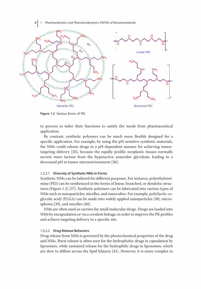
1.2.2.1 Diversity of Synthetic NMs in Forms
Synthetic NMs can be tailored for different purposes. For instance, polyethylenei-mine (PEI) can be synthesized in the forms of linear, branched, or dendritic struc-tures (Figure 1.2) [37]. Synthetic polymers can be fabricated into various types ofNMs such as nanoparticles, micelles, and nanocubes. For example, poly(lactic-co-glycolic acid) (PLGA) can be made into widely applied nanoparticles [38], micro-spheres [39], and micelles [40].NMs are often used as carriers for small molecular drugs. Drugs are loaded into
NMs by encapsulation or via a covalent linkage, in order to improve the PKprofilesand achieve targeting delivery to a specific site.
1.2.2.2 Drug Release Behaviors
Drug release from NMs is governed by the physiochemical properties of the drugand NMs. Burst release is often seen for the hydrophobic drugs in capsulation byliposomes, while sustained release for the hydrophilic drugs in liposomes, whichare slow to diffuse across the lipid bilayers [41]. However, it is more complex to
1.2 Commonly Utilized NMs in Pharmaceutical Research 5
investigate the drug release profile whenNMs are injected to the body, and knowl-edge of the in vitro in vivo correlation (IVIVC) is still insufficient.In order to reduce the unwanted drug exposure, a number of strategies have
been developed to achieve a site-specific release of the loaded cargos.
pH-sensitive NMsThe slightly acidic tumor environment and endosome’s even lower pH haveattracted extensive interests in the application for designing NMs with theability to respond to pH changes during the delivery. This strategy has beenintensively explored by employing the polycationic dendrimers such as PEIand PAMAM. Acidic pH could cause the electrostatic repulsions betweenside chains in these polyamines because of the protonation of amino groups.As a result, the dendrimers swell in response to the acidic condition and theabrupt pH drop – the so-called “proton sponge” effect [42]. After engulfedby the cells, the swelling of NMs can lead to endosome rupture, and thus isfavorable for intracellular drug release.Another strategy is to use pH-sensitive linkage (e.g., hydrazone bonds) forcross-linking and building the NMs [43]. The NMs disassemble in a pH-dependent pattern, thus triggering drug release in acidic environments.
Redox-sensitive NMsBesides the decreased pH, the rapidly growing tumor is also characterizedby the intracellular reducibility due to the increased level of glutathione(GSH) [42]. The redox-sensitive NMs (e.g., NMs built via disulfide linkage)can display an accelerated drug release once entering the tumor cells.
Enzyme-sensitive NMsTumor-associated proteases have been widely investigated for their appli-cation as biomarkers in cancer diagnosis, prognosis, and therapy. Overex-pression of tumor-associated proteases (e.g.,MMP-2 [44],MMP-9 [45], andlegumain [46]) in tumors provides ideal targets for the design of “smart”NMs with controlled release. A general strategy is to use a specific substratepeptide to modify the NMs, and the cleavage of the peptide would triggerdrug release or cellular uptake.
Thermo/radio wave-sensitive NMsOther stimulus-responsive NMs can respond to external physical stimu-lation (such as localized heating and electromagnetic radiation) and havebeen applied in drug delivery [47]. Specificity of this strategy is largelydependent on the precise control of the applied stimuli at the target sites.
1.2.3Inorganic NMs
Inorganic NMs are distinguished from the organic NMs (soft matters) with thehard cores. In order to avoid aggregation in aqueousmedia, the inorganic cores aretypically modified with surfactants or hydrophilic polymers to form a core–shellstructure [48]. Of note, the in vivo biofate of inorganic NMs is greatly affected bythe surface characteristics of the coating materials [49].

6 1 Pharmacokinetics and Pharmacodynamics (PK/PD) of Bionanomaterials
Inorganic NMs exhibit many unique physical properties – for example –fluorescence (quantum dots), superparamagnetism (iron oxide nanoparticles),photothermal effect (gold nanorods, carbon nanotubes), or special opticalproperties (silver nanoparticles) [13, 50]. These properties of the inorganic NMshave been utilized in cancer diagnosis and treatment applications.
1.2.4Other NMs
Together with the rapid development of NMs, knowledge of NMs has accumu-lated. Proteins, in terms of the size, can also be viewed as bionanomaterials, whichare rich in the body. As a case in point, albumin (MW 67 kDa) with a diameteraround 7 nm [51] can serve as a unique “protein carrier” for drugs. Anotherexample is the red blood cells (RBCs), with diameter from several to tens ofmicrometers [15], whichmay be regarded as a type of “microliposomes” to delivertherapeutic macromolecules. Moreover, even the protein capsids of a virus (size<100 nm) can be used as a “nanocapsule” [52]. Inspired by biomimetics, thesephysiologically originated NMs have been explored as novel carriers for drugdelivery.Moreover, there is another form of nanodrugs – the nanocrystals of hydropho-
bic drugs [53]. Such nanodosage forms can solve the solubility problem, and fur-thermore improve the PK profiles.
1.3In vivo Biodistribution and the Evolving Targeting Principles for NMs
The targeting strategies have been mostly employed in cancer therapy areas. Ingeneral, there are two types of targeting strategies: the passive targeting via EPReffect and the active targeting mediated by antibodies or ligands that can specif-ically bind with receptors on cancer cells. Recently, with the growing knowledgeof tumor physiology, the tumor microenvironments (e.g., acidic pH and the over-expressed proteases) have been used as a target for cancer drug delivery [6]. Acombination of the targeting strategies by using multifunctional NMs has alsobeen investigated for achieving improved specific targeted delivery and controlledrelease [54]. The ultimate goal is to increase drug concentration in tumor whilereducing its exposure to the healthy organs.
1.3.1Organ Distribution versus Cell-Specific Targeting
Ideally, NMs should be able to deliver the cargo drugs specifically into a targetsite. The in vivo fate of NMs is determined by a combination of multiple factors,including particle size, shape, and surface characteristics [55]. It is thus difficult toassess the overall targeting efficiency from in vitro data (e.g., cellular uptake by a

1.3 In vivo Biodistribution and the Evolving Targeting Principles for NMs 7
target cell line) for a given NM. A biodistribution study of the NMs is necessaryto investigate the preferential accumulation site of the administrated NMs. If acertainNM is highly accumulated in a specific organ, it could be used as a potentialdrug carrier for targeting the organ.However, organ-specific accumulation may not ensure the satisfactory treat-
ment outcomes. For example, in brain cancer, an ideal delivery should be a“dual-targeting” mode, that is, the chemotherapeutics could not only prefer-entially accumulate in brain, but also selectively act on the cancer cells, whilesparing the normal cells in the central nervous system and thus preventingadverse effects.
1.3.2Targeting Delivery Strategies
With growing understanding of the pathology and diseases, different targetingstrategies have been developed.
Passive targetingEPR effect is one of the major mechanisms responsible for cancernanomedicine. However, due to nonspecific capture by the RES andretention by organs such as liver, spleen, and lung, the vast majority (>95%)of administered nanomedicine distributes into the undesired organs [56].The overall benefit of the targeting efficiency by EPR effect for clinical useis still not clear [57]. Much effort has been made on surface modificationwith hydrophilic polymers such as PEG to evade the RES uptake [58].Furthermore, the extended blood circulation facilitates extravasation forincreased tumor accumulation.
Targeting to membrane-overexpressed receptorsMany diseases are related to gene mutations that generally lead to phe-notypic changes and overexpression of certain membrane receptors. Theoverexpressed membrane receptors may serve as “zip code” and facilitatesite-specific drug delivery. The drug carriers modified with targetingligands (e.g., antibody and peptide), like “postmen,” would recognize the“address” and achieve precise delivery. Moreover, some receptors canalso induce receptor-mediated endocytosis after binding [59]. An idealtargeting receptor should be specifically expressed in the pathological site,but does not exist in normal tissues or cells.
Tumor-overexpressed enzyme-mediated targetingTumor-overexpressed enzymes provide another group of promisingtargets for site-specific drug release. A typical design is that drugs arecross-linked with NMs by peptide substitutes, or encapsulated into NMsthat are featured by the protease-triggered cell penetration, thus achievingtumor-specific drug release or cell entry. Furthermore, if the fluorochromeis linked with its quencher by using a protease substrate peptide, FRET(fluorescence resonance energy transfer) effect can be produced, leading tofluorescence quench. Such type of FRET-based probe can be specifically

8 1 Pharmacokinetics and Pharmacodynamics (PK/PD) of Bionanomaterials
Tumor cells
Endothelial cells
Quenched QDs
Activated QDs
Blood flow
QD
LH
PEP
QSY21
Tumor enzyme
Figure 1.3 Schematic illustrations of activat-able nanoprobe. The nanoprobe comprisestwo components (the fluorescence donorQD-LH and the FRET quencher QSY21),
which are cross-linked via a chimeric pep-tide comprising LP and a legumain-cleavablesequence [60]. (Reprinted with permissionsfrom John Wiley & Sons, Copyright 2014.)
activated by the tumor-overexpressed proteases, and thus serve as apotential method for tumor detection and imaging (Figure 1.3) [60].
Tumor microenvironment-responsive NMsThe slightly acidic tumormicroenvironment [36] and high level of GSH [61]have been extensively explored for controlled drug release. For example,amphoteric polymers are neutral or negatively charged at pH 7.4, butbecome positively charged at slightly acidic condition [62]. The chargeconversion facilitates NMs to be preferentially engulfed by the tumor cellsbecause of the lowered extracellular pH in tumor tissue.High level of GSH in cancer cells is the key factor for reduction-responsivedrug delivery. For example, NMs that are built via disulfide bond can disas-semble when exposed to the increased intracellular GSH, leading to burstrelease of the encapsulated drugs. Another interesting application is the cell-penetrating peptide (CPP)-mediated intracellular drug delivery. CPPs areeffective in transporting macromolecules across the cellular membranes,but the delivery through the cell membrane could be an in-and-out bidirec-tional pattern [63]. The potential solution to entrap the cargo drugs insidethe cytoplasm is to conjugate CPP with the drugs via disulfide bond. Oncethe linker is cleaved in the presence of high level of intracellular GSH, thecargo drugs will thus be left in the cytoplasm [15].
Physical targetingThe superparamagnetism of iron oxide nanoparticles with size of 3–50 nmhas been applied for magnetic targeted drug delivery. In this strategy, drugsare normally encapsulated in the coating layers on the magnetic cores, orcross-linked onto the surface of the NMs [64].In addition, photothermal effects of gold NMs have been exploredfor thermal-triggered drug release at the target site. For instance, gold

1.4 Processing NMs by the Biological Systems 9
N N
N
N
N
N
N
N
N
N
N
N
N
N
NN
N
N
N
N
N
NN N
N
N
N
N
NN
NN
NN
N
NN
NN
NN
NN
NN N
NN N N
N
N
N
NN
N
NN
NNN
N
Normal tissuepH >7 Tumor
pH<6.5
N N
N
N
N
N
N
PAMAM pH sensitive polymer PEG FA Drug
N N
N
N
N
N
N
N
N
N
N
N
N
N
NN
N
N
N
N
N
NN N
N
N
N
N
NN
NN
NN
N
NN
NN
NN
NN
N
N NN
N N N
N
N
N
NN
N
NN
NNN
N
Figure 1.4 Schematic illustration of folate-modified pH-sensitive PAMAM. (Reprinted withpermissions from Elsevier, Copyright 2012.)
nanorods are coated with a thermal sensitive layer, which could be degradedby heating of the inorganic cores under external light radiation [65].Physical targeting delivery could be relatively precisely controlled byfine-tuning the external stimulations. However, there is still much room toimprove for the precise focus of the external energy on the target sites.
Combination of different targeting strategies to achieve a “smart delivery”The different targeting strategies can be combined to prepare the “smart”NMs by taking advantage of multiple characteristics of the tumor microen-vironment to achieve a synergetic effect of drug delivery. For example,the pH-sensitive NMs can be further modified with a targeting moiety. Ithas been reported that a multifunctional drug delivery system was devel-oped for enhanced tumor targeting via multiple mechanisms: (1) acidicmicroenvironment-triggered drug release, (2) folate modification forreceptor-mediated active targeting, and (3) PEGylation-induced longcirculation for passive targeting through EPR effect (Figure 1.4) [66].
1.4Processing NMs by the Biological Systems
The body has evolved with a conservative mechanism of dealing with the bio-logical “invaders” [67]: first, the circulating NMs will encounter serum proteinsthat tend to bind on to the surface of NMs and form a protein “corona” [68], andthus be identified and captured by the circulating leukocytes [69]; second, themajority of the circulating NMs will be trapped by RES organs and kidneys withfenestrated vasculatures, resulting in macrophage-mediated metabolism or renal

10 1 Pharmacokinetics and Pharmacodynamics (PK/PD) of Bionanomaterials
excretion [55]; third, the remaining NMs that survive from blood clearance willgain a chance to reach the target organs, where the retaining NMs will degrade,with drugs releasing into the extracellular space and releasing to the cells [70].Therefore, once entering the blood circulation, NMs overcome multiple physio-logical barriers before reaching their target cells or subcellular organelles.
1.4.1Anatomic Basis of NMs’ in vivo Biodistribution Behavior
EPR effect is the important physiological basis for NMs’ application in tumor.Anatomically, the neovasculatures in rapidly growing tumor are impaired withendothelial cells, thus generating nanosized gaps (400–600 nm) [71]. The leakyvessels facilitate extravasation of NMs to the tumor site [72]. Besides, the tumorsoverexpress a number of membrane receptors such as EGFRs [73], αγβ3 integrinreceptor [74], HER-2 [73], folate receptor [75], and transferrin receptor [76],which have been applied for mediating active targeting via interactions with theligand-modified NMs and triggering receptor-induced endocytosis of NMs. Bycontrast, some overexpressed membrane proteins, such as CD-20 that not induceendocytosis [77], are less frequently used for targeting delivery. Moreover, thetumor-overexpressed enzymes, such as legumain,MMPs, and hyaluronidase [78],can serve as the stimulus factors for the enzyme-sensitive NMs with controlledrelease behavior.However, the overall transport efficiency to tumor via blood circulation is still
below satisfaction, because themajority of the circulatingNMs is retained by otherorgans [79]. A better anatomical understanding of in vivo procession of NMs willbe helpful for rational design of a highly efficient system.In blood circulation, interactions between NMs and blood cells and serum pro-
teins greatly affect the biofate of the circulating NMs. The composition of the“protein corona” on the NMs is determined by the protein/NM binding affinityand regional concentrations of the proteins [80]. Albumin, lipoprotein, and com-plements are the major constitutions of the “corona.” Nevertheless, the proteinabsorption is a competitive process, and the difference in the composition leadsto different biofate of the NMs [81]. Albumin is the most abundant serum pro-tein with a plasma half-life of about 19–22 days. Albumin absorption onto thesurface of NMs will prevent from further interaction with other types of proteins,and benefit prolonged circulation. On the contrary, the lipoprotein-bound NMstend to accumulate in the tissues rich in lipoprotein receptors, while for example,albumin bindingwith complements usually induces clearance by themacrophages[81].Tissues and organs serve as “filters,” playing important roles in drug retention
and metabolism as well (Figure 1.5). Due to anatomic differences (e.g., bloodperfusion rate, vascular structure, and resident macrophage density), the tissuesprocess the NMs in different mechanisms [81]. For example, the RES organs (e.g.,liver, spleen, and the lung) with rich blood flow are the traps for NMs, whereabout 90–95% of the injected NMs are accumulated and metabolized [82]. Other

1.4 Processing NMs by the Biological Systems 11
CNS
Lungs
5800 mL min−1
(100%)
5800 mL min−1
(100%)
1100 mL min−1
(20%)
Spleen
Liver
Gut
700 mL min−1
(13%)
240 mL min−1
(4%) 1240 mL min−1
(22%)
77 mL min−1
(1.4%)
300 mL min−1
(5%)
Portalvein:
1150 mL min−1
(21%)
1310 mL min−1
(23%)
Muscle, skin, and adipose tissues
Intravenous
injection
Kidneys
Heart
Figure 1.5 Distribution of blood flow in the pulmonary and systemic circulations [81].(Reprinted with permissions from Elsevier, Copyright 2012.)
organs with rich blood flow such as heart and brain, on the contrary, do not showsignificant retention of the NMs, because of the rapid blood perfusion or tightjunctions of the vasculature [83].
1.4.2Factors Affecting in vivo Biodistribution of NMs
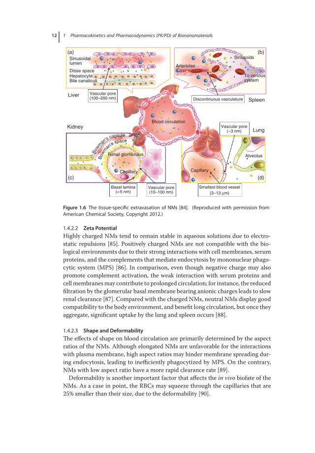
1.4.2.1 SizeThe “filtration efficiency” in various organs is closely associated with the “cutoffsize” of NMs (Figure 1.6). As for the liver, the size of the endothelial fenestratedcapillaries ranges from 100 to 200 nm, and inside the sinusoid capillaries they arerich in kupffer cells that account for 80–90% of the body’s resident macrophageswith [81].Therefore, the NMs size 100–200 nm will be engulfed by the sinusoidalresident macrophages; the spleen is apt to entrap the NMs larger than 200 nmdue to the existence of discontinuous arterioles [84]. Although vascular pores ofthe lungs restrain extravasation of NMs larger than 3 nm, NM aggregates or largeparticles could be easily captured by the lungs in the extremely small diameter ofthe capillaries (3–13 μm) [81].Tight junctions of renal basal lamina only allow the NMs smaller than 5 nm to
pass through and be eliminated via urine [81].
12 1 Pharmacokinetics and Pharmacodynamics (PK/PD) of Bionanomaterials
Sinusoidallumen
Arterioles
Blood circulation
Renal glomerulus
Capillary Capillary
Alveolus
Sinusoids
To venoussystem
(a)
LiverSpleen
Lung
(b)
(d)(c)
Kidney
Disse space
Vascular pore(100–200 nm)
Vascular pore(10–100 nm)
Vascular pore(~3 nm)
Smallest blood vessel
(3–13 μm)
Basal lamina(<5 nm)
Discontinuous vasculature
HepatocyteBile canaliculi
Bow
m
an’s
capsule
Bow
m
an’s s
pace
Figure 1.6 The tissue-specific extravasation of NMs [84]. (Reproduced with permission fromAmerican Chemical Society, Copyright 2012.)
1.4.2.2 Zeta PotentialHighly charged NMs tend to remain stable in aqueous solutions due to electro-static repulsions [85]. Positively charged NMs are not compatible with the bio-logical environments due to their strong interactions with cell membranes, serumproteins, and the complements that mediate endocytosis by mononuclear phago-cytic system (MPS) [86]. In comparison, even though negative charge may alsopromote complement activation, the weak interaction with serum proteins andcellmembranesmay contribute to prolonged circulation; for instance, the reducedfiltration by the glomerular basal membrane bearing anionic charges leads to slowrenal clearance [87]. Compared with the charged NMs, neutral NMs display goodcompatibility to the body environment, and benefit long circulation, but once theyaggregate, significant uptake by the lung and spleen occurs [88].
1.4.2.3 Shape and DeformabilityThe effects of shape on blood circulation are primarily determined by the aspectratios of the NMs. Although elongated NMs are unfavorable for the interactionswith plasma membrane, high aspect ratios may hinder membrane spreading dur-ing endocytosis, leading to inefficiently phagocytized by MPS. On the contrary,NMs with low aspect ratio have a more rapid clearance rate [89].Deformability is another important factor that affects the in vivo biofate of the
NMs. As a case in point, the RBCs may squeeze through the capillaries that are25% smaller than their size, due to the deformability [90].
1.5 Rational Design of Long-Circulating NMs 13
1.4.2.4 Hydrophilicity and HydrophobicityHydrophilicity and hydrophobicity also greatly affect the NMs’ pharmacokineticbehaviors [91]. In the physiological fluids, the hydrophobic NMs show high affin-ity to the nonpolar domains of the plasma proteins, promoting opsonization andleading to the decreased circulation time. Therefore, surface modification withhydrophilic polymers is a useful strategy for improving the stability and PK pro-files of the hydrophobic NMs.
1.4.3Metabolism and Elimination of NMs
1.4.3.1 CommonMetabolismThe different biodistribution and disposition of the NMs will lead to differentmetabolism in the body. In general, themajority of the administratedNMs are cap-tured by the RES organs, where the tissue-residentmacrophages serve as the scav-enger for degradation and clearance of the NMs. Apart from leukocyte-mediatedclearance, superfine NMs and the degraded debris of NMs with size smaller than5 nm will be excreted by the kidney; and the liver-accumulated NMs will be par-tially processed in the biliary pathway and excreted via feces [92].
1.4.3.2 Degradable versus Nondegradable NMsDegradable NMs have been well accepted in drug delivery because of their bio-compatibility, and nondegradable, inorganic NMs also have been applied withbenefits of stability and robustness in resisting to enzyme digestion. However, itis a potential risk that nondegradable NMs may retain in the body for months,and the NMs that are deposited in the host organs may be released and lead toredistribution [93]. There are unknown threats about the excessive exposure ofthe NMs to themajor organs such as the brain and heart.Therefore, the long-termnanotoxicity should be placed under scrutiny.
1.4.3.3 Free Drug versus Drug Encapsulated by NMsOnce encapsulated by NMs, the metabolism and elimination of drugs change,together with the altered biodistribution of NMs into different target organs. Thedrug-loaded NMs are apt to accumulate in macrophages, which express low levelof drug metabolic enzymes [94], thereby potentially leading to a decreased rate ofdrug metabolism. On the contrary, the activities of the enzymes may be up- ordownregulated by the NMs, giving rise to diverse effect on metabolism of thereleased drugs [95].
1.5Rational Design of Long-Circulating NMs
In the body, MPS serves as scavenger to clear up the circulating exogenousparticles. This self-protection nature, meanwhile, creates an extra barrier against
14 1 Pharmacokinetics and Pharmacodynamics (PK/PD) of Bionanomaterials
effective drug delivery by using the NM carriers. In fact, the drug deliveryefficiency mediated by NMs is often under expectation, because the majority ofthe administrated NMs are entrapped in the undesired organs, leading to a rapidclearance from blood. The off-target effect raises side-toxicity concerns.In order to address this problem, a number of methods have been investigated
to reduce nonspecific retention by the RES, extend circulation time, and improvethe delivery efficiency.
1.5.1NMs with Optimal Physicochemical Characters
The nanosized entities should be built with optimal physicochemical parameterssuch as size, zeta potential, and shape, to achieve long circulation.In general, superfine NMs less than 5 nm, or the nanoparticles with a size range
of 10–20 nm, are disadvantageous for long circulation due to the potentiallyrapid renal excretion or hepatic uptake. However, if the NMs are designed todeliver drugs across capillaries with tight junctions such as the blood–brainbarrier, where the gaps between endothelial cells are estimated to be less than3 kDa (2.3 nm) [83], the superfine nanoparticles would have advantages. Usually,NMs between 20 and 200 nm have been mostly applied because of decreasedhepatic retention.The shape of the NMs should be tailored to achieve an extended circulation.
For example, their shape significantly affects the interaction of NMs with the ves-sel wall. The nanorods, compared with nanospheres, may have a higher chance ofbinding with the wall due to larger length and tumbling motion [96]. The largercontact area usually produces stronger adhesion to the wall, displaying higherresistance to blood perfusion.
1.5.2Surface Modification to Improve the Intrinsic Features of NMs
Surface modification plays an important role in determining the in vivo fate ofthe NMs, because it changes the physical characters of NMs such as size, zetapotential, and the bind affinity to plasma proteins [49].PEGylation is the most commonly used method for surface modification of
NMs. The coated hydrophilic and flexible PEG chains protect the NMs fromadsorption of various plasma proteins, resulting in a decreased uptake of NMs bythe MPS. Of note, the shielding effect of PEGylation is governed by the surfacedensity of the coated PEG [58] and the length of the PEG chains [97]. In general,proper density PEG coatings in the form of brush- or mushroom-like structure iscrucial in protecting the NMs from opsonization [98].Apart from PEG, a number of materials such as surfactants, polymers, pro-
teins, or even plasma membranes, have been utilized as coating materials. Somematerials can provide special functions. For example, surface coating with the sur-factants, such as Tween-80 and Pluronic, is helpful to promote the NMs to cross
1.6 Mathematic Simulation of NM-Mediated Cancer Drug Delivery 15
the blood–brain barrier [99]. RBCs have a long half-life [100], and surface coat-ing with RBC membranes will increase the circulation time of the NMs due tothe biomimetic effect [101]. Serum albumin also offers a promising material forsurface modification. It has been reported that human serum albumin was usedto modify the hydrophobic PLGA nanoparticles, thus yielding the hydrophilic,biocompatible surface [102].
1.6Mathematic Simulation of NM-Mediated Cancer Drug Delivery
1.6.1Progress: From Experiment to Simulation
The pharmacological effects correlate with the local concentration of drugs deliv-ered by the NMs [103]. Researchers have worked on how to predict the tumorconcentration of NMs (PK) and the treatment effects (PD) and establish a reliabletheoretical model depicting the dynamic processes. However, the difficulties lie inthe vast diversity of theNMs in the compositions and physicochemical properties,as well as the highly variable drug release behaviors in the body. Extra parametersand variations will make the simulation too complicated and less precise.
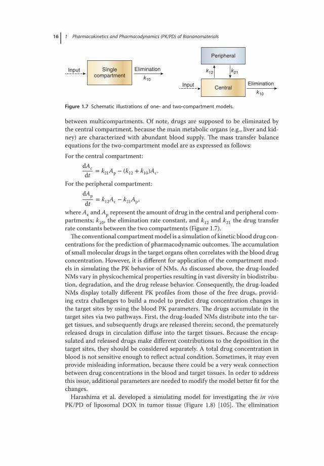
1.6.2Compartment Models for PK Assessment of NMs
Simulations have been widely applied in drug development and safety assessmentby describing the absorption, distribution, metabolism, and excretion (ADME) ofsmall molecular drugs by using the compartment models [104]. Tissues/organsare classified into different theoretical compartments, and the drug distributionin each compartment is treated as in an equilibrium state, with homogeneous drugconcentration.When the blood carries drugswith indiscriminate distribution intoall the organs, the whole body can be viewed as a single compartment.Meanwhile,the drugs undergo first-order elimination. Taken together, mass balance equationfor one compartment model after a single intravenously administration can beexpressed as follows:
dAcdt
= −k10Ac,
where Ac and k10 represent drug amount in the single compartment and the first-order clearance rate constant, respectively. In general, the single-compartmentmodel is one of the most simplified simulations based on the assumption thatall the organs/tissues are homogeneous in blood flow, drug partition coefficient,as well as elimination rate. However, in most cases, great flow diversity existsamong different organs/tissues. Under this circumstance, the organs with slowerblood flow are categorized as peripheral compartment, and drug transport occurs
16 1 Pharmacokinetics and Pharmacodynamics (PK/PD) of Bionanomaterials
Input
Input
Singlecompartment
Elimination
Elimination
Peripheral
Central
k10
k10
k12 k21
Figure 1.7 Schematic illustrations of one- and two-compartment models.
between multicompartments. Of note, drugs are supposed to be eliminated bythe central compartment, because the main metabolic organs (e.g., liver and kid-ney) are characterized with abundant blood supply. The mass transfer balanceequations for the two-compartment model are as expressed as follows:For the central compartment:
dAcdt
= k21Ap − (k12 + k10)Ac.
For the peripheral compartment:dAp
dt= k12Ac − k21Ap,
where Ac and Ap represent the amount of drug in the central and peripheral com-partments; k10, the elimination rate constant, and k12 and k21 the drug transferrate constants between the two compartments (Figure 1.7).The conventional compartmentmodel is a simulation of kinetic blood drug con-
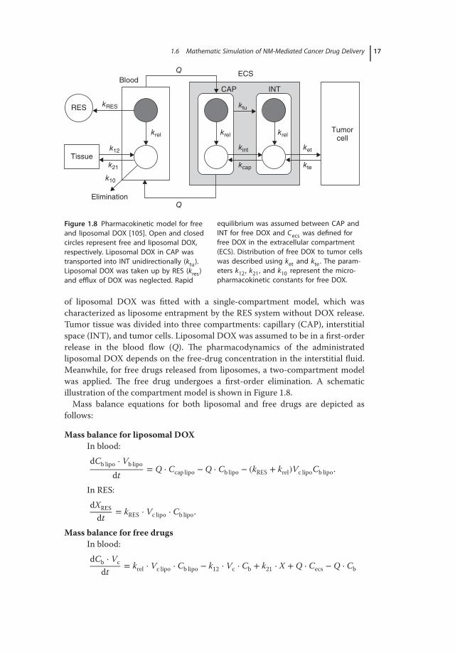
centrations for the prediction of pharmacodynamic outcomes. The accumulationof small molecular drugs in the target organs often correlates with the blood drugconcentration. However, it is different for application of the compartment mod-els in simulating the PK behavior of NMs. As discussed above, the drug-loadedNMs vary in physicochemical properties resulting in vast diversity in biodistribu-tion, degradation, and the drug release behavior. Consequently, the drug-loadedNMs display totally different PK profiles from those of the free drugs, provid-ing extra challenges to build a model to predict drug concentration changes inthe target sites by using the blood PK parameters. The drugs accumulate in thetarget sites via two pathways. First, the drug-loaded NMs distribute into the tar-get tissues, and subsequently drugs are released therein; second, the prematurelyreleased drugs in circulation diffuse into the target tissues. Because the encap-sulated and released drugs make different contributions to the deposition in thetarget sites, they should be considered separately. A total drug concentration inblood is not sensitive enough to reflect actual condition. Sometimes, it may evenprovide misleading information, because there could be a very weak connectionbetween drug concentrations in the blood and target tissues. In order to addressthis issue, additional parameters are needed to modify the model better fit for thechanges.Harashima et al. developed a simulating model for investigating the in vivo
PK/PD of liposomal DOX in tumor tissue (Figure 1.8) [105]. The elimination
1.6 Mathematic Simulation of NM-Mediated Cancer Drug Delivery 17
RES
BloodECS
CAP INT
Tumorcell
Tissue
EliminationQ
Q
kRES
krel krel krel
ktu
kint
kcap
ket
kte
k12
k21
k10
Figure 1.8 Pharmacokinetic model for freeand liposomal DOX [105]. Open and closedcircles represent free and liposomal DOX,respectively. Liposomal DOX in CAP wastransported into INT unidirectionally (ktu).Liposomal DOX was taken up by RES (kres)and efflux of DOX was neglected. Rapid
equilibrium was assumed between CAP andINT for free DOX and Cecs was defined forfree DOX in the extracellular compartment(ECS). Distribution of free DOX to tumor cellswas described using ket and kte. The param-eters k12, k21, and k10 represent the micro-pharmacokinetic constants for free DOX.
of liposomal DOX was fitted with a single-compartment model, which wascharacterized as liposome entrapment by the RES system without DOX release.Tumor tissue was divided into three compartments: capillary (CAP), interstitialspace (INT), and tumor cells. Liposomal DOX was assumed to be in a first-orderrelease in the blood flow (Q). The pharmacodynamics of the administratedliposomal DOX depends on the free-drug concentration in the interstitial fluid.Meanwhile, for free drugs released from liposomes, a two-compartment modelwas applied. The free drug undergoes a first-order elimination. A schematicillustration of the compartment model is shown in Figure 1.8.Mass balance equations for both liposomal and free drugs are depicted as
follows:
Mass balance for liposomal DOXIn blood:dCb lipo ⋅ Vb lipo
dt= Q ⋅ Ccap lipo − Q ⋅ Cb lipo − (kRES + krel)Vc lipoCb lipo.
In RES:dXRESdt
= kRES ⋅ Vc lipo ⋅ Cb lipo.
Mass balance for free drugsIn blood:dCb ⋅ Vc
dt= krel ⋅ Vc lipo ⋅ Cb lipo − k12 ⋅ Vc ⋅ Cb + k21 ⋅ X + Q ⋅ Cecs − Q ⋅ Cb

18 1 Pharmacokinetics and Pharmacodynamics (PK/PD) of Bionanomaterials
In tissue:dXdt
= k12 ⋅ Vc ⋅ Cb − k21 ⋅ X
In extracellular compartment (ECS):
dCecs ⋅ Vecsdt
= Q ⋅ Cb + kte ⋅ Vtu ⋅ Ctu − Q ⋅ Cecs − ket ⋅ Vecs ⋅ Cecs
+ krel(Vcap ⋅ Ccap lipo + Vint ⋅ Cint lipo)
In tumor cell:dCtu ⋅ Vtu
dt= ket ⋅ Vecs ⋅ Cecs − kte ⋅ Vtu ⋅ Ctu.
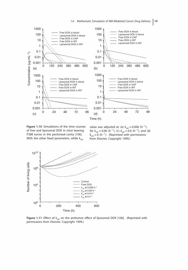
By using this model, the drug release rate and nonspecific uptake of liposomesby the RES systems could be assessed by krel and kres values (Figure 1.9).The tumorDox retention generally benefits from decreased RES uptake and a relatively fasterdrug release.By taking advantage of this model, PK/PD profiles of the long-circulating lipo-
somal DOX were simulated in a peritoneal leukemia mouse model. It was foundthat AUC of free DOX in interstitial space could be optimized by adjusting kelvalue at 0.06 h−1 (Figure 1.9). The results were further confirmed by the PD simu-lation, of which krel set as 0.06 h−1 displayed the most proficient antitumor effect(Figures 1.10 and 1.11) [106].
1600 0.6 4
3
2
1
0
0.4
0.2
0.0
1200
800
400
0
(a) (b) (c)
(d) (e) (f)
0.0 0.2 0.4
Kres
(h−1)
Cm
ax (μg
mL
−1)
80
60
40
20
0
T max (
h1)
80100
80
60
40
20
0
60
40
20
0
T max (
h1)
T max (
h1)
Cm
ax (μg
mL
−1)
Cm
ax (μg
mL
−1)
0.6 0.0 0.2 0.4
Kres
(h−1)
0.6 0.0 0.2 0.4
Kres
(h−1)
0.6
a: Cint
lipo
b: Cecs
dox
c: Ctu
dox
d: Tint
lipo
e: Tecs
dox
f: Ttu
dox
0.0 0.2 0.4
Kres
(h−1)
0.6 0.0 0.2 0.4
Kres
(h−1)
0.6 0.0 0.2 0.4
Kres
(h−1)
0.6
Krel
= 0.006
Krel
= 0.06
Krel
= 0.6
Krel
= 0.006
Krel
= 0.06
Krel
= 0.6
Figure 1.9 Changes of Cmax and Tmax in dif-ferent compartments with different K rel andK res. The parameters Cint lipo, Cecs dox, andCtu dox denote liposomal dox in the inter-stitial space, free dox in the ECS, and free
dox in the tumor site, respectively; T int lipo,Tecs dox, and T tu dox, the time at which Cmaxwas reached. (Reproduced from Table 2,with permissions from Elsevier, Copyright1999.)
1.6 Mathematic Simulation of NM-Mediated Cancer Drug Delivery 19
1000Free DOX in blood
Free DOX in blood
Free DOX in CAP
Free DOX in INT
Liposomal DOX in blood
Liposomal DOX in INT
Free DOX in CAP
Free DOX in INT
Liposomal DOX in blood
Liposomal DOX in INT
Free DOX in blood
Free DOX in CAP
Free DOX in INT
Liposomal DOX in blood
Liposomal DOX in INT
Free DOX in blood
Free DOX in CAP
Free DOX in INT
Liposomal DOX in blood
Liposomal DOX in INT
100
10
1
0.1
0.01
0.001
(a) (b)
(c) (d)
0 120 240 360 480 600
1000
100
10
1
0.1
0.01
0.0010 120 240 360 480 600
1000
Co
nce
ntr
atio
n (μg
·mL
−1)
100
10
1
0.1
0.01
0.0010 24 48 72 96 0 24 48 72 96
1000
100
10
1
0.1
0.01
0.001
Time (h)
Figure 1.10 Simulations of the time coursesof free and liposomal DOX in mice bearingP388 tumor in the peritoneal cavity [106].With the other fixed parameters, while krel
value was adjusted at: (a) krel = 0.006 (h−1),(b) krel = 0.06 (h−1), (c) krel = 0.6 (h−1), and (d)krel = 6 (h−1). (Reprinted with permissionsfrom Elsevier, Copyright 1999.)
108
104
100
0 200 400
Control
Free DOX
krel
at 0.006 h–1
krel
at 0.06 h–1
krel
at 0.6 h–1
krel
at 6 h–1
Time (h)
Num
ber
of liv
ing c
ells
600
1012
Figure 1.11 Effect of krel on the antitumor effect of liposomal DOX [106]. (Reprinted withpermissions from Elsevier, Copyright 1999.)

20 1 Pharmacokinetics and Pharmacodynamics (PK/PD) of Bionanomaterials
The model is useful, especially in predicting how drugs release and thenonspecific distribution affects the delivery efficiency of the encapsulated drugs.However, this model does not take other important factors into account, suchas physicochemical characters of the NMs (size, shape, zeta potential, etc.),NM-mediated tumor retention, site-specific drug release/drug delivery, and thephysiological differences among organs. These factors often play important rolesin NMs’ ADME. Therefore, specific models should be developed for differentcases in the PK/PD assessment of the drug-loaded NMs.
1.6.3Physiologically Based Compartment Models
The development of physiologically based compartment models has provided asolid basis for the establishment of physiologically based PK (PBPK) models forNMs [107, 108]. In thismethod, each organ or tissue of the bodywith anatomicallydistinct structure is defined as an independent compartment. These compart-ments are connected with blood flow, through which the NMs enter the compart-ment in a perfusion- or membrane-limited pattern. Therefore, the model can besubsequently divided into different types (Figure 1.12). For the perfusion-limitedmodel, NMs are assumed to be able to diffuse into the compartment rapidly, andthe transportation is mainly governed by the local blood supply. The mass trans-fer balance for a certain tissue is described as follows (annotations are listed inFigure 1.12):
dCTdt
=QTVT
⋅ CART-QT
VT ⋅ RT⋅ CVEN-CLT ⋅ CT.
Lungs
Spleen
Liver
Gl
Gl lumen
Kidney
Body
(a) (c)
(b)
Membrane-limited tissue
Metabolism
Excretion
Excretion
Vein
s
Art
erie
s
Absorption
Blood flow-limited tissue
Blood capillary
Intracellular space Tissue interstitial space
QT, CART QT, CVEN
KP,T
CT, VT
CLT
KT,P
QT, CART Qi, CVEN
KP,T
CT, VT
CLT
KT,P
Figure 1.12 (a) A typical blood flow-limited physiologically based pharmacokineticmodel structure; (b) a membrane-limited tissue; and (c) a blood flow-limited tissue [108].(Reprinted with permissions from American Chemical Society, Copyright 2010.)

1.6 Mathematic Simulation of NM-Mediated Cancer Drug Delivery 21
Table 1.1 Drug transport process equtions [109].
Specified process Equations
Capillary blood flow Navier–Stokes equation: 𝜌𝜕tuv − 𝜇𝛻2uv + 𝜌(uv ⋅ 𝛻)uv + 𝛻Pv = 0Tumor interstitial blood flow Darcy’s law: ui − V ⋅ [KPi] = 0Transmural blood flow Starling′s law: Jv = Lp(Pv − Pi) − 𝜎d(𝜋v − 𝜋i)Diffusion drug transport Diffusion convection reaction equation: 𝜕tc + 𝛻 ⋅ (−D𝛻c + uc) − R = 0Transmural drug transport Kedem–Katchalsky equation: JF = P(cv − ci) + (1 − 𝜎f )Jvc
For themembrane-limitedmodel, the rate-limiting step for transportation is theprocess of membrane penetration, and the mass balance equation is described asfollows:
dCTdt
= KP,T ⋅ CART −KT,P
RT⋅ CT − CLT ⋅ CT.
1.6.3.1 Protocols of Building a PBPKModel for NMsLi and coworkers proposed a three-step protocol for building PBPK models forassessing the PK profile of NMs [108]. First, specific analysis of each componentin themodel, including the description of characteristics of the physiological com-partments and NMs. Second, development of equations to describe the processof NMs/body interactions. Some equations reflecting the transport process havebeen summarized inTable 1.1. Generally,mass transfer balance equations are usedto describe transportation of the NMs and drug release. Third, calculation of thePBPK parameters. The experimental data are input into the established equationsfor calculating the model parameters. The robustness of the model could also beverified by comparing the simulated data with themeasured values. An optimizedmodel should be able to predict the ADME of NMs with minimum deviation.
1.6.3.2 ExamplesSize is an important factor affecting the NMs’ in vivo distribution (e.g., the bloodcirculation and transmembrane processes). Bachler and coworkers developed aPBPK model to assess the biodistribution of TiO2 nanoparticles based on theirability to cross the capillary and to be phagocytosed in the MPS [110]. The effectsof the fenestrated vasculatures on capture ofNMs could be divided into five groupsbased on the permeation of epithelial wall (Table 1.2).In the case of excretion, high permeation of the biologically stable nano-TiO2
into the liver resulted inmore pronounced biliary excretion than the urinary path-way. The biliary/urinary ratio of excretion was estimated to be 70 : 1. The ADMEprocesses are separately described later, and the mass balance equations can beexpressed as follows:
Absorption
mabsorption = nano-TiO2exposure ⋅ f cabsorption.

22 1 Pharmacokinetics and Pharmacodynamics (PK/PD) of Bionanomaterials
Table 1.2 Category and feature of capillary wall from different organs [110].
Category Features Organs
CT1 Nonsinusoidal, nonfenestrated capillary wall Brain, heart, lungCT2 Nonsinusoidal, fenestrated capillary Intestines, kidneys, stomachCT3 Sinusoidal capillary with pores> 15 nm Liver, spleenCT4 Myeloid bone marrow sinusoidal capillary BoneCT5 Other compartment that cannot be assigned to a CT group Hair, fat
CT, capillary wall type.
Distribution:In blood:dAblood
dt= mabsorption +
∑organsd
(ktrans-organ-blood ⋅ Aorgan)
−∑
organsa
(ktrans-blood-organ ⋅ Ablood).
In organ compartment:
dAorgan
dt= ktrans-blood-organ ⋅ Ablood − ktrans-organ-blood ⋅ Aorgan − kdeposition ⋅ Aorgan,
where the transportation coefficient from blood to the organs was definedas:
ktrans-blood-organ = btrans-constant-organ ⋅Qorgan-blood
Vblood
Organ deposition:
dAdeposition
dt= kdepositon ⋅ Aorgan − kclearance ⋅ Aorgan
Excretion:Biliary excretion:
dAbile eldt
= ktrans-blood-organ ⋅ Ablood +∑
organsa
(kclearance ⋅ Adeposition).
Urinary excretion:
dAurinary el
dt= ktrans-blood-organ ⋅ Ablood.
It was found that in a size range of 15–150 nm, the size of nano-TiO2 had aminor impact on the biodistribution. Furthermore, with high-dose exposure, theadministrated nanoparticles tended to agglomerate and bemainly captured by themacrophages in MPS.
1.6 Mathematic Simulation of NM-Mediated Cancer Drug Delivery 23
–K*el Cb
–K*el Cb
–K*21 CT
–K*21 CT
–K*12 Cb
AP
(Cr–
Cb)
AP
(Cr–
Cb)
–K*12 Cb
CT
CT
VT
VT
Vb
Vb
VrVrCrCr
Drug release
Blood
Blood
GI GI
GI
Tissues
Tissues
Figure 1.13 Schematic representation of drug RADME process in a polymer formulation[111]. (Reprinted with permissions from American Chemical Society, Copyright 2010.)
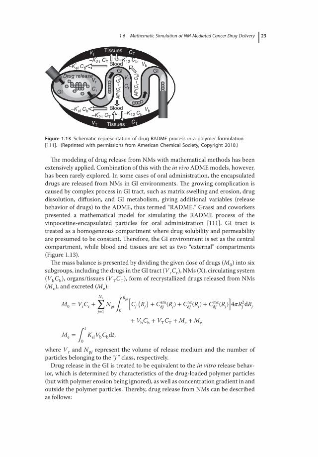
The modeling of drug release from NMs with mathematical methods has beenextensively applied. Combination of this with the in vivo ADMEmodels, however,has been rarely explored. In some cases of oral administration, the encapsulateddrugs are released from NMs in GI environments. The growing complication iscaused by complex process in GI tract, such as matrix swelling and erosion, drugdissolution, diffusion, and GI metabolism, giving additional variables (releasebehavior of drugs) to the ADME, thus termed “RADME.” Grassi and coworkerspresented a mathematical model for simulating the RADME process of thevinpocetine-encapsulated particles for oral administration [111]. GI tract istreated as a homogeneous compartment where drug solubility and permeabilityare presumed to be constant. Therefore, the GI environment is set as the centralcompartment, while blood and tissues are set as two “external” compartments(Figure 1.13).The mass balance is presented by dividing the given dose of drugs (M0) into six
subgroups, including the drugs in theGI tract (V rCr), NMs (X), circulating system(V bCb), organs/tissues (VTCT), form of recrystallized drugs released from NMs(Mc), and excreted (Me):
M0 = VrCr +Nc∑j=1
Npj ∫Rpj
0
[Cj
(Rj)+ Cam
dj (Rj) + Cncdj (Rj) + Cmc
dj (Rj)]4𝜋R2
j dRj
+ VbCb + VTCT + Mc + Me
Me = ∫t
0KelVbCbdt,
where V r and Npj represent the volume of release medium and the number ofparticles belonging to the “j ” class, respectively.Drug release in the GI is treated to be equivalent to the in vitro release behav-
ior, which is determined by characteristics of the drug-loaded polymer particles(but with polymer erosion being ignored), as well as concentration gradient in andoutside the polymer particles. Thereby, drug release from NMs can be describedas follows:
24 1 Pharmacokinetics and Pharmacodynamics (PK/PD) of Bionanomaterials
20
18
16
14
12
10
8
6
4
2
00 50 100
Experimental (Dose = 5 mg)
Experimental 5v
Experimental 3v
Model best fitting
Experimental (Dose = 2.5 mg)
Model best fitting
Model prediction
t (min) t (h)
Cr (μ
g c
m−
3)
Cb (
ng
cm
−3)
150 200 0 1 2 3 4 5 6 7
250
(b)(a)
200
150
100
50
0
Figure 1.14 Comparison of simulated vin-pocetine concentration with experimentaldata both in vitro (a, showing Cr –time profilein GI) and in vivo (b, showing Cb –time pro-file in blood). In both sets, solid lines refer to
the simulated data, while open or filled cir-cles represent experimental data from differ-ent tests [111]. (Reprinted with permissionsfrom American Chemical Society, Copyright2010.)
𝜕Cpj
𝜕t= 1
R2j
𝜕
𝜕Rj
(D𝜕Cj
𝜕RjR2
j
)−
(𝜕Cam
dj
𝜕t+
𝜕Cncdj
𝜕t+
𝜕Cmcdj
𝜕t
)
j = 1, 2, 3,…Nc,
where Cj is the concentration of the dissolved drug at radius Rj inside the par-ticles of the jth class;Cam
dj , Cncdj , and Cmc
dj are, respectively, the concentrations ofthe nondissolved drug in the amorphous, nanocrystalline, and macrocrystallinestates at radius Rj inside the particles of the jth class; and D is the drug diffusioncoefficient (depending on Cpj(Rj)).Blood concentration Cb changes as a result of the combined contribution of
drug influx from GI tract fluid, distribution into (or redistribution from) theorgans, and elimination. Thus the differential equation is defined as
VbdCbdt
= AP(Cr − Cb) − K∗elCb − K∗
12Cb + K∗21CT.
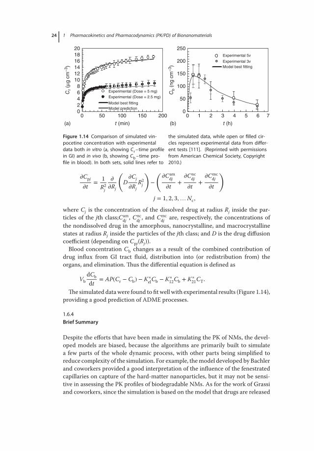
The simulated datawere found to fitwell with experimental results (Figure 1.14),providing a good prediction of ADME processes.
1.6.4Brief Summary
Despite the efforts that have been made in simulating the PK of NMs, the devel-oped models are biased, because the algorithms are primarily built to simulatea few parts of the whole dynamic process, with other parts being simplified toreduce complexity of the simulation. For example, themodel developed by Bachlerand coworkers provided a good interpretation of the influence of the fenestratedcapillaries on capture of the hard-matter nanoparticles, but it may not be sensi-tive in assessing the PK profiles of biodegradable NMs. As for the work of Grassiand coworkers, since the simulation is based on the model that drugs are released

1.7 Experimental PK Data of the Applied NMs 25
fromNMs before absorption by the GI tract, the model is not applicable when thedrugs are loaded onto NMs via specifically designed covalent bonds. The draw-back of the biased simulation is that the model only responds to changes of theparameters that are built in the functions.However, the PK of drug-loaded NMs is a very complicated process with each
component varying distinctly depending on different design. In order to makethe model feasible to a wide application, additional factors concerning features ofNMs (opsonization, receptor-mediated binding and internalization, site-specificrelease, etc.) and the body (receptor density, blood perfusion, enzyme activity,physiological volume of the organs, etc.) should be taken into account. On thecontrary, the incorporation of additional parameters to the functions will dra-matically increase the complexity of the model. As a solution, the transportationparameters that derive from a series of interrelated parameters are introduced. Forexample, the multiple properties of NMs, such as size, zeta potential, and PEGyla-tion, could be transformed into an individual parameter by a certain function (i.e.,linear multivariate regression) [112].Furthermore, establishment of a PBPK model for NMs is an interdisciplinary
work that includes characterization of the physicochemical properties of NMs,specification of physiological systems, and mathematical simulation. Therefore,it requires collaboration of professionals from different fields such as physiology,physical chemistry, nanotechnology, mathematics, and computer science. Impor-tantly, the robustness of the model is needed to be improved, which requires agreat amount of high-quality training data [113].
1.7Experimental PK Data of the Applied NMs
Owing to the development of nanotechnology, a variety of NMs are currentlyavailable for market and clinical trials in drug delivery. Here, we summarize thePK data of a number of NM-loaded drugs including paclitaxel [114–116], doxoru-bicin [117–121], curcumin [122–124], and large molecules such as siRNA [125]and insulin (Tables 1.3 and 1.4) [126, 127, 133]. By comparing the PK behaviorsof drugs encapsulated by different NMs, it would be helpful to get a better under-standing on how the physiochemical changes of NMs affect the ADME:
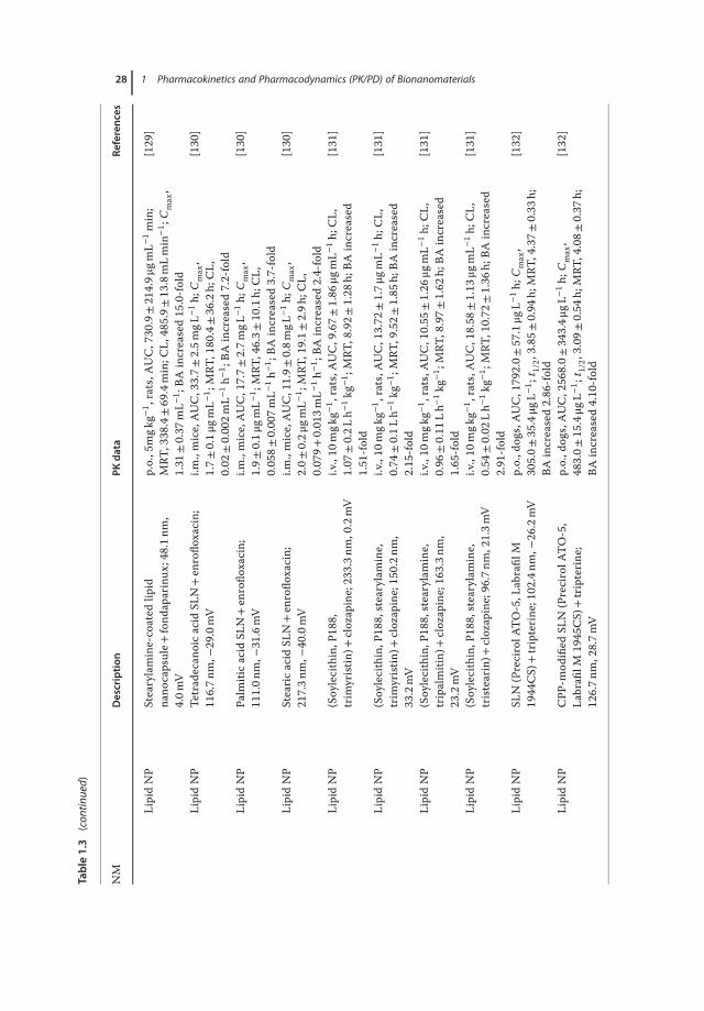
1. Size. In a study using PAMAMdendrimer for calcitonin delivery, it was foundthat the pharmacological effect was enhanced when large PAMAMwas used:the pharmacological availability (PA) was increased from 22.0% to 27.6% in acomparison between the G0 polymer and G3 dendrimer [141]. However, inthe case of orally administered drugs, small NMsmay be superior to the largeones in transporting across the intestinal membrane. Similarly, PEGylationthat help prevent NMs from aggregation under physiological conditions wasdemonstrated to be effective to increase the oral bioavailability of the encap-sulated drugs [121].
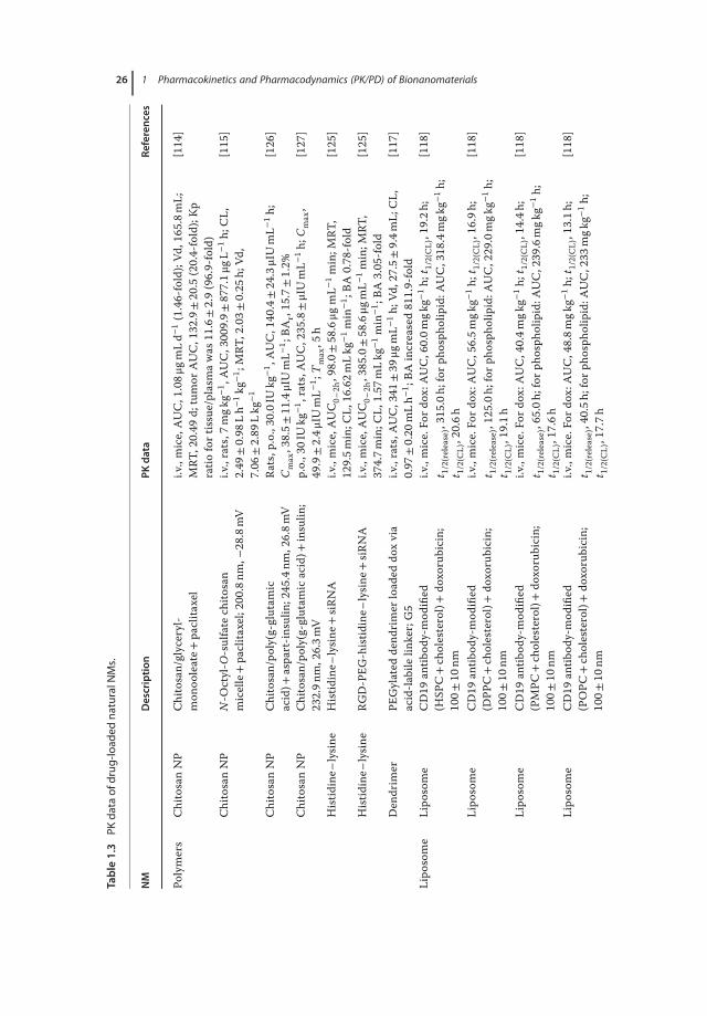
26 1 Pharmacokinetics and Pharmacodynamics (PK/PD) of Bionanomaterials
Table1.3
PKda
taof
drug
-load
edna
turalN
Ms.
NM
Description
PKdata
References
Polymers
ChitosanNP
Chitosan/glyceryl-
mon
ooleate+
paclita
xel
i.v.,mice,AUC,1.08μ
gmLd−
1(1.46-fold);Vd
,165
.8mL;
MRT
,20.49
d;tumor
AUC,132.9±20
.5(20.4-fold);Kp
ratio
fortissue/plasmawas
11.6±2.9(96.9-fold)
[114
]
ChitosanNP
N-O
ctyl-O
-sulfate
chito
san
micelle+paclita
xel;20
0.8n
m,−
28.8mV
i.v.,rats,7
mgk
g−1 ,AUC,3009.9±
877.1μ
gL−1h;
CL,
2.49
±0.98
Lh−
1kg
−1 ;MRT
,2.03±
0.25
h;Vd
,7.06
±2.89
Lkg
−1
[115
]
ChitosanNP
Chitosan/po
ly(g-glutamic
acid)+
aspart-in
sulin
;245
.4nm
,26.8m
VRa
ts,p
.o.,30
.0IU
kg−1 ,AUC,140.4±24.3μIUmL−
1h;
Cmax,38.5±
11.4μIUmL−
1 ;BA
r,15.7±1.2%
[126
]
ChitosanNP
Chitosan/po
ly(g-glutamicacid)+
insulin
;232.9n
m,26.3m
Vp.o.,30IUkg
−1 ,rats,A
UC,235.8±μIUmL−
1h;
Cmax,
49.9±2.4μ
IUmL−
1 ;Tmax,5
h[127
]
Histid
ine–
lysin
eHistid
ine–
lysin
e+siR
NA
i.v.,mice,AUC0–
2h,98.0±
58.6μg
mL−
1min;M
RT,
129.5m
in;C
L,16.62m
Lkg
−1min
−1 ;BA
0.78
-fold
[125
]
Histid
ine–
lysin
eRG
D-PEG
-histid
ine–
lysin
e+siR
NA
i.v.,mice,AUC0–
2h,385.0±58.6μg
mL−
1min;M
RT,
374.7m
in;C
L,1.57
mLkg
−1min
−1 ;BA
3.05
-fold
[125
]
Den
drim
erPE
Gylated
dend
rimer
loaded
doxvia
acid-la
bilelin
ker;G5
i.v.,rats,A
UC,341
±39
μgmL−
1h;
Vd,27.5±
9.4m
L;CL,
0.97
±0.20
mLh−
1 ;BA
increased81
1.9-fold
[117
]
Lipo
some
Lipo
some
CD19
antib
ody-mod
ified
(HSP
C+ch
olesterol)+do
xorubicin;
100±
10nm
i.v.,mice.Fo
rdox
:AUC,60.0m
gkg−
1h;
t 1/2(C
L),19.2h
;t 1/2(release),31
5.0h
;for
phosph
olipid:A
UC,318
.4mgk
g−1h;
t 1/2(C
L),20.6h
[118
]
Lipo
some
CD19
antib
ody-mod
ified
(DPP
C+ch
olesterol)+do
xorubicin;
100±
10nm
i.v.,mice.Fo
rdox
:AUC,56.5m
gkg−
1h;
t 1/2(C
L),16.9h
;t 1/2(release),12
5.0h
;for
phosph
olipid:A
UC,229
.0mgk
g−1h;
t 1/2(C
L),19.1h
[118
]
Lipo
some
CD19
antib
ody-mod
ified
(PMPC
+ch
olesterol)+do
xorubicin;
100±
10nm
i.v.,mice.Fo
rdox
:AUC,40.4m
gkg−
1h;
t 1/2(C
L),14.4h
;t 1/2(release),65
.0h;
forp
hospho
lipid:A
UC,239
.6mgk
g−1h;
t 1/2(C
L),17.6h
[118
]
Lipo
some
CD19
antib
ody-mod
ified
(POPC
+ch
olesterol)+do
xorubicin;
100±
10nm
i.v.,mice.Fo
rdox
:AUC,48.8m
gkg−
1h;
t 1/2(C
L),13.1h
;t 1/2(release),40
.5h;
forp
hospho
lipid:A
UC,233
mgk
g−1h;
t 1/2(C
L),17.7h
[118
]

1.7 Experimental PK Data of the Applied NMs 27
Lipo
some
CD19
antib
ody-mod
ified
(DOPC
+ch
olesterol)+do
xorubicin;
100±
10nm
i.v.,mice.Fo
rdox
:AUC,19.5m
gkg−
1h;
t 1/2(C
L),7.6h
t 1/2(release),1.9h
;for
phosph
olipid:A
UC,182
.0mgk
g−1h;
t 1/2(C
L),15.7h
[118
]
Lipo
some
Stealth
liposom
e+do
xorubicin;
100±
20nm
i.v.,mice,AUC,141
7.6μ
molmL−
1h;
CL,
0.08
mLh−
1 ;Vd
,1.7m
L;MRT
,19.9h
[128
]
Lipo
some
Fvfragmento
fCD19
antib
ody-mod
ified
stealth
liposom
e+do
xorubicin;
100±
20nm
i.v.,mice,AUC,997
.7μm
olmL−
1h;
CL,0.12
mLh−
1 ;Vd
,1.7m
L;MRT
,14.2h
[128
]
Lipo
some
Fab’fragmento
fCD19
antib
ody-mod
ified
stealth
liposom
e+do
xorubicin;
100±
20nm
i.v.,mice,AUC,129
1.2μ
molmL−
1h;
CL,
0.10
mLh−
1 ;Vd
,1.8m
L;MRT
,18.1h
[128
]
Lipo
some
Who
leCD19
antib
ody-mod
ified
stealth
liposom
e+do
xorubicin;
100±
20nm
i.v.,mice,AUC,292
.6μm
olmL−
1h;
CL,
0.41
mLh−
1 ;Vd
,2.3m
L;MRT
,5.6h
[128
]
Lipo
some
PEGylated
liposom
e+do
xi.v.,rats.A
UC,1786±
367μ
gmL−
1h;
Vd,6.6±1.5m
L;CL,
0.19
±0.02
mLh−
1 ;BA
increased42
52.4-fold
[117
]
Lipo
some
PEG-lipo
some+
dox;100.7n
m,−
20.1mV
AUC,2183±
460μ
gmLh;
MRT
,26.96
±4.01
h;t 1/2𝛽,
25.32±
4.89
h;CL0.69
±0.07
mLh−
1 ;BA
increased8.3-fold
[119
]
Lipo
some
RGD-PEG
-lipo
some+
dox;114.1n
m,
−24.9mV
AUC,1679±
189μ
gmLh;
MRT
,21.88
±0.97
h;t 1/2𝛽,
24.10±
2.42
h;CL1.10
±0.05
mLh−
1 ;BA
increased6.4-fold
[119
]
Lipo
some
PEGylated
liposom
e+do
xi.v.,rats6m
gkg−
1 ,Mice,AUC,225
7.5μ
gmL−
1h;
Tmax,3
h;Cmax,2.18μ
gmL−
1 ;BA
increased14
61-fold
[120
]
SLNs
LipidNP
SLN+do
x;28
0.2n
m,b
utmay
increased
to46
2.3n
mafterinc
ubated
with
intestinalflu
ids
p.o.,rats,
Cmax,1.90μ
gmL−
1 ;Tmax,1.5h,
AUC0–
t,17.79μ
gmL−
1h;
MRT
,22.87
h;Ke,0.2h
−1 ;BA
377.7%
[121
]
LipidNP
PEGylated
SLN+do
x;150.2n
m,and
remains
stablein
intestinalflu
ids
p.o.,rats,
Cmax,2.26μ
gmL−
1 ;Tmax,0.5,4
h,AUC0–
t,35.4μg
mL−
1h;
MRT
,33.03
h;Ke,0.07
h−1 ;BA
751.6%
[121
]
LipidNP
CTA
B-coated
lipid
nano
capsule+
fond
aparinux
;58.2n
m,
42.9mV
p.o.,rats5
mgk
g−1 ,rats,A
UC,336.5±187.3μ
gmL−
1min;
MRT
,177.1±82.6min;C
L,450.7±
7.1m
Lmin
−1 ;
Cmax,
0.98
±0.33
mL−
1 ;BA
increased6.9-fold
[129
]
(con
tinue
dov
erle
af)
28 1 Pharmacokinetics and Pharmacodynamics (PK/PD) of BionanomaterialsTable1.3
(con
tinued)
NM
Description
PKdata
References
LipidNP
Stearylamine-coated
lipid
nano
capsule+
fond
aparinux
;48.1n
m,
4.0m
V
p.o.,5mgk
g−1 ,rats,A
UC,730.9±214.9μ
gmL−
1min;
MRT
,338.4±69.4min;C
L,485.9±
13.8mLmin
−1 ;
Cmax,
1.31
±0.37
mL−
1 ;BA
increased15
.0-fold
[129
]
LipidNP
Tetradecan
oicacid
SLN+en
roflo
xacin;
116.7n
m,−
29.0mV
i.m.,mice,AUC,33.7±
2.5m
gL−1h;
Cmax,
1.7±
0.1μ
gmL−
1 ;MRT
,180.4±36.2h;
CL,
0.02
±0.002m
L−1h−
1 ;BA
increased7.2-fold
[130
]
LipidNP
Palm
iticacid
SLN+en
roflo
xacin;
111.0n
m,−
31.6mV
i.m.,mice,AUC,17.7±
2.7m
gL−1h;
Cmax,
1.9±
0.1μ
gmL−
1 ;MRT
,46.3±
10.1h;
CL,
0.058±
0.007m
L−1h−
1 ;BA
increased3.7-fold
[130
]
LipidNP
Stearicacid
SLN+en
roflo
xacin;
217.3n
m,−
40.0mV
i.m.,mice,AUC,11.9±
0.8m
gL−1h;
Cmax,
2.0±
0.2μ
gmL−
1 ;MRT
,19.1±
2.9h
;CL,
0.079+
0.013m
L−1h−
1 ;BA
increased2.4-fold
[130
]
LipidNP
(Soylecithin,P
188,
trim
yristin
)+clozapine;233.3n
m,0.2mV
i.v.,10
mgk
g−1 ,rats,A
UC,9.67±
1.86
μgmL−
1h;
CL,
1.07
±0.2L
h−1kg
−1 ;MRT
,8.92±
1.28
h;BA
increased
1.51
-fold
[131
]
LipidNP
(Soylecithin,P
188,stearylamine,
trim
yristin
)+clozapine;150.2n
m,
33.2mV
i.v.,10
mgk
g−1 ,rats,A
UC,13.72
±1.7μ
gmL−
1h;
CL,
0.74
±0.1L
h−1kg
−1 ;MRT
,9.52±
1.85
h;BA
increased
2.15
-fold
[131
]
LipidNP
(Soylecithin,P
188,stearylamine,
tripalmitin)
+clozapine;163.3n
m,
23.2mV
i.v.,10
mgk
g−1 ,rats,A
UC,10.55
±1.26
μgmL−
1h;
CL,
0.96
±0.11
Lh−
1kg
−1 ;MRT
,8.97±
1.62
h;BA
increased
1.65
-fold
[131
]
LipidNP
(Soylecithin,P
188,stearylamine,
tristearin)+
clozapine;96.7nm
,21.3m
Vi.v.,10
mgk
g−1 ,rats,A
UC,18.58
±1.13
μgmL−
1h;
CL,
0.54
±0.02
Lh−
1kg
−1 ;MRT
,10.72
±1.36
h;BA
increased
2.91
-fold
[131
]
LipidNP
SLN
(PrecirolA
TO-5,L
abrafil
M19
44CS)
+tripterine
;102.4nm
,−26.2mV
p.o.,d
ogs,AUC,179
2.0±
57.1μg
L−1h;
Cmax,
305.0±
35.4μg
L−1 ;
t 1/2,3.85±
0.94
h;MRT
,4.37±
0.33
h;BA
increased2.86
-fold
[132
]
LipidNP
CPP
-mod
ified
SLN
(PrecirolA
TO-5,
Labrafi
lM19
45CS)
+tripterine
;126.7n
m,28.7m
V
p.o.,d
ogs,AUC,256
8.0±
343.4μ
gL−1h;
Cmax,
483.0±
15.4μg
L−1 ;
t 1/2,3.09±
0.54
h;MRT
,4.08±
0.37
h;BA
increased4.10
-fold
[132
]

1.7 Experimental PK Data of the Applied NMs 29
Table1.4
PKda
taof
drug
-load
edsynthe
ticNMs.
NM
Description
PKdata
References
NPs
PLGANP
PEGylated
PLGA+insulin
;189.6nm
,−34.0mV
p.o.,10IUkg
−1 ,rats.5672±
4006
μIUmL−
1min
[133]
PLGANP
L-R8
-mod
ified
PEGylated
PLGA+insulin
;196.8nm
,14.7m
Vp.o.,10IUkg
−1 ,rats.135
70±8059
μIUmL−
1min
[133]
PLGANP
R-R8
-mod
ified
PEGylated
PLGA+insulin
;200.1nm
,15.3m
Vp.o.,10IUkg
−1 ,rats.191
45±10
876μ
IUmL−
1min
[133]
Polyan
hydride
NP
NP+paclita
xel;17
7nm,−
44.2mV
p.o.,rats,AUC,7.4±0.9μ
gmL−
1h;
Cmax,
0.2±
0.0μ
gmL−
1 ;MRT
,31±
1h;t
1/2,27
±1h
[134
]
Polyan
hydride
NP
PEG2k-N
P+paclita
xel;17
8nm,
−40.3mV
p.o.,rats,AUC,56±
3μgm
L−1h;
Cmax,2.1±0.1μ
gmL−
1 ;MRT
,26±
1h;t
1/2,9.3±
0.5h
[134
]
Polyan
hydride
NP
PEG6k-N
P+paclita
xel;18
0nm,
−39.5mV
p.o.,rats,AUC,32±
2μgm
L−1h;
Cmax,1.9±0.1μ
gmL−
1 ;MRT
,27±
1h;t
1/2,6.2±
0.3h
[134
]
Polyan
hydride
NP
PEG10k-NP+paclita
xel;18
8nm,
−41.1mV
p.o.,rats,AUC,13±
1μgm
L−1h;
Cmax,1.4±0.1μ
gmL−
1 ;MRT
,18±
1h;t
1/2,29
±2h
[134
]
PLGANP
PLGANP+gentam
icin;P
LGA50
:50,
13.7kD
a,1.84
±0.70
μmi.v.,mice,liv
er:A
UC,468
.3±58.7μg
g−1d(390
.3-fold);
MRT
,13.7d
;spleen:
AUC,416.0±168μ
gg−1d(462
.2-fold);
MRT
,45.0d
;kidne
y:AUC,109.3±3.3μ
gg−1d(7.3-fold);
MRT
,1.7d
[135
]
PLGANP
PLGANP+gentam
icin;P
LGA75
:25,
25kD
a,2.5±
0.17
μmi.v.,mice,liv
er:A
UC,425
.0±55
μgg−
1d(354
.2-fold);M
RT,
13.9d;
spleen
:AUC,745.3±55.7μg
g−1d(828
.1-fold);
MRT
,60.4d
;kidne
y:AUC,221.7±30.7μg
g−1d(14.8-fold);
MRT
,1.6d
[135
]
PLGANP
CylindricalshapeNP+do
cetaxel;
200×
200n
m(d
×h);−
3.4m
Vi.v.,mice,AUC0–
72h,1344
29ng
mL−
1h;
CL,
72mLkg
−1h;
Vd,257
mLkg
−1 ;BA
increased23
.1-fold;
fortum
or:
AUC0–
24h,79
644n
gmL−
1h;
Cmax,4413±
323n
gmL−
1 ;BA
increased1.3-fold
[136
]
(con
tinue
dov
erle
af)

30 1 Pharmacokinetics and Pharmacodynamics (PK/PD) of Bionanomaterials
Table1.4
(con
tinued)
NM
Description
PKdata
References
PLGANP
CylindricalshapeNP+do
cetaxel;
80×320n
m(d
×h);−
3.2m
Vi.v.,mice,AUC0–
72h,1334
62ng
mL−
1h;
CL,
73mLkg
−1h;
Vd,474
mLkg
−1 ;BA
increased23
.0-fold;
fortum
or:
AUC0–
24h,95
516n
gmL−
1h;
Cmax,4187±
23ng
mL−
1 ;BA
increased1.5-fold
[136
]
PLGA
PLGANP+curcum
in;161
.9nm
p.o.,50m
gkg−
1 ,rats,A
UC,134.25±
3.45
ngmL−
1h;
Cmax,
11.78±
0.45
ngmL;
Tmax,2
h;Ke,0.18
±0.02
h−1 ;Vd
,2073.66±
352.61
Lkg
−1 ;CL,
365.19
±37.35L
kg−1h−
1 ;BA
increased15
.6-fold
[122
]
PLGA
PEG-PLG
A+curcum
in;152
.4nm
p.o.,50m
gkg−
1 ,rats,A
UC,447.80±
64.03n
gmL−
1h;
Cmax,29.78
±4.63
ngmL;
Tmax,3
h;Ke,0.18
±0.02
h−1 ;Vd
,900.54
±225.77
Lkg
−1 ;CL,
103.679±
10.90L
kg−1h-2;BA
increased55
.5-fold
[122
]
PolymericNP
Polystyren
ebeads;30
0nm
Oral,mice,BD
atday1:brain14
.3%,h
eart12
.8%,kidne
y28
.2%,lun
g3.4%
,liver
41.3%,spleen0.2%
;BDat
day7:
brain13
.6%,h
eart10
.5%,kidne
y18
%,lun
g2.9%
,liver
53%,
spleen
2%
[137
]
PolymericNP
Rhod
amineNP;
374n
m,−
(10–
18)m
VOral,mice,BD
atday1:brain16
.4%,h
eart21
.3%,kidne
y25
.9%,lun
g4.2%
,liver
41.3%,spleen1.7%
;BDat
day7:
brain10
.1%,h
eart15
.7%,kidne
y18
.3%,lun
g11
.4%,liver
38.1%,spleen6.4%
[137
]
PolymericNP
Cou
marineNP;
375n
m,−
(10–
18)m
VOral,mice,BD
atday1:brain10
.8%,h
eart9.4%
,kidne
y23
.1%,lun
g2.7%
,liver
52.9%,spleen1.1%
;BDat
day7:
brain18
.1%,h
eart14
%,kidne
y27
.1%,lun
g4.7%
,liver
33.4%,spleen2.7%
[137
]
Micelles
Cop
olym
eric
micelle
PLGA-PEG
-PLG
Amicelle+curcum
in;
26.3nm
,−0.71
mV
I.V.,10
mgk
g−1 ,Mice.AUC,621
.6±24.7μg
L−1h;
Cmax,
2.17
±0.10
μgmL−
1 ;MRT
,0.90d
d±0.11
h;CL,
16.11±
0.66
mL−
1h−
1 ;BA
increased1.31
-fold;
liver:A
UC,
546.1μ
gL−1h(3.85-fold);spleen
:AUC,213
.7μg
L−1h
(0.34-fold);lung
:AUC,692
2.8μ
gL−1h(5.56-fold)
[123
]

1.7 Experimental PK Data of the Applied NMs 31
Polymeric
micelle
Polyethylene
glycol
poly(glutamicacid)
blockcopo
lymers+
cisplatin
;30n
mi.v.,rats,A
UC,132
5.9±
77.9
mL−
1h(64.7-fold);CL,
3.77
±0.21
mLh−
1kg
−1 ;MRT
,10.67
±0.15
h;Vs
s,40
±2.3m
Lkg
−1
[138
]
Polymeric
micelle
PCL-PE
G-PEG
+curcum
in;62.4n
m,
−4.25
mV
i.v.,rats,A
UC,277
2.5μ
gmL−
1h−
1 ;CL,
5mLh−
1kg
−1 ;
MRT
,30.07
h;Vs
s,0.216L
kg−1 ;BA
increased4.15
-fold
[124
]
Polymeric
micelle
PEG-polyaspartate
micelle+paclita
xel,
85nm
i.p.,mice,AUC,191
±32.1μg
mL−
1h;
BAincreased
127.3-fold,com
paredwith
crem
opho
rELform
ulation
[116
]
Polymeric
micelle
PEG-poly(metha
crylicacid)+
cisplatin
;110n
m,−
12mV
i.v.,mice,AUC,707
.03μ
gmL−
1h;
Vss,1.69
Lkg
−1 ;MRT
,12
.37h
;BAincreased21
.68-fold;tissueAUC:for
kidn
ey,
372μ
gg−1h(1.39-fold);forliver,229
1μgg
−1h(9.75-fold);
fors
pleen,
12115μ
gg−1h(178
.16-fold);fortum
or,
170μ
gg−1h(3.15-fold)
[139
]
Den
drim
erPA
MAM
G4PA
MAM
+indo
metha
cin
i.v.,3.3m
gkg−
1 ,rats,A
UC,125.9±22.3μg
mL−
1h;
MRT
,5.72
±0.47
h;BA
increased1.37
-fold;
AUCfortargeto
rgan
,396.4±
18.7μg
mL−
1h(1.91)
[140
]
PAMAM
Folic
acid-m
odified
G4
PAMAM
+indo
metha
cin;
folic
acid:
PAMAM
=3.86
i.v.,3.3m
gkg−
1 ,rats,A
UC,127.6±25.2μg
mL−
1h;
MRT
,6.60
±0.01
h;BA
increased1.39
-fold;
AUCfortarget
organ,57
9.0±
19.3μg
mL−
1h(2.79)
[140
]
PAMAM
Folic
acid-m
odified
G4
PAMAM
+indo
metha
cin;
folic
acid:
PAMAM
=11.9
i.v.,3.3m
gkg−
1 ,rats,A
UC,121.2±1.9μ
gmL−
1h;
MRT
,7.77
±1.70
h;BA
increased1.32
-fold;
AUCfortargeto
rgan
,602.9±
22.2μg
mL−
1h(2.91)
[140
]
PAMAM
Folic
acid-m
odified
G4
PAMAM
+indo
metha
cin;
folic
acid:
PAMAM
=21.4
i.v.,3.3m
gkg−
1 ,rats,A
UC,166.6±0.2μ
gmL−
1h;
MRT
,7.03
±0.07
h;BA
increased1.81
-fold;
AUCfortargeto
rgan
,850.7±
20.6μg
mL−
1h(4.10)
[140
]
PAMAM
PAMAM
G3+
insulin
;1%PA
MAM
Pulm
onaryabsorptio
n,rats.A
UC,
27916.8±
2140.8mUmL−
1min;B
A%,40.4±
3.1%
(4.2-fold)
[141
]
PAMAM
PAMAM
G0+
calcito
nin;
1%PA
MAM
Pulm
onaryabsorptio
n,rats.PA%,22.0±
0.8%
(2.3-fold)
[141
]
(con
tinue
dov
erle
af)
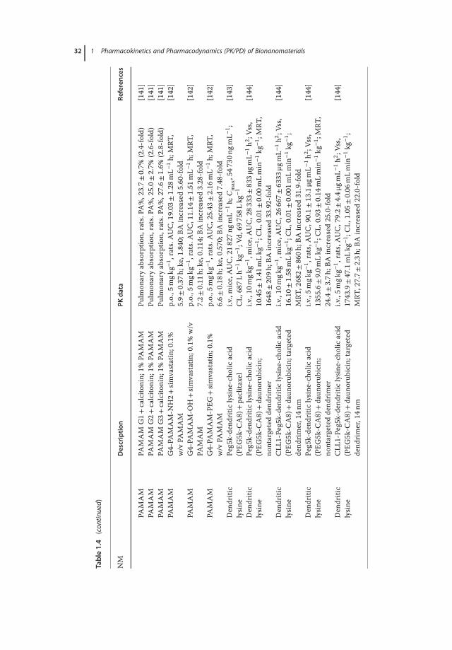
32 1 Pharmacokinetics and Pharmacodynamics (PK/PD) of Bionanomaterials
Table1.4
(con
tinued)
NM
Description
PKdata
References
PAMAM
PAMAM
G1+
calcito
nin;
1%PA
MAM
Pulm
onaryabsorptio
n,rats.PA%,23.7±
0.7%
(2.4-fold)
[141
]PA
MAM
PAMAM
G2+
calcito
nin;
1%PA
MAM
Pulm
onaryabsorptio
n,rats.PA%,25.0±
2.7%
(2.6-fold)
[141
]PA
MAM
PAMAM
G3+
calcito
nin;
1%PA
MAM
Pulm
onaryabsorptio
n,rats.PA%,27.6±
1.6%
(2.8-fold)
[141
]PA
MAM
G4-PA
MAM-N
H2+
simvastatin;0.1%
w/v
PAMAM
p.o.,5
mgk
g−1 ,rats.A
UC,19.03
±1.28
mL−
1h;
MRT
,5.9±
0.37
h;ke,1.840
;BAincreased5.60
-fold
[142
]
PAMAM
G4-PA
MAM-O
H+simvastatin;0.1%w/v
PAMAM
p.o.,5
mgk
g−1 ,rats.A
UC,11.14
±1.51
mL−
1h;
MRT
,7.2±
0.11
h;ke,0.114
;BAincreased3.28
-fold
[142
]
PAMAM
G4-PA
MAM-PEG
+simvastatin;0.1%
w/v
PAMAM
p.o.,5
mgk
g−1 ,rats.A
UC,25.43
±2.16
mL−
1h;
MRT
,6.6±
0.18
h;ke,0.570
;BAincreased7.48
-fold
[142
]
Den
dritic
lysine
Peg5k-dend
ritic
lysin
e-ch
olicacid
(PEG
5k-C
A8)
+paclita
xel
i.v.,mice,AUC,218
27ng
mL−
1h;
Cmax,547
30ng
mL−
1 ;CL,
687L
h−1kg
−1 ;Vd
,697
58Lkg
−1
[143
]
Den
dritic
lysine
Peg5k-dend
ritic
lysin
e-ch
olicacid
(PEG
5k-C
A8)
+daun
orub
icin;
nontargeteddend
rimer
i.v.,10
mgk
g−1 ,mice,AUC,283
33±833μ
gmL−
1h2;V
ss,
10.45±
1.41
mLkg
−1 ;CL,
0.01
±0.00
mLmin
−1kg
−1 ;MRT
,1648
±20
9h;B
Aincreased33
.92-fold
[144
]
Den
dritic
lysine
CLL
1-Pe
g5k-dend
ritic
lysin
e-ch
olicacid
(PEG
5k-C
A8)
+daun
orub
icin;targeted
dend
rimer,14n
m
i.v.,10
mgk
g−1 ,mice,AUC,266
67±6333
μgmL−
1h2;V
ss,
16.10±
1.58
mLkg
−1 ;CL,
0.01
±0.001m
Lmin
−1kg
−1 ;
MRT
,2682±
860h
;BAincreased31
.9-fold
[144
]
Den
dritic
lysine
Peg5k-dend
ritic
lysin
e-ch
olicacid
(PEG
5k-C
A8)
+daun
orub
icin;
nontargeteddend
rimer
i.v.,5m
gkg−
1 ,rats,A
UC,90.1±
13.1μg
mL−
1h2;V
ss,
1355.6±9.0m
Lkg
−1 ;CL,
0.93
±0.14
mLmin
−1kg
−1 ;MRT
,24.4±3.7h
;BAincreased25
.0-fold
[144
]
Den
dritic
lysine
CLL
1-Pe
g5k-dend
ritic
lysin
e-ch
olicacid
(PEG
5k-C
A8)
+daun
orub
icin;targeted
dend
rimer,14n
m
i.v.,5m
gkg−
1 ,rats,A
UC,79.2±
4.4μ
gmL−
1h2;V
ss,
1743.9±47.1mLkg
−1 ;CL,
1.05
±0.06
mLmin
−1kg
−1 ;
MRT
,27.7±
2.3h
;BAincreased22
.0-fold
[144
]
1.7 Experimental PK Data of the Applied NMs 33
2. Components. By adjusting the constituents, the features of NMs, such as size,zeta potential, and hydrophobicity, can be changed, thereby leading to differ-ent blood PK of the loading drugs [130, 131, 135].
3. Shape. In line with the findings that NMs with high aspect ratio wereless-efficiently phagocytized by MPS, cylinder PLGA nanoparticles with lowaspect ratio (200 nm× 200 nm, d × h) were more readily captured by the RESorgans, compared with elongated ones (80 nm× 320 nm, d × h), and thus wasmore effective in improving the BA of encapsulated drugs [136].
4. Surface coating. PEGylation is effective in improving systemic circulation ofNMs. For example, the blood clearance rate (CL) and mean resident time(MRT) of Dox [121] and curcumin [122] in the nanoformulations were greatlyextended with PEGylation. Surfactant coating was also effective in adjustingthe surface characteristics of NMs, and thus changed blood PK of the drugs[129].
5. Ligand modification. Generally, ligand modification promotes preferentialuptake by the target organ. In order to increase the targeting efficiency forindomethacin delivery, PAMAMs modified with different amounts of folicacid were investigated. The BA of the drugs in the target organ increasedwhen the dendrimer was coupled with more folate molecules. However,ligand modification may also cause unfavorable changes of the NMs and leadto accelerated elimination of NMs from the blood [119], or decreased bloodconcentration due to increased volume of apparent distribution (Vd) [144],which may account for the decreased blood BA. As a potential solution, smalland active fragments may be utilized. For example, the liposomes modifiedwith the Fab′, or Fv fragment fromCD 19 antibody can lower CL and Vd, with3.4-fold increase in blood AUC of the encapsulated dox compared with thosemodified with the whole antibody (1291.2 μgmL−1 h vs 292.6 μgmL−1 h)[128].
6. CPP modification. CPP modification enhances transport efficiency of NMs,and has been applied for oral drug delivery. It was demonstrated that the oralBA of tripterine [132] and insulin [133] was increased when NM loading wasmodified with CPPs.
It should be mentioned that the PK profiles are also affected by many other fac-tors such as intrinsic features of the drugs and routes of administration. All thesefactors contribute to diversity of the drug biofate.
1.7.1PK Data of NMsWithout Drugs
Dendrimers or metallic nanoparticles are useful tools for investigating NMs’ PKparameters, because the NMs can be conveniently tailored with tunable size ormodified with different ligands. For example, by using dendritic polylysines, it wasfound that the blood clearance rate decreased from 194 to 0.4mLh−1 when theMW increased from 6 to 68 kDa [97]. Furthermore, the incorporation of lipophilic

34 1 Pharmacokinetics and Pharmacodynamics (PK/PD) of Bionanomaterials
12.5
10.0
7.5
5.0
2.5
0.0
2 4 6 50
Size (nm)
Tis
su
e c
on
ce
ntr
atio
n o
f A
uN
P (
%/g
)
100 150 200
Liver
Spleen
Blood
Lungs
BrainKidney
Figure 1.15 Organ content of triphenylphosphine-coated AuNP with different size.
drugs can increase the surface hydrophobicity of the dendrimer, leading to a rapidblood clearance [145]. Although the RES organs have different “appetites” on theuptake of particles, PK results of triphenylphosphine-modified gold nanoparti-cles of size 1.4–200 nm revealed that the liver remained the most preferentialtarget of the injected NMs [146]. By plotting the tissue concentration against theparticle size, it was further depicted that the particle retention by the liver andspleen increased together with the increase of particle size, whereas renal contentdecreased as the particle size was increased to 5 nm, in line with the reported “cut-off” size of renal clearance (Figure 1.15). Other factors, such as zeta potential andsurface coating, could also be adjusted and lead to different PK profiles, which aresummarized in Table 1.5.
1. Long-chain PEGs are more effective than their short-chains counterpartsto create dense surface polymer coating [145]. Besides, the brush- andmushroom-like PEG confirmations are the most effective forms in protectingNMs from rapid clearance [98].
2. Renal clearance is a major way for elimination of ultrafine particles, but itdecreases once the NMs’ size surpasses the renal “cutoff.” Uptake by the RESorgan becomes the major clearance route for the large NMs.
1.7.2PK Differences Between Drugs Encapsulated by Different NMs
PK analysis of different formulations for a certain drug enables the selection ofthe suitable NMs for efficient delivery. Table 1.6 lists the PK data of paclitaxel

1.7 Experimental PK Data of the Applied NMs 35
Table1.5
PKda
taof
someNMs.
NM
Description
PKdata
References
Den
drim
ers
PEG-ly
sine
lys8(PEG
200)16
;6kD
ai.v.,5m
gkg,
Rats;C
L19
4±5.4m
Lh−
1 ;%
3 Hdo
sein
urine:
69.0±17.7%at
8h;82.2±
6.4%
atday7
[97]
PEG-ly
sine
lys16(PE
G20
0)32
;11.1k
Da
i.v.,5m
gkg,Ra
ts;C
L11
5±4.9m
Lh−
1 ;%
3 Hdo
sein
urine:
51.2±44.4%at
8h;80.4±
13.5%at
day7
[97]
PEG-ly
sine
lys16(PE
G57
0)32
,22.4k
Da
i.v.,5m
gkg,Ra
ts;C
L5.2±
0.8m
Lh−
1 ;%
3 Hdo
sein
urine:
27.4±3.4%
at8h
;42.9±
2.7%
atday7
[97]
PEG-ly
sine
lys8(PEG
2000
)16,34
.1kD
ai.v.,5m
gkg,Ra
ts;C
L0.9±
0.1m
Lh−
1 ;%
3 Hdo
sein
urine:4.0±
0.5%
at8h
;26.0±
4.6%
atday7
[97]
PEG-ly
sine
lys16(PE
G20
00)32;68
kDa
i.v.,5m
gkg,Ra
ts;C
L0.4±
0.1m
Lh−
1 ;%
3 Hdo
sein
urine:
0.6±
0.3%
at24
h;3.1±
1.9%
atday7
[97]
PEG-ly
sine
G4:(PEG
570)32
;MW,22.2k
Da
i.v.,mice,Vc
,13.8±0.9m
L;CL,
2.7±
0.1m
Lh−
1 ;%
3 Hdo
sein
urine:32
.7±7.8
[145
]
PEG-ly
sine
G5:(PEG
570)64
;MW,47.5k
Da
i.v.,mice,Vc
,13.0±0.6m
L;CL,
0.4±
0.0m
Lh−
1 ;%
3 Hdo
sein
urine:5.7±
0.5
[145
]
PEG-m
etho
trexate
G3:(PEG
570)8(MTX)8;M
W,10.5k
Da
i.v.,mice,Vc
,16.2±
0.7m
L;CL,
133±
19mLh−
1 ;%
3 Hdo
sein
urine:55
.7±8.6
[145
]
PEG-m
etho
trexate
G3:(PEG
1100)8(M
TX)8;M
W,14.8k
Da
i.v.,mice,Vc
,19.6±6.1m
L;CL,
98.5±29.3mLh−
1 ;%
3 Hdo
sein
urine:64
.1±4.7
[145
]
PEG-m
etho
trexate
G4:(PEG
570)16(M
TX)16;MW,21.1k
Da
i.v.,mice,Vc
,23.3±1.4m
L;CL,
51.9±2.3m
Lh−
1 ;%
3 Hdo
seexcreted
inurine:29
.0±3.4
[145
]
PEG-m
etho
trexate
G4:(PEG
1100)16(MTX)16;MW,
29.6kD
ai.v.,mice,Vc
,14.4±
1.7m
L;CL,
1.4±
0.1m
Lh−
1 ;%
3 Hdo
sein
urine:23
.5±17.7
[145
]
PEG-m
etho
trexate
G4:(PEG
2200)16(MTX)16;MW,
47.2kD
ai.v.,mice,Vc
,13.3±
0.6m
L;CL,
0.6±
0.1m
Lh−
1 ;%
3 Hdo
sein
urine:8.4±
1.4
[145
]
PEG-m
etho
trexate
G5:(PEG
570)32(M
TX)32;MW,42.2k
Da
i.v.,mice,Vc
,16.2±0.1m
L;CL,
1.5±
0.1m
Lh−
1 ;%
3 Hdo
sein
urine:2.4±
0.5
[145
]
PEG-m
etho
trexate
G5:(PEG
1100)32(MTX)32;MW,
59.2kD
ai.v.,mice,Vc
,16.0±
1.1m
L;CL,
0.6±
0.0m
Lh−
1 ;%
3 Hdo
sein
urine:1.2±
0.1
[145
]
(con
tinue
dov
erle
af)
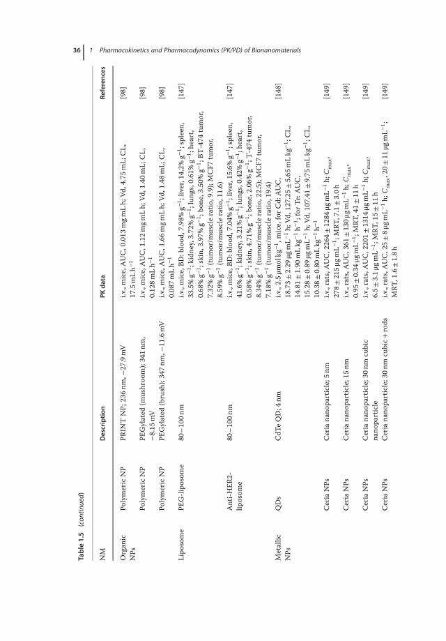
36 1 Pharmacokinetics and Pharmacodynamics (PK/PD) of Bionanomaterials
Table1.5
(con
tinued)
NM
Description
PKdata
References
Organ
icNPs
PolymericNP
PRIN
TNP;
236n
m,−
27.9mV
i.v.,mice,AUC,0.013
mgm
Lh;
Vd,4.75m
L;CL,
17.5mLh−
1[98]
PolymericNP
PEGylated
(mushroo
m);341n
m,
−8.15
mV
i.v.,mice,AUC,1.12m
gmLh;
Vd,1.40m
L;CL,
0.128m
Lh−
1[98]
PolymericNP
PEGylated
(brush);347n
m,−
11.6mV
i.v.,mice,AUC,1.66m
gmLh;
Vd,1.48m
L;CL,
0.087m
Lh−
1[98]
Lipo
some
PEG-lipo
some
80–100n
mi.v.,mice,BD
:blood
,7.98%
g−1 ;liv
er,14.2%
g−1 ;spleen
,33.5%g−
1 ;kidn
ey,3.72%
g−1 ;lung
s,0.61%g−
1 ;he
art,
0.68%g−
1 ;skin,3.97%
g−1 ;bo
ne,3.50%
g−1 ;BT
-474
tumor,
7.32%g−
1(tu
mor/m
uscleratio
,9.9);MCF7
tumor,
8.59%g−
1(tu
mor/m
uscleratio
,11.6)
[147
]
Anti-H
ER2-
liposom
e80
–10
0nm
i.v.,mice,BD
:blood
,7.04%
g−1 ;liv
er,15.6%
g−1 ;spleen
,41.6%g−
1 ;kidn
ey,3.21%
g−1 ;lung
s,0.42%g−
1 ;he
art,
0.58%g−
1 ;skin,4.71%
g−1 ;bo
ne,2.06%
g−1 ;T-474
tumor,
8.34%g−
1(tu
mor/m
uscleratio
,22.5);M
CF7
tumor,
7.18%g−
1(tu
mor/m
uscleratio
,19.4)
[147
]
Metallic
NPs
QDs
CdT
eQD;4
nmi.v.,2.5μ
molkg
−1 ,mice,forC
d:AUC,
18.73±
2.29
μgmL−
1h;
Vd,127.25±
5.65
mLkg
−1 ;CL,
14.81±
1.90
mLkg
−1h−
1 ;forT
e:AUC,
15.28±
0.89
μgmL−
1h;
Vd,107.41±
9.75
mLkg
−1 ;CL,
10.38±
0.80
mLkg
−1h−
1
[148
]
CeriaNPs
Ceriana
noparticle;5
nmi.v.,rats,A
UC,226
4±1284
μgmL−
1h;
Cmax,
278±
215μ
gmL−
1 ;MRT
,7.1±3.0h
[149
]
CeriaNPs
Ceriana
noparticle;15n
mi.v.,rats,A
UC,361
±130μ
gmL−
1h;
Cmax,
0.95
±0.34
μgmL−
1 ;MRT
,41±
11h
[149
]
CeriaNPs
Ceriana
noparticle;30n
mcubic
nano
particle
i.v.,rats,A
UC,220
1±1314
μgmL−
1h;
Cmax,
6.5±
3.1μ
gmL−
1 ;MRT
,15±
11h
[149
]
CeriaNPs
Ceriana
noparticle;30n
mcubic+
rods
i.v.,rats,A
UC,25±
8μgm
L−1h;
Cmax,20±
11μg
mL−
1 ;MRT
,1.6±1.8h
[149
]

1.7 Experimental PK Data of the Applied NMs 37
GoldNPs
Ceriana
noparticle;50n
mi.v.,rats,A
UC,1.5±0.5μ
gmL−
1h;
Cmax,
0.78
±0.12
μgmL−
1 ;MRT
,1.5±0.1h
[149
]
GoldNPs
Citrate-coated
gold
nano
particle;
22.4nm
,−44.7±7.5m
Vi.v.,rats,B
Dat
24h:
liver,57.94
±2.57
%;spleen,
1.34
±0.23%;lun
g,0.72
±0.44
%;b
lood
,1.27±
0.43
%;h
eart,
0.04
±0.01
%;kidne
y,0.08
±0.02%
[150
]
GoldNPs
MUA-coatedgo
ldna
noparticle;33.2n
m,
−37.3±8.4m
Vi.v.,rats,B
Dat
24h:
liver,59.37
±4.08
%;spleen,
2.26
±0.52%;lun
g,0.44
±0.19
%;b
lood
,1.13±
0.71
%;h
eart,
0.01
±0.01
%;kidne
y,0.03
±0.03%
[150
]
GoldNPs
CALN
N-coatedgo
ldna
noparticle;
22.4nm
,−47.1±6.0m
Vi.v.,rats,B
Dat
24h:
liver,86.11
±7.13
%;spleen,
2.79
±0.30%;lun
g,0.53
±0.06
%;b
lood
,0.32±
0.13
%;h
eart,
0.01
±0.00
%;kidne
y,0.20
±0.07%
[150
]
GoldNPs
CALN
D-coatedgo
ldna
noparticle;
22.4nm
,−43.6±9.2m
Vi.v.,rats,B
Dat
24h:
liver,77.47
±2.52
%;spleen,
2.51
±0.59%;lun
g,0.88
±0.6%
;blood
,0.19±
0.08
%;h
eart,
0.02
±0.01
%;kidne
y,0.05
±0.02%
[150
]
GoldNPs
CALN
S-coated
gold
nano
particle;
22.4nm
,−40.5±7.0m
Vi.v.,rats,B
Dat
24h:
liver,73.66
±4.84
%;spleen,
1.88
±0.13%;lun
g,0.52
±0.09
%;b
lood
,0.38±
0.07
%;h
eart,
0.01
±0.00
%;kidne
y,0.01
±0.00%
[150
]
GoldNPs
Triphe
nylpho
sphine
-mod
ified
AuN
P;1.4n
m,−
20.6mV
i.v.,3.1μ
g/rat,NPconcentrationat
24h:
liver,5.1%g−
1 ;spleen
,2.3%g−
1 ;lung
s,0.2%
g−1 ;kidn
ey,1.9%g−
1 ;brain,
0.0064%g−
1 ;bloo
d,0.11%g−
1
[146
]
GoldNPs
Triphe
nylpho
sphine
-mod
ified
AuN
P;5n
m,−
21.1mV
i.v.,43
.7μg/rat,N
Pconcentrationat
24h:
liver,10.0%
g−1 ;
spleen
,2.7%g−
1 ;lung
s,0.035%
g−1 ;kidn
ey,0.037%g−
1 ;brain,
0.00006%
g−1 ;bloo
d,0.02
4%g−
1
[146
]
GoldNPs
Triphe
nylpho
sphine
-mod
ified
AuN
P;18
nm,−
22.8mV
i.v.,2.9μ
g/rat,NPconcentrationat
24h:
liver,10.0%
g−1 ;
spleen
,1.6%g−
1 ;lung
s,0.038%
g−1 ;kidn
ey,0.013%g−
1 ;brain,
0.000031%g−
1 ;bloo
d,0.01
6%g−
1
[146
]
GoldNPs
Triphe
nylpho
sphine
-mod
ified
AuN
P;80
nm,−
22.3mV
i.v.,18
.5μg/rat,N
Pconcentration:
liver,11%
g−1 ;spleen
,1.9%
g−1 ;lung
s,0.027%
g−1 ;kidn
ey,0.016%g−
1 ;brain,
0.00029%
g−1 ;bloo
d,0.00
16%g−
1
[146
]
(con
tinue
dov
erle
af)

38 1 Pharmacokinetics and Pharmacodynamics (PK/PD) of Bionanomaterials
Table1.5
(con
tinued)
NM
Description
PKdata
References
GoldNPs
Triphe
nylpho
sphine
-mod
ified
AuN
P;200n
m,−
41.3mV
i.v.,19
.8μg/rat,N
Pconcentration:
liver,12%
g−1 ;spleen
,3.6%
;lun
gs,0.037%g−
1 ;kidn
ey,0.0025%
g−1 ;brain,
0.000021%g−
1 ;bloo
d,0.00
086%
g−1
[146
]
GoldNPs
Thioglycolicacid-m
odified
AuN
P;ne
gativ
ei.v.,1.6μ
g/rat,NPconcentration:
liver,9.2%g−
1 ;spleen
,8.9%
g−1 ;lung
s,1.2%
g−1 ;kidn
ey,0.4%g−
1 ;brain,
0.0018%g−
1 ;bloo
d,0.11%g−
1
[146
]
GoldNPs
Cysteam
ine-mod
ified
AuN
P;po
sitive
i.v.,29
.0μg/rat,N
Pconcentration:
liver,7.9%g−
1 ;spleen
,13%g−
1 ;lung
s,1.2%
g−1 ;kidn
ey,0.48%
g−1 ;brain,
0.0028%g−
1 ;bloo
d,0.09
5%g−
1
[146
]
Silver
NPs
Aqu
eous
AgN
P;20.3nm
,23.8μ
gmL−
1i.v.,23
.8μg/rat,N
Pconcentrationat
2d:b
lood
,602±
98ng
g−1 ;liv
er,232
6±676n
gg−1 ;lung
s,31
±11
ngg−
1 ;spleen
,38±
5ngg
−1 ;brain,
8±5n
gg−1 ;
heart,7±
5ngg
−1 ;kidn
ey,110
±45
ngg−
1 ;total
tissue-accumulated
Ag,13
±3%
[151
]
Silver
NPs
Aqu
eous
AgN
P;79.8nm
,26.4μ
gmL−
1i.v.,26
.4μg/rat,N
Pconcentrationat
2d:b
lood
,896±
303n
gg−1 ;liv
er,696
8±3231
ngg−
1 ;lung
s,603±
356n
gg−1 ;spleen
,110
±22
ngg−
1 ;brain,
10±3n
gg−1 ;he
art,63
±11
ngg−
1 ;kidn
ey,9
±1n
gg−1 ;
totaltissue-accumulated
Ag,33
±10%
[151
]
Silver
NPs
Aqu
eous
AgN
P;112.6n
m,27.6μ
gmL−
1i.v.,27
.6μg/rat,N
Pconcentrationat
2d:b
lood
,582±
358n
gg−1 ;liv
er,133
81±567n
gg−1 ;lung
s,129±
60ng
g−1 ;spleen
,859
±34
ngg−
1 ;brain,
8±1n
gg−1 ;
heart,11
±3n
gg−1 ;kidn
ey,83±
27ng
g−1 ;total
tissue-accumulated
Ag,55
±0%
[151
]
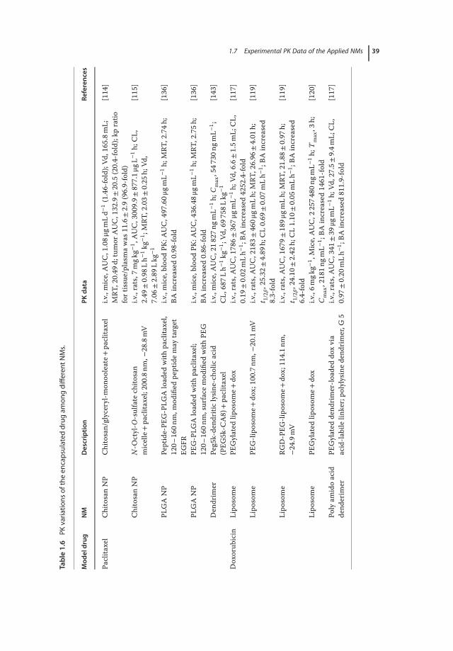
1.7 Experimental PK Data of the Applied NMs 39
Table1.6
PKvaria
tions
oftheen
capsulateddrug
amon
gdifferen
tNMs.
Mod
eldrug
NM
Description
PKdata
References
Paclita
xel
ChitosanNP
Chitosan/glyceryl-m
onoo
leate+
paclita
xel
i.v.,mice,AUC,1.08μ
gmLd−
1(1.46-fold);Vd
,165
.8mL;
MRT
,20.49
d;tumor
AUC,132.9±20
.5(20.4-fold);kp
ratio
fortissue/plasmawas
11.6±2.9(96.9-fold)
[114
]
ChitosanNP
N-O
ctyl-O
-sulfate
chito
san
micelle+paclita
xel;20
0.8n
m,−
28.8mV
i.v.,rats,7
mgk
g−1 ,AUC,3009.9±
877.1μ
gL−1h;
CL,
2.49
±0.98
Lh−
1kg
−1 ;MRT
,2.03±
0.25
h;Vd
,7.06
±2.89
Lkg
−1
[115
]
PLGANP
Peptide-PE
G-PLG
Aloaded
with
paclita
xel,
120–
160n
m,m
odified
peptidemay
target
EGFR
i.v.,mice,bloo
dPK
:AUC,497
.60μ
gmL−
1h;
MRT
,2.74h
;BA
increased0.98
-fold
[136
]
PLGANP
PEG-PLG
Aloaded
with
paclita
xel;
120–
160n
m,surface
mod
ified
with
PEG
i.v.,mice,bloo
dPK
:AUC,436
.48μ
gmL−
1h;
MRT
,2.75h
;BA
increased0.86
-fold
[136
]
Den
drim
erPe
g5k-dend
ritic
lysin
e-ch
olicacid
(PEG
5k-C
A8)
+paclita
xel
i.v.,mice,AUC,218
27ng
mL−
1h;
Cmax,547
30ng
mL−
1 ;CL,
687L
h−1kg
−1 ;Vd
,697
58Lkg
−1
[143
]
Doxorub
icin
Lipo
some
PEGylated
liposom
e+do
xi.v.,rats,A
UC,1786±
367μ
gmL−
1h;
Vd,6.6±1.5m
L;CL,
0.19
±0.02
mLh−
1 ;BA
increased42
52.4-fold
[117
]
Lipo
some
PEG-lipo
some+
dox;100.7n
m,−
20.1mV
i.v.,rats,A
UC,2183±
460μ
gmLh;
MRT
,26.96
±4.01
h;t 1/2𝛽,25.32
±4.89
h;CL0.69
±0.07
mLh−
1 ;BA
increased
8.3-fold
[119
]
Lipo
some
RGD-PEG
-lipo
some+
dox;114.1n
m,
−24.9mV
i.v.,rats,A
UC,167
9±189μ
gmLh;
MRT
,21.88
±0.97
h;t 1/2𝛽,24.10
±2.42
h;CL1.10
±0.05
mLh−
1 ;BA
increased
6.4-fold
[119
]
Lipo
some
PEGylated
liposom
e+do
xi.v.,6m
gkg−
1 ,Mice,AUC,2
2574
80ng
mL−
1h;
Tmax,3
h;Cmax,2181n
gmL−
1 ;BA
increased14
61-fold
[120
]
Polyam
idoacid
dend
erim
erPE
Gylated
dend
rimer-lo
aded
doxvia
acid-la
bilelin
ker;po
lylysine
dend
rimer,G
5i.v.,rats,A
UC,341
±39
μgmL−
1h;
Vd,27.5±
9.4m
L;CL,
0.97
±0.20
mLh−
1 ;BA
increased81
1.9-fold
[117
]

40 1 Pharmacokinetics and Pharmacodynamics (PK/PD) of Bionanomaterials
and doxorubicin encapsulated by different NMs. It should be noted that conclu-sions should be carefully drawn based the comparison because the experimentaldifferences exist in drug dose, administration route, animal species, sampling pro-tocol, PK model, and the method of data analysis. A “standard protocol” shouldbe designed to acquire the high-quality data in analyzing PK profiles of differ-ent NMs.
1.7.3Reciprocal Blood and Tissue PK
There could be a poor correlation between drug concentrations in blood and thetarget organs for NM-based drug delivery. In this case, the discrepancy betweenblood PK and PD cannot be precisely predicted by using the conventional PK/PDmodel. Therefore, it is important to compare both the blood and tissue concen-trations when analyzing PK of drug-loaded NMs. Table 1.7 summarizes both theblood and tissue PK from a number of NMs. It was demonstrated that the NMswith a relatively longer blood resident time were usually less efficiently taken bythe major organs [152–154]. As for tumor-targeting NMs, there often was notmuch difference between the ligand-modified and nonmodified particles in bloodPK [136], probably because of the small proportion of tumor-retained drugs.
1.7.4PK Differences Between Different Components of the Drug-NM System
In the case of drug co-encapsulation, the PK profiles also show different behaviorsfor two drugs (Table 1.8). For example, the t1/2 of the paclitaxel was less than halfof the co-loaded EGCG (26.5 h vs 56.1 h) due to the intrinsic differences betweenthe two drugs [155]. Furthermore, variations also existed between NMs and theirencapsulated drugs (Table 1.9). It was found that blood PK of encapsulated Doxwas different from that of the loading phospholipid, and drug t1/2 depended onthe release kinetics of the drugs from the liposomes; the liposomes with slowerdrug release had higher AUC [118].
1.7.5PK Variations Among Different Routes of Administration
For intravenous administration, biodistribution of NMs is size-dependent, andrapid uptake of smallNMsby theRESorgans leads to a fast clearance of the loadingdrugs from the blood. When other types of administration (such as p.o., i.p., s.c.,and i.m.) are applied, the given NMs have to penetrate a physiological membranebarrier before entering blood circulation, thereby leading to the changed PK pro-files [158–160]. For the routes other than intravenous (i.v.) injection, NMs needto be carefully tailored by adopting effective strategies such as small size [134],PEGylation [121, 122], and CPP modification [132], so as to improve the bloodPK of the loaded drugs (Table 1.10).
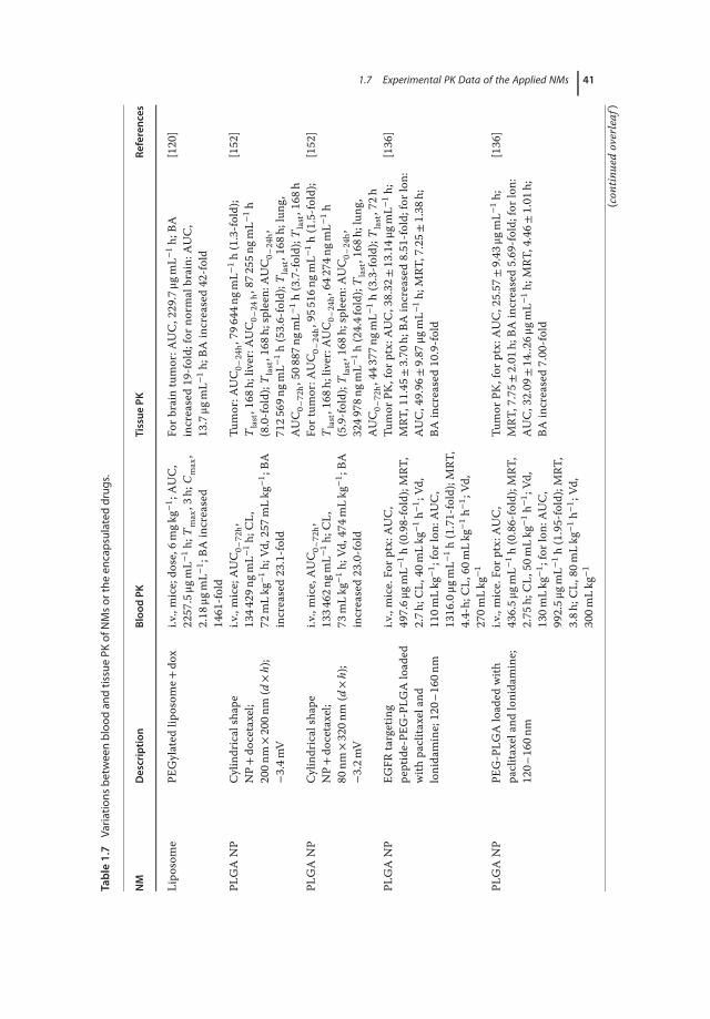
1.7 Experimental PK Data of the Applied NMs 41
Table1.7
Varia
tions
betweenblood
andtissuePK
ofNMsor
theen
capsulateddrug
s.
NM
Description
Blood
PKTissue
PKRe
ferences
Lipo
some
PEGylated
liposom
e+do
xi.v.,mice;do
se,6
mgk
g−1 ;AUC,
2257.5μg
mL−
1h;
Tmax,3
h;Cmax,
2.18
μgmL−
1 ;BA
increased
1461
-fold
Forb
rain
tumor:A
UC,229.7μg
mL−
1h;
BAincreased19
-fold;
forn
ormalbrain:
AUC,
13.7μg
mL−
1h;
BAincreased42
-fold
[120
]
PLGANP
Cylindricalshape
NP+do
cetaxel;
200n
m×200n
m(d
×h);
−3.4m
V
i.v.,mice;AUC0–
72h,
1344
29ng
mL−
1h;
CL,
72mLkg
−1h;
Vd,257
mLkg
−1 ;BA
increased23
.1-fold
Tumor:A
UC0–
24h,79
644n
gmL−
1h(1.3-fold);
Tlast,168
h;liv
er:A
UC0–
24h,87
255n
gmL−
1h
(8.0-fold);T
last,168
h;spleen
:AUC0–
24h,
7125
69ng
mL−
1h(53.6-fold);
Tlast,168
h;lung
,AUC0–
72h,50
887n
gmL−
1h(3.7-fold);T
last,168
h
[152
]
PLGANP
Cylindricalshape
NP+do
cetaxel;
80nm
×320n
m(d
×h);
−3.2m
V
i.v.,mice,AUC0–
72h,
1334
62ng
mL−
1h;
CL,
73mLkg
−1h;
Vd,474
mLkg
−1 ;BA
increased23
.0-fold
Fortum
or:A
UC0–
24h,95
516n
gmL−
1h(1.5-fold);
Tlast,168
h;liv
er:A
UC0–
24h,64
274n
gmL−
1h
(5.9-fold);T
last,168
h;spleen
:AUC0–
24h,
3249
78ng
mL−
1h(24.4fold);T
last,168
h;lung
,AUC0–
72h,44
377n
gmL−
1h(3.3-fold);T
last,72h
[152
]
PLGANP
EGFR
targeting
peptide-PE
G-PLG
Aloaded
with
paclita
xeland
lonidamine;120–
160n
m
i.v.,mice.Fo
rptx:A
UC,
497.6μ
gmL−
1h(0.98-fold);MRT
,2.7h
;CL,
40mLkg
−1h−
1 ;Vd
,110m
Lkg
−1 ;forlon
:AUC,
1316.0μg
mL−
1h(1.71-fold);MRT
,4.4-h;
CL,
60mLkg
−1h−
1 ;Vd
,270m
Lkg
−1
Tumor
PK,for
ptx:AUC,38.32
±13.14μ
gmL−
1h;
MRT
,11.45
±3.70
h;BA
increased8.51
-fold;
forlon
:AUC,49.96
±9.87
μgmL−
1h;
MRT
,7.25±
1.38
h;BA
increased10
.9-fold
[136
]
PLGANP
PEG-PLG
Aloaded
with
paclita
xeland
lonidamine;
120–
160n
m
i.v.,mice.Fo
rptx:A
UC,
436.5μ
gmL−
1h(0.86-fold);MRT
,2.75
h;CL,
50mLkg
−1h−
1 ;Vd
,130m
Lkg
−1 ;forlon
:AUC,
992.5μ
gmL−
1h(1.95-fold);MRT
,3.8h
;CL,
80mLkg
−1h−
1 ;Vd
,300m
Lkg
−1
Tumor
PK,for
ptx:AUC,25.57
±9.43
μgmL−
1h;
MRT
,7.75±
2.01
h;BA
increased5.69
-fold;
forlon
:AUC,32.09
±14..26μ
gmL−
1h;
MRT
,4.46±
1.01
h;BA
increased7.00
-fold
[136
]
(con
tinue
dov
erle
af)

42 1 Pharmacokinetics and Pharmacodynamics (PK/PD) of Bionanomaterials
Table1.7
(con
tinued)
NM
Description
Blood
PKTissue
PKRe
ferences
Nan
oemulsio
nNE+ch
lorambu
cil;
soybeanoil,egglecithin,
cholesterol
i.v.10m
gkg−
1 ,mice.AUC,
32.4±0.1μ
gmL−
1h;
MRT
,1.3±
0.04
h;CL,
309±
16mLkg
−1h−
1 ;Vs
s,409±
15mLkg
−1 ;BA
increased
1.91
-fold
AUC:liver,7.4±0.02
g−1h(0.90-fold);kidn
ey,
8.0±
0.03
g−1h(0.72-fold);he
art,8.2±
0.1g
−1h
(0.75-fold);lung
s,12
.0±0.2g
−1h(0.92-fold);
spleen
,5.2±0.3g
−1h(1.24-fold)
[153
]
Nan
oemulsio
nPE
G-N
E+ch
lorambu
cil;
soybeanoil,egglecithin,
cholesterol,
DSP
E-PE
G2000
i.v.10m
gkg−
1 ,mice.AUC,
45±30
μgmL−
1h;
MRT
,1.9±
0.1h
;CL,
221±
1.4m
Lkg
−1h−
1 ;Vs
s,339±
5.3m
Lkg
−1 ;BA
increased
2.65
-fold
AUC:liver
,3.1±0.02
g−1h(0.38-fold);kidn
ey,
7.1±
0.02
g−1h(0.64-fold);he
art,8.0±
0.1g
−1h
(0.73-fold);lung
s,5.0±
0.1g
−1h(0.38-fold);spleen
,1.0±
0.1g
−1h(0.24-fold)
[153
]
Cyclodextran
inclusioncomplex
HP-β-CDan
ditracon
azole
inclusioncomplex
Oral,10
mgk
g−1 ,rats.A
UC,
87.29±
5.6m
gL−1h;
MRT
,1.75
±0.09
h;V,
0.54
±0.18
Lkg
−1 ;
CL,
0.12
±0.01
Lkg
−1h−
1;i.v.,
mice,AUC,114
05.3μg
L−1h;
MRT
,3.4h
i.v.,Mice,he
art:AUC,938
.3μg
L−1h;
MRT
,3.1h;
liver:A
UC,600
0.2μ
gL−1h;
MRT
,2.1h;
spleen
:AUC,930.4μg
L−1h;
MRT
,4.4h;
lung
:AUC,
960.7μ
gL−1h;
MRT
,4.0h;
kidn
ey:A
UC,
3263.5μg
L−1h;
MRT
,6.0h
[154
]
Lipo
some
Lipo
somalitracon
azole;
264.5n
m,−
73.6mV
Oral,10
mgk
g−1 ,rats.A
UC,
155.5±
13.8mgL
−1h;
MRT
,3.98
±0.32
h;V,
0.38
±0.05
Lkg
−1 ;
CL,
0.06
±0.01
Lkg
−1h−
1 ;i.v.,
mice,AUC,186
10.9μg
L−1h;
MRT
,9.1h
i.v.,Mice,he
art:AUC,299
.1μg
L−1h;
MRT
,2.0h;
liver:A
UC,116
06.7μg
L−1h;
MRT
,2.9h;
spleen
:AUC,3230.7μ
gL−1h;
MRT
,9.9h;
lung
:AUC,
940.7μ
gL−1h;
MRT
,4.1h;
kidn
ey:A
UC,
1306.1μg
L−1h;
MRT
,4.8h
[154
]
PEG-lipo
some
80–100n
mi.v.,mice,BD
:blood
,7.98%
g−1
Liver,14
.2%g−
1 ;spleen
,33.5%
g−1 ;kidn
ey,
3.72%g−
1 ;lung
s,0.61%g−
1 ;he
art,0.68%g−
1 ;skin,
3.97%g−
1 ;bo
ne,3.50%
g−1 ;BT
-474
tumor,
7.32%g−
1(tu
mor/m
uscleratio
,9.9);MCF7
tumor,
8.59%g−
1(tu
mor/m
uscleratio
,11.6)
[147
]

1.7 Experimental PK Data of the Applied NMs 43
Anti-H
ER2-
liposom
e80
–10
0nm
i.v.,Mice,BD
:blood
,7.04%
g−1
Liver,15
.6%g−
1 ;spleen
,41.6%
g−1 ;kidn
ey,
3.21%g−
1 ;lung
s,0.42%g−
1 ;he
art,0.58%g−
1 ;skin,
4.71%g−
1 ;bo
ne,2.06%
g−1 ;T-474
tumor,8.34%
g−1
(tumor/m
uscleratio
,22.5);M
CF7
tumor,7.18%
g−1
(tumor/m
uscleratio
,19.4)
[147
]
LipidNP
(Soylecithin,P
188,
trim
yristin
)+clozapine;
233.3n
m,0.2mV
i.v.,10
mgk
g−1 ,rats,A
UC,
9.67
±1.86
μgmL−
1h;
CL,
1.07
±0.2L
h−1kg
−1 ;MRT
,8.92
±1.28
h;BA
increased
1.51
-fold
AUC:kidne
y,52
.44μ
gg−1h(1.94);liver,
56.86μ
gg−1h(2.08);spleen,
70.85μ
gg−1h(4.38);
heart,44.01μ
gg−1h(1.65);b
rain:73.69
μgg−
1h
(4.05)
[131
]
LipidNP
(Soylecithin,P
188,
stearylamine,
trim
yristin
)+clozapine;
150.2n
m,33.2m
V
i.v.,10
mgk
g−1 ,rats,A
UC,
13.72±
1.7μ
gmL−
1h;
CL,
0.74
±0.1L
h−1kg
−1 ;MRT
,9.52
±1.85
h;BA
increased
2.15
-fold
AUC:kidne
y,37
.16μ
gg−1h(1.38);liver,
45.50μ
gg−1h(1.67);spleen,
48.35μ
gg−1h(2.99);
heart,36.42μ
gg−1h(1.36);b
rain:95.78
μgg−
1h
(5.27)
[131
]
Polymeric
dend
rimer
G4
PAMAM
+indo
metha
cin
i.v.,3.3m
gkg−
1 ,rats,A
UC,
125.9±
22.3μg
mL−
1h;
Cmax,
34.6±0.6μ
gmL−
1 ;CL,
0.40
±0.03
mLmin
−1kg
−1 ;MRT
,5.72
±0.47
h;BA
increased
1.37
-fold
AUC:b
rain,107.7μg
mL−
1h(0.77);h
eart,
225.4μ
gmL−
1h(0.59);kidne
y,71
.9μg
mL−
1h
(0.75);liver,150
.3μg
mL−
1h(0.85);lun
g,198.2μ
gmL−
1h(0.87);spleen,
189.0μ
gmL−
1h
(0.87);for
target
organ,
396.4μ
gmL−
1h(1.91)
[140
]
Polymeric
dend
rimer
Folic
acid-m
odified
G4
PAMAM
+indo
metha
cin;
folic
acid
:PAMAM
=3.86
i.v.,3.3m
gkg−
1 ,rats,A
UC,
127.6±
25.2μg
mL−
1h;
Cmax,
28.3±5.5μ
gmL−
1 ;CL,
0.40
±0.04
mLmin
−1kg
−1 ;MRT
,6.60
±0.01
h;BA
increased
1.39
-fold
AUC:b
rain,79.0μ
gmL−
1h(0.57);h
eart,
341.5μ
gmL−
1h(0.90);kidne
y,76
.6μg
mL−
1h
(0.80);liver,183
.6μg
mL−
1h(1.04);lun
g,229.0μ
gmL−
1h(1.00);spleen,
176.8μ
gmL−
1h
(0.81);for
target
organ,
579.0μ
gmL−
1h(2.79)
[140
]
(con
tinue
dov
erle
af)

44 1 Pharmacokinetics and Pharmacodynamics (PK/PD) of Bionanomaterials
Table1.7
(con
tinued)
NM
Description
Blood
PKTissue
PKRe
ferences
Polymeric
dend
rimer
Folic
acid-m
odified
G4
PAMAM
+indo
metha
cin;
folic
acid:PAMAM
=11.9
i.v.,3.3m
gkg−
1 ,rats,A
UC,
121.2±
1.9μ
gmL−
1h;
Cmax,
18.8±9.2μ
gmL−
1 ;CL,
0.46
±0.01
mLmin
−1kg
−1 ;MRT
,7.77
±1.70
h;BA
increased
1.32
-fold
AUC:b
rain,90.0μ
gmL−
1h(0.65);h
eart,
353.7μ
gmL−
1h(0.93);kidne
y,14
3.2μ
gmL−
1h
(1.50);liver,195
.2μg
mL−
1h(1.11);lun
g,205.3μ
gmL−
1h(0.90);spleen,16
2.9μ
gmL−
1h
(0.75);for
target
organ,
602.9μ
gmL−
1h(2.91)
[140
]
Polymeric
dend
rimer
Folic
acid-m
odified
G4
PAMAM
+indo
metha
cin;
folic
acid:PAMAM
=21.4
i.v.,3.3m
gkg−
1 ,rats,A
UC,
166.6±
0.2μ
gmL−
1h;
Cmax,
28.4±1.5μ
gmL−
1 ;CL,
0.33
±0.00
mLmin
−1kg
−1 ;MRT
,7.03
±0.07
h;BA
increased
1.81
-fold
AUC:b
rain,172.0μg
mL−
1h(1.30);h
eart,
229.9μ
gmL−
1h(0.60);kidne
y,76
.6μg
mL−
1h
(0.80);liver,155
.0μg
mL−
1h(0.88);lun
g,182.0μ
gmL−
1h(0.80);spleen,
119.2μ
gmL−
1h
(0.55);for
target
organ,
850.7μ
gmL−
1h(4.10)
[140
]
Metallic
NP
Triphe
nylpho
sphine
-mod
ified
AuN
P;1.4n
m,
−20.6mV
i.v.,3.1μ
g/rat,NPconcentrationat
24h:
bloo
d,0.11
%g−
1NPconcentrationat
24h:
liver,5.1%g−
1 ;spleen
,2.3%
g−1 ;lung
s,0.2%
g−1 ;kidn
ey,1.9%g−
1 ;brain,
0.0064%g−
1
[146
]
Metallic
NP
Triphe
nylpho
sphine
-mod
ified
AuN
P;5n
m,
−21.1mV
i.v.,43
.7μg/rat,N
Pconcentration
at24
h:bloo
d,0.02
4%g−
1NPconcentrationat
24h:
liver,10.0%
g−1 ;spleen
,2.7%
g−1 ;lung
s,0.035%
g−1 ;kidn
ey,0.037%g−
1 ;brain,
0.00006%
g−1
[146
]
Metallic
NP
Triphe
nylpho
sphine
-mod
ified
AuN
P;18
nm,
−22.8mV
i.v.,2.9μ
g/rat,NPconcentrationat
24h:
bloo
d,0.01
6%g−
1NPconcentrationat
24h:
liver,10.0%
g−1 ;spleen
,1.6%
g−1 ;lung
s,0.038%
g−1 ;kidn
ey,0.013%g−
1 ;brain,
0.000031%g−
1
[146
]
Metallic
NP
Triphe
nylpho
sphine
-mod
ified
AuN
P;80
nm,
−22.3mV
i.v.,18
.5μg/rat,N
Pconcentration:
bloo
d,0.00
16%g−
1NPconcentration:
liver,11%
g−1 ;spleen
,1.9%g−
1 ;lung
s,0.027%
g−1 ;kidn
ey,0.016%g−
1 ;brain,
0.00029%
g−1
[146
]
Metallic
NP
Triphe
nylpho
sphine
-mod
ified
AuN
P;200n
m,
−41.3mV
i.v.,19
.8μg/rat,N
Pconcentration:
bloo
d,0.00
086%
g−1
NPconcentration:
liver,12%
g−1 ;spleen
,3.6%;
lung
s,0.037%
g−1 ;kidn
ey,0.0025%
g−1 ;brain,
0.000021%g−
1
[146
]
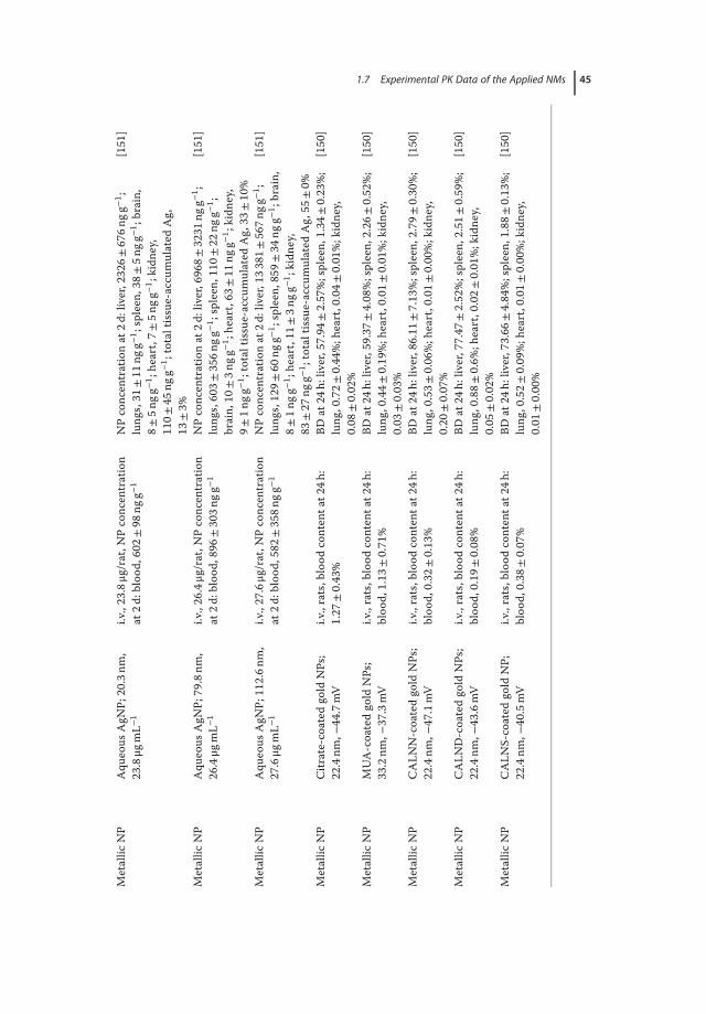
1.7 Experimental PK Data of the Applied NMs 45
Metallic
NP
Aqu
eous
AgN
P;20.3nm
,23.8μg
mL−
1i.v.,23
.8μg/rat,N
Pconcentration
at2d
:blood
,602
±98
ngg−
1NPconcentrationat
2d:liver,232
6±676n
gg−1 ;
lung
s,31
±11
ngg−
1 ;spleen
,38±
5ngg
−1 ;brain,
8±5n
gg−1 ;he
art,7±
5ngg
−1 ;kidn
ey,
110±
45ng
g−1 ;totaltissue-accumulated
Ag,
13±3%
[151
]
Metallic
NP
Aqu
eous
AgN
P;79.8nm
,26.4μg
mL−
1i.v.,26
.4μg/rat,N
Pconcentration
at2d
:blood
,896
±303n
gg−1
NPconcentrationat
2d:liver,696
8±3231
ngg−
1 ;lung
s,603±
356n
gg−1 ;spleen
,110
±22
ngg−
1 ;brain,
10±3n
gg−1 ;he
art,63
±11
ngg−
1 ;kidn
ey,
9±1n
gg−1 ;totaltissue-accumulated
Ag,33
±10%
[151
]
Metallic
NP
Aqu
eous
AgN
P;11
2.6n
m,
27.6μg
mL−
1i.v.,27
.6μg/rat,N
Pconcentration
at2d
:blood
,582
±358n
gg−1
NPconcentrationat
2d:liver,133
81±567n
gg−1 ;
lung
s,129±
60ng
g−1 ;spleen
,859
±34
ngg−
1 ;brain,
8±1n
gg−1 ;he
art,11
±3n
gg−1 ;kidn
ey,
83±27
ngg−
1 ;totaltissue-accumulated
Ag,55
±0%
[151
]
Metallic
NP
Citrate-coated
gold
NPs;
22.4nm
,−44.7mV
i.v.,rats,b
lood
contenta
t24h
:1.27
±0.43%
BDat
24h:
liver,57.94
±2.57
%;spleen,
1.34
±0.23
%;
lung
,0.72±
0.44
%;h
eart,0.04±
0.01
%;kidne
y,0.08
±0.02%
[150
]
Metallic
NP
MUA-coatedgo
ldNPs;
33.2nm
,−37.3mV
i.v.,rats,b
lood
contenta
t24h
:bloo
d,1.13
±0.71%
BDat
24h:
liver,59.37
±4.08
%;spleen,
2.26
±0.52
%;
lung
,0.44±
0.19
%;h
eart,0.01±
0.01
%;kidne
y,0.03
±0.03%
[150
]
Metallic
NP
CALN
N-coatedgo
ldNPs;
22.4nm
,−47.1mV
i.v.,rats,b
lood
contenta
t24h
:bloo
d,0.32
±0.13%
BDat
24h:
liver,86.11
±7.13
%;spleen,
2.79
±0.30
%;
lung
,0.53±
0.06
%;h
eart,0.01±
0.00
%;kidne
y,0.20
±0.07%
[150
]
Metallic
NP
CALN
D-coatedgo
ldNPs;
22.4nm
,−43.6mV
i.v.,rats,b
lood
contenta
t24h
:bloo
d,0.19
±0.08%
BDat
24h:
liver,77.47
±2.52
%;spleen,
2.51
±0.59
%;
lung
,0.88±
0.6%
;heart,0.02±
0.01
%;kidne
y,0.05
±0.02%
[150
]
Metallic
NP
CALN
S-coated
gold
NP;
22.4nm
,−40.5mV
i.v.,rats,b
lood
contenta
t24h
:bloo
d,0.38
±0.07%
BDat
24h:
liver,73.66
±4.84
%;spleen,
1.88
±0.13
%;
lung
,0.52±
0.09
%;h
eart,0.01±
0.00
%;kidne
y,0.01
±0.00%
[150
]

46 1 Pharmacokinetics and Pharmacodynamics (PK/PD) of Bionanomaterials
Table1.8
PKvaria
tions
oftheen
capsulateddrug
sinthedrug
-NMsystem
s.
NM
Description
PKdata
References
Drug1
Drug2
PLGANP
Casein-coated
PLGA
NP+paclita
xeland
ECCG;
190n
m,w
ithPL
GAcore
loaded
with
paclita
xel,an
dcoated
casein
loaded
with
ECCG
i.v.,rats,for
ptx:AUC,
179.3±
34.2μg
mL−
1h;
Cmax,
1.48
±0.65
μgmL−
1 ;MRT
,207.7±45.2h;
t 1/2,26.5±
2.4h
;BAincreased8.0-fold
ForE
GCG:A
UC,
6861.5±169.6μ
gmL−
1h;
Cmax,
119.4±
6.5μ
gmL−
1 ;MRT
,110.7±33.0h;
t 1/2,56.1±
11.6h;
BAincreased17
.6-fold
[155
]
PLGANP
Peptide-PE
G-PLG
Aloaded
with
paclita
xeland
lonidamine;
120–
160n
m,m
odified
peptide
may
target
EGFR
i.v.,mice,forp
tx:A
UC,
497.60
±17.46μ
gmL−
1h(0.98-fold);
MRT
,2.74h
;CL,
40mLkg
−1h−
1 ;Vd
,110m
Lkg
−1 ;tumor
PK:A
UC,
38.32±
13.14μ
gmL−
1h(8.51-fold);MRT
,11.45h
Forlon
:AUC,1316.01
±33.07μ
gmL−
1h
(1.71-fold);MRT
,4.38h
;CL,
60mLkg
−1h−
1 ;Vd
,270
mLkg
−1 ;tumor
PK:A
UC,49.96
±9.87
μgmL−
1h
(10.9-fold);MRT
,7.25h
[136
]
PLGANP
PEG-PLG
Aloaded
with
paclita
xel
andlonidamine;12
0–16
0nm,
surfacemod
ified
with
PEG
i.v.,mice.Fo
rptx:A
UC,436
.48μ
gmL−
1h
(0.86-fold);MRT
,2.75h
;CL,
50mLkg
−1h−
1 ;Vd
,130
mLkg
−1 ;tumor
PK:A
UC,25.57
±9.43
μgmL−
1h
(5.69-fold);MRT
,7.75h
Forlon
:AUC,992.44±
35.46μ
gmL−
1h
(1.95-fold);MRT
,3.77h
;CL,
80mLkg
−1h−
1 ;Vd
,300
mLk;
tumor
PK:
AUC,32.09
μgmL−
1h(7.0-fold);M
RT,
4.46
h
[136
]
Micelle
PEG,D
SPE/TPG
Smixed
micelle+paclita
xeland
tane
spim
ycin;∼
10nm
i.v.,mice,forp
tx:A
UC,
4216.8±195.0μ
Mmin
(10.2-fold);CL t,
5.6m
Lkg
−1min
−1 ;Vd
,311
.2mLkg
−1
Fortan
espimycin:A
UC,
3316.6±174.1μ
Mmin
(3.0-fold);C
L t,
19.3mLkg
−1min
−1 ;Vd
,468
.6mLkg
−1
[156
]
PLGANP
PEG-coatedPL
GA+rifampicin
andison
iazid;
229–
382n
m,
12.45±
4.53
mV
Oral,mice,AUC,498
.5μg
mL−
1min
(0.44fold);C
max,50.2μ
gmL−
1 ;CL,
2.8m
Lmin
−1 ;MRT
,13.4m
in
Oral,mice,AUC,503
.1μg
mL−
1min
(3.36-fold);
Cmax,29.7μ
gmL−
1 ;CL,
8.2m
Lmin
−1 ;MRT
,17.8m
in
[157
]

1.7 Experimental PK Data of the Applied NMs 47
Table1.9
PKvaria
tions
betweenen
capsulateddrug
san
dothe
rcom
pon
entsinthedrug
-NMsystem
s.
NM
Description
PKdata
References
Drug
NMcompon
ent
Lipo
some
CD19
antib
ody-mod
ified
(HSP
C+ch
olesterol)+
doxorubicin;
100±
10nm
i.v.,mice.Fo
rdox
:AUC,60.0m
gkg−
1h;
t 1/2(C
L),19.2h
;t1/2(release),315.0h
Forp
hospho
lipid:A
UC,318
.4mgk
g−1h;
t 1/2(C
L),
20.6h
[118
]
Lipo
some
CD19
antib
ody-mod
ified
(DPP
C+ch
olesterol)+
doxorubicin;
100±
10nm
i.v.,mice.Fo
rdox
:AUC,56.5m
gkg−
1h;
t 1/2(C
L),16.9h
;t1/2(release)125.0h
Forp
hospho
lipid:A
UC,229
.0mgk
g−1h;
t 1/2(C
L),
19.1h
[118
]
Lipo
some
CD19
antib
ody-mod
ified
(PMPC
+ch
olesterol)+
doxorubicin;
100±
10nm
i.v.,mice.Fo
rdox
:AUC,40.4m
gkg−
1h;
t 1/2(C
L),14.4h
;t1/2(release)65.0h
Forp
hospho
lipid:A
UC,239
.6mgk
g−1h;
t 1/2(C
L),
17.6h
[118
]
Lipo
some
CD19
antib
ody-mod
ified
(POPC
+ch
olesterol)+
doxorubicin;
100±
10nm
i.v.,mice.Fo
rdox
:AUC,48.8m
gkg−
1h;
t 1/2(C
L),13.1h
;t1/2(release)40.5h
Forp
hospho
lipid:A
UC,233
mgk
g−1h;
t 1/2(C
L),
17.7h
[118
]
Lipo
some
CD19
antib
ody-mod
ified
(DOPC
+ch
olesterol)+
doxorubicin;
100±
10nm
i.v.,mice.Fo
rdox
:AUC,19.5m
gkg−
1h;
t 1/2(C
L),7.6h;
t 1/2(release),1.9h
Forp
hospho
lipid:A
UC,182
.0mgk
g−1h;
t 1/2(C
L),
15.7h
[118
]
QDs
CdT
eQD;4
nmi.v.,2.5μ
molkg
−1 ,mice,Cd:
AUC,
18.73μ
gmL−
1h;
Vd,127.25m
Lkg
−1 ;CL,
14.81m
Lkg
−1h−
1
Te:A
UC,15.28
μgmL−
1h;
Vd,107.41m
Lkg
−1 ;
CL,
10.38m
Lkg
−1h−
1[148
]
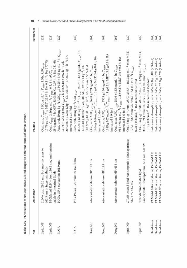
48 1 Pharmacokinetics and Pharmacodynamics (PK/PD) of BionanomaterialsTable1.10
PKvariations
ofNMs(ore
ncap
sulateddrug
s)viadifferen
trou
tesof
administration.
NM
Description
PKdata
References
LipidNP
SLN+do
x;28
0.2n
m,b
utmay
increase
to46
2.3n
min
intestinalflu
ids
Oral,rats,C
max,1.90μ
gmL−
1 ;Tmax,1.5h,
AUC0–
t,17.79μ
gmL−
1h;
MRT
,22.87
h;Ke,0.2h
−1 ;BA
377.7%
[121
]
LipidNP
PEGylated
SLN+do
x;150.2n
m,and
remains
stablein
intestinalflu
ids
Oral,
Cmax,2.26μ
gmL−
1 ;Tmax,0.5,4
h,AUC0–
t,35.4μg
mL−
1h;
MRT
,33.03
h;Ke,0.07
h−1 ;BA
751.6%
[121
]
PLGA
PLGANP+curcum
in;161
.9nm
Rats,o
ral,50
mgk
g−1 ;AUC0–
t,134.25±
3.45
ngmL−
1h;
Cmax,
11.78±
0.45
ngmL;
Tmax,2
h;Ke,0.18
±0.02
h−1 ;Vd
,2073.66±
352.61
Lkg
−1 ;CL365.19
±37.35L
kg−1h−
1 ;BA
increased15
.6-fold
[122
]
PLGA
PEG-PLG
A+curcum
in;152
.4nm
Rats,o
ral,50
mgk
g−1 ;AUC0–
t,447.80
±64.03n
gmL−
1h;
Cmax,29.78
±4.63
ngmL;
Tmax,3
h;Ke,0.18
±0.02
h−1 ;Vd
,900
.54±
225.77
Lkg
−1 ;CL
103.679±
10.90L
kg−1h−
1 ;BA
increased55
.5-fold
[122
]
DrugNP
Atorvastatin
calcium
NP;
123n
mOral,rats;A
UC0–
12h,3688
±269n
gmL−
1h;
Cmax,
1454
±166n
gmL−
1 ;Tmax,1.0±0h
;MRT
,3.4±0.4h
;BA
increased2.1-fold
[161
]
DrugNP
Atorvastatin
calcium
NP;
181n
mOral,rats;A
UC0–
12h,3305
±136n
gmL−
1h;
Cmax,
1145
±107n
gmL−
1 ;Tmax,1.1±0.2h
;MRT
,3.6±0.3h
;BA
increased1.9-fold
[161
]
DrugNP
Atorvastatin
calcium
NP;
453n
mOral,rats;A
UC0–
12h,2846
±133n
gmL−
1h;
Cmax,
986±
64ng
mL−
1 ;Tmax,1.2±0.3h
;MRT
,3.6±0.3h
;BA
increased1.6-fold
[161
]
LipidNP
CTA
B-coated
lipid
nano
capsule+
fond
aparinux
;58.2nm
,42.9m
VOral,5m
gkg−
1 ,rats.A
UC,336.5±187.3μ
gmL−
1min;M
RT,
177.1±
82.6min;C
L,450.7±
7.1m
Lmin
−1 ;
Cmax,
0.98
±0.33
mL−
1 ;BA
increased6.9-fold
[129
]
LipidNP
Stearylamine-coated
lipid
nano
capsule+
fond
aparinux
;48.1n
m,4.0mV
Oral,5m
gkg−
1 ,rats.A
UC,730.9±214.9μ
gmL−
1min;M
RT,
338.4±
69.4min;C
L,485.9±
13.8mLmin
−1 ;
Cmax,
1.31
±0.37
mL−
1 ;BA
increased15
.0-fold
[129
]
Den
drim
erPA
MAM
G0+
calcito
nin;
1%PA
MAM
Pulm
onaryabsorptio
n,rats.PA%,22.0±
0.8%
(2.3-fold)
[141
]Den
drim
erPA
MAM
G1+
calcito
nin;
1%PA
MAM
Pulm
onaryabsorptio
n,rats.PA%,23.7±
0.7%
(2.4-fold)
[141
]Den
drim
erPA
MAM
G2+
calcito
nin;
1%PA
MAM
Pulm
onaryabsorptio
n,rats.PA%,25.0±
2.7%
(2.6-fold)
[141
]
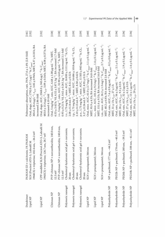
1.7 Experimental PK Data of the Applied NMs 49
Den
drim
erPA
MAM
G3+
calcito
nin;
1%PA
MAM
Pulm
onaryabsorptio
n,rats.PA%,27.6±
1.6%
(2.8-fold)
[141
]LipidNP
SLN
(PrecirolA
TO-5,L
abrafil
M19
44CS)
+tripterine
;102.4nm
,−26.2mV
Oral,do
gs,A
UC,179
2.0±
57.1μg
L−1h;
Cmax,
305.0±
35.4μg
L−1 ;
t 1/2,3.85±
0.94
h;MRT
,4.37±
0.33
h;BA
increased2.86
-fold
[132
]
LipidNP
CPP
-mod
ified
SLN
(PrecirolA
TO-5,L
abrafil
M19
45CS)
+tripterine
;126.7nm
,28.7m
VOral,do
gs,A
UC,256
8.0±
343.4μ
gL−1h;
Cmax,
483.0±
15.4μg
L−1 ;
t 1/2,3.09±
0.54
h;MRT
,4.08±
0.37
h;BA
increased4.10
-fold
[132
]
ChitosanNP
PVP-ch
itosanNP+no
rcan
tharidin;140.0nm
,12.3mV
Oral,5m
gkg−
1 ,rats;A
UC,6.68±
1.08
μgmL−
1h;
MRT
,6.53
±3.42
h;Vd
,6.52±
3.87
Lkg
−1 ;BA
increased1.7-fold
[158
]
ChitosanNP
PVP-ch
itosanNP+no
rcan
tharidin;140.0nm
,12.3mV
i.v.,5m
gkg−
1 ,rats;A
UC,32.90
±4.63
μgmL−
1h;
MRT
,0.93
±0.21
h;Vd
,0.62±
0.26
Lkg
−1 ;BA
increased3.1-fold
[158
]
Polymericna
nogel
Cho
lesteryl-hyaluronicacid
gel+
curcum
in;
20nm
i.v.,1.75
mgk
g−1 ,mice.AUC,168
30±1111
ngmL−
1h;
CL,
2.08
±0.14
mLh−
1 ;MRT
,8.51±
0.52
h[159
]
Polymericna
nogel
Cho
lesteryl-hyaluronicacid
gel+
curcum
in;
20nm
i.p.,1.75
mgk
g−1 ,mice.AUC,166
86±1018
ngmL−
1h;
CL,
2.10
±0.13
mLh−
1 ;MRT
,8.58±
0.62
h[159
]
Polymericna
nogel
Cho
lesteryl-hyaluronicacid
gel+
curcum
in;
20nm
p.o.,1.75m
gkg−
1 ,mice.AUC,115
03±843n
gmL−
1h;
CL,
3.17
±0.24
mLh−
1 ;MRT
,8.95±
0.51
h[159
]
LipidNP
SLN+praziquantel;344
nmOral,mice.AUC,61.0±2.7m
gL−1h;
Cmax,1.1±0.1μ
gmL−
1 ;MRT
,95.9±
4.3h
;BAincreased14
.8-fold
[160
]
LipidNP
SLN+praziquantel;344
nms.c
.,mice.AUC,101
.6±9.2m
gL−1h;
Cmax,1.4±0.1μ
gmL−
1 ;MRT
,151.6±9.3h
;BAincreased16
.0-fold
[160
]
LipidNP
SLN+praziquantel;344
nmi.m
.,mice.AUC,26.2±
1.3m
gL−1h;
Cmax,2.7±0.2μ
gmL−
1 ;MRT
,48.2±
6.8h
;BAincreased2.6-fold
[160
]
Polyan
hydrideNP
NP+paclita
xel;17
7nm,−
44.2mV
Oral,rats,A
UC,7.4±0.9μ
gmL−
1h;
Cmax,0.2±0.0μ
gmL−
1 ;MRT
,31±
1h;t
1/2,27
±1h
[134
]
Polyan
hydrideNP
PEG2k-N
P+paclita
xel;17
8nm,−
40.3mV
Oral,rats,A
UC,56±
3μgm
L−1h;
Cmax,2.1±0.1μ
gmL−
1 ;MRT
,26±
1h;t
1/2,9.3±
0.5h
[134
]
Polyan
hydrideNP
PEG6k-N
P+paclita
xel;18
0nm,−
39.5mV
Oral,rats,A
UC,32±
2μgm
L−1h;
Cmax,1.9±0.1μ
gmL−
1 ;MRT
,27±
1h;t
1/2,6.2±
0.3h
[134
]
Polyan
hydrideNP
PEG10
k-NP+paclita
xel;18
8nm,−
41.1mV
Oral,rats,A
UC,13±
1μgm
L−1h;
Cmax,1.4±0.1μ
gmL−
1 ;MRT
,18±
1h;t
1/2,29
±2h
[134
]

50 1 Pharmacokinetics and Pharmacodynamics (PK/PD) of Bionanomaterials
1.8Perspectives
1.8.1Development of NMs
The development of NMs has raised great attentions. NMs can improve the drugstability and protect drugs from enzymatic digestion, and enhance the bioavail-ability. The targeting delivery and controlled release of the NM-encapsulateddrugs are also beneficial for cancer treatment and diagnosis.However, there are many biological barriers in the human body against exoge-
nous particles, and the majority of the administrated NMs will end up in the RESsystem.One of the design strategies is to escape the surveillance of the RES systemby using surfacemodification.Moreover, targeting ligands are also used tomodifythe NMs to increase the specific binding with the desired organs/cells. Althoughmuch effort has been made, the overall efficiency is still under satisfaction forclinical use. The efficiency of the NM-based delivery systems needs to be furtherimproved.
1.8.2Pharmacokinetic Study andModel Development
The currently available pharmacokinetic studies of NMs focus on the biodistri-bution models. The drawback lies in the fact that the parameters such as tissuedrug concentrations are hard to be detected continuously. As consequence, thelimited, discontinuous data make it difficult to depict the detailed and precise PKprofiles for the NMs. Compared with organ distribution, blood concentration ismore convenient to determine, but the results from blood PK analysis are lesssuitable for NM-based systems, which are intended for an organ-specific delivery.Furthermore, the diversity of intrinsic features of NMs, modification, and speciesvariation contribute to deviations of the overall PK behaviors of NMs. There-fore, “standard protocols” for PK experiments, including administration, dosing,sampling, and data processing, would be helpful to acquire high-quality parame-ters to develop PK models. A potential solution to the NM-based drug PK modelcould be the establishment of a theoretical “body,” which is composed of differ-ent “digital” compartments simulating the physiological features of each organ.The model would help us to give better predictions on the in vivo behaviour ofthe NMs.
References
1. Feynman, R.P. (1960) “There’s plentyof room at the bottom” an invitationto open up a new field of physics. Eng.Sci., 23, 22–36.
2. Roduner, E. (2006) Size mat-ters: why nanomaterials aredifferent. Chem. Soc. Rev., 35,583–592.

References 51
3. Matsumura, Y. and Maeda, H. (1986)A new concept for macromoleculartherapeutics in cancer chemother-apy: mechanism of tumoritropicaccumulation of proteins and theantitumor agent smancs. Cancer Res.,46, 6387–6392.
4. Maeda, H., Takeshita, J., and Kanamaru,R. (1979) A lipophilic derivative of neo-carzinostatin. A polymer conjugation ofan antitumor protein antibiotic. Int. J.Pept. Protein Res., 14, 81–87.
5. Tanaka, T., Shiramoto, S., Miyashita,M., Fujishima, Y., and Kaneo, Y. (2004)Tumor targeting based on the effect ofenhanced permeability and retention(EPR) and the mechanism of receptor-mediated endocytosis (RME). Int. J.Pharm., 277, 39–61.
6. Byrne, J.D., Betancourt, T., andBrannon-Peppas, L. (2008) Activetargeting schemes for nanoparticle sys-tems in cancer therapeutics. Adv. DrugDelivery Rev., 60, 1615–1626.
7. Shaikh, J., Ankola, D.D., Beniwal, V.,Singh, D., and Kumar, M.N. (2009)Nanoparticle encapsulation improvesoral bioavailability of curcumin by atleast 9-fold when compared to cur-cumin administered with piperine asabsorption enhancer. Eur. J. Pharm.Sci., 37, 223–230.
8. Wong, C., Stylianopoulos, T., Cui,J., Martin, J., Chauhan, V.P., Jiang,W., Popovic, Z., Jain, R.K., Bawendi,M.G., and Fukumura, D. (2011) Mul-tistage nanoparticle delivery systemfor deep penetration into tumor tis-sue. Proc. Natl. Acad. Sci. U.S.A., 108,2426–2431.
9. Siissalo, S., de Waard, H., de Jager,M.H., Hayeshi, R., Frijlink, H.W.,Hinrichs, W.L., Dinter-Heidorn, H., vanDam, A., Proost, J.H., Groothuis, G.M.,and de Graaf, I.A. (2013) Nanopar-ticle formulation of a poorly solublecannabinoid receptor 1 antagonistimproves absorption by rat and humanintestine. Drug Metab. Dispos., 41,1557–1565.
10. Fahr, A. and Liu, X. (2007) Drug deliv-ery strategies for poorly water-solubledrugs. Expert Opin. Drug Delivery, 4,403–416.
11. Mager, D.E., Mody, V., Xu, C., Forrest,A., Lesniak, W.G., Nigavekar, S.S.,Kariapper, M.T., Minc, L., Khan, M.K.,and Balogh, L.P. (2012) Physiologicallybased pharmacokinetic model for com-posite nanodevices: effect of charge andsize on in vivo disposition. Pharm. Res.,29, 2534–2542.
12. Kibria, G., Hatakeyama, H., Ohga, N.,Hida, K., and Harashima, H. (2013)The effect of liposomal size on thetargeted delivery of doxorubicin toIntegrin alphavbeta3-expressing tumorendothelial cells. Biomaterials, 34,5617–5627.
13. Gao, Y. and Tang, Z. (2011) Design andapplication of inorganic nanoparticlesuperstructures: current status andfuture challenges. Small, 7, 2133–2146.
14. Murakami, T. and Tsuchida, K.(2008) Recent advances in inorganicnanoparticle-based drug deliverysystems. Mini Rev. Med. Chem., 8,175–183.
15. Kwon, Y.M., Chung, H.S., Moon, C.,Yockman, J., Park, Y.J., Gitlin, S.D.,David, A.E., and Yang, V.C. (2009)L-Asparaginase encapsulated intacterythrocytes for treatment of acutelymphoblastic leukemia (ALL). J. Con-trolled Release, 139, 182–189.
16. Huang, B., Tabata, Y., and Gao, J.Q.(2012) Mesenchymal stem cells as ther-apeutic agents and potential targetedgene delivery vehicle for brain diseases.J. Controlled Release, 162, 464–473.
17. Fujihara, A., Kurooka, M., Miki, T.,and Kaneda, Y. (2008) Intratumoralinjection of inactivated Sendai virusparticles elicits strong antitumoractivity by enhancing local CXCL10expression and systemic NK cell activa-tion. Cancer Immunol. Immunother., 57,73–84.
18. Ziegler, A., Nervi, P., Durrenberger, M.,and Seelig, J. (2005) The cationic cell-penetrating peptide Cpp(TAT) derivedfrom the HIV-1 protein TAT is rapidlytransported into living fibroblasts:optical, biophysical, and metabolic evi-dence. Biochemistry-US, 44, 138–148.
19. Cai, S., Yang, Q.H., Bagby, T.R., andForrest, M.L. (2011) Lymphatic drugdelivery using engineered liposomes

52 1 Pharmacokinetics and Pharmacodynamics (PK/PD) of Bionanomaterials
and solid lipid nanoparticles. Adv. DrugDelivery Rev., 63, 901–908.
20. Guo, Q.Q., Wang, H.Y., Zhao, Y.X.,Wang, H.X., Zeng, F., Hua, H.Y., Xu, Q.,and Huang, Y.Z. (2013) Cell-penetratingalbumin conjugates for enhanced dox-orubicin delivery. Polym. Chem.-UK , 4,4584–4587.
21. Cormode, D.P., Fisher, E.A., Stroes,E.S.G., Mulder, W.J.M., and Fayad, Z.A.(2013) High-density lipoprotein is ananoparticle, but not all nanoparticlesare high-density lipoprotein. Proc. Natl.Acad. Sci. U.S.A., 110, E3548.
22. Zhang, Z.H., Wang, X.P., Ayman,W.Y., Munyendo, W.L.L., Lv, H.X., andZhou, J.P. (2013) Studies on lactoferrinnanoparticles of gambogic acid for oraldelivery. Drug Delivery, 20, 86–93.
23. Budin, I., Prwyes, N., Zhang, N., andSzostak, J.W. (2014) Chain-length het-erogeneity allows for the assembly offatty acid vesicles in dilute solutions.Biophys. J., 107, 1582–1590.
24. Jaworska, M., Sikora, E., andOgonowski, J. (2014) Nanoemul-sions. Characteristics and methodsfor preparation. Przem. Chem., 93,1087–1092.
25. YashRoy, R.C. (1994) Destabilisationof lamellar dispersion of thylakoidmembrane lipids by sucrose. Biochim.Biophys. Acta, 1212, 129–133.
26. Mehnert, W. and Mäder, K. (2001)Solid lipid nanoparticles: production,characterization and applications. Adv.Drug Delivery Rev., 47, 165–196.
27. Joseph, S. and Bunjes, H. (2012) Prepa-ration of nanoemulsions and solid lipidnanoparticles by premix membraneemulsification. J. Pharm. Sci.-US, 101,2479–2489.
28. Qiao, N., Liu, Q., Meng, H., and Zhao,D.Y. (2014) Haemolytic activity andadjuvant effect of soyasaponins andsome of their derivatives on theimmune responses to ovalbumin inmice. Int. Immunopharmacol., 18,333–339.
29. Analoui, M. (2011) Nanomedicineand NanoDiagnostics: Innovation,Regulation, and Investment.
30. Sripriyalakshmi, S., Anjali, C.H.,George, P.D., Rajith, B., and Ravindran,
A. (2014) BSA nanoparticle loadedatorvastatin calcium – a new facet foran old drug. PLoS One, 9, e86317.
31. Du, C., Deng, D., Shan, L., Wan, S.,Cao, J., Tian, J., Achilefu, S., and Gu, Y.(2013) A pH-sensitive doxorubicinprodrug based on folate-conjugatedBSA for tumor-targeted drug delivery.Biomaterials, 34, 3087–3097.
32. Karewicz, A., Bielska, D., andNowakowska, M. (2012) Modifiedpolysaccharides as versatile materialsin controlled delivery of antidegen-erative agents. Curr. Pharm. Des., 18,2518–2535.
33. Nakai, T., Hirakura, T., Sakurai, Y.,Shimoboji, T., Ishigai, M., and Akiyoshi,K. (2012) Injectable hydrogel for sus-tained protein release by salt-inducedassociation of hyaluronic acid nanogel.Macromol. Biosci., 12, 475–483.
34. Song, Y.B., Zhou, Y., and Chen, L.Y.(2012) Wood cellulose-based poly-electrolyte complex nanoparticles asprotein carriers. J. Mater. Chem., 22,2512–2519.
35. Wang, Y., Chen, H., Liu, Y., Wu, J.,Zhou, P., Wang, Y., Li, R., Yang, X.,and Zhang, N. (2013) pH-sensitivepullulan-based nanoparticle carrier ofmethotrexate and combretastatin A4for the combination therapy againsthepatocellular carcinoma. Biomaterials,34, 7181–7190.
36. Kato, Y., Ozawa, S., Miyamoto, C.,Maehata, Y., Suzuki, A., Maeda, T.,and Baba, Y. (2013) Acidic extracellularmicroenvironment and cancer. CancerCell Int., 13, 89.
37. Wiseman, J.W., Goddard, C.A.,McLelland, D., and Colledge, W.H.(2003) A comparison of linear andbranched polyethylenimine (PEI) withDCChol/DOPE liposomes for genedelivery to epithelial cells in vitro andin vivo. Gene Ther., 10, 1654–1662.
38. Chen, H.L., Liu, R., Nan, W.B., Wang,Y.X., Wang, Y.S., Yang, W.C., and Yang,W.C. (2013) Surface modification ofepirubicin-loaded PLGA nanoparticlewith biotinylated chitosan enhancesanti-cancer efficacy in breast cancercells. Cancer Res., 73, 5659.

References 53
39. Li, J.Q., Pu, Y.D., Wang, S.L., Ding,M.Z., Chen, D.L., and Zhu, M.D. (2013)Pharmacokinetic study and effec-tiveness evaluation of slow-releasePLGA-5-fluorouracil microsphere.Cancer Chemother. Pharmacol., 71,351–359.
40. Ling, T., Yu, M.F., Weng, W.J., Wang,H.M., Cheng, K., Lin, J., and Du, P.Y.(2013) Improvement of drug elution inthin mineralized collagen coatings withPLGA-PEG-PLGA micelles. J. Biomed.Mater. Res. Part A, 101, 3256–3265.
41. Nounou, M.M., El-Khordagui, L.K.,Khalafallah, N.A., and Khalil, S.A.(2006) In vitro release of hydrophilicand hydrophobic drugs from liposomaldispersions and gels. Acta Pharm., 56,311–324.
42. Freeman, E.C., Weiland, L.M., andMeng, W.S. (2013) Modeling the protonsponge hypothesis: examining protonsponge effectiveness for enhancingintracellular gene delivery throughmultiscale modeling. J. Biomater. Sci.,Polym. Ed., 24, 398–416.
43. Bandyopadhyay, S., Xia, X., Maiseiyeu,A., Mihai, G., Rajagopalan, S., andBong, D. (2012) Z-group ketone chaintransfer agents for RAFT polymernanoparticle modification via hydra-zone conjugation. Macromolecules, 45,6766–6773.
44. Shi, M., Yu, B., Gao, H., Mu, J., and Ji,C. (2013) Matrix metalloproteinase 2overexpression and prognosis in col-orectal cancer: a meta-analysis. Mol.Biol. Rep., 40, 617–623.
45. Reis, S.T., Leite, K.R., Piovesan, L.F.,Pontes-Junior, J., Viana, N.I., Abe, D.K.,Crippa, A., Moura, C.M., Adonias, S.P.,Srougi, M., and Dall’Oglio, M.F. (2012)Increased expression of MMP-9 andIL-8 are correlated with poor prognosisof Bladder Cancer. BMC Urol., 12, 18.
46. Liu, Y., Bajjuri, K.M., Liu, C., andSinha, S.C. (2012) Targeting cell sur-face alpha(v)beta(3) integrin increasestherapeutic efficacies of a legumainprotease-activated auristatin prodrug.Mol. Pharmaceutics, 9, 168–175.
47. Yang, J. (2012) Stimuli-responsive drugdelivery systems. Adv. Drug DeliveryRev., 64, 965–966.
48. Tan, L., Huang, C., Peng, R., Tang,Y., and Li, W. (2014) Developmentof hybrid organic–inorganic surfaceimprinted Mn-doped ZnS QDs andtheir application as a sensing materialfor target proteins. Biosens. Bioelectron.,61, 506–511.
49. Wong, O.A., Hansen, R.J., Ni, T.W.,Heinecke, C.L., Compel, W.S.,Gustafson, D.L., and Ackerson, C.J.(2013) Structure–activity relationshipsfor biodistribution, pharmacokinetics,and excretion of atomically precisenanoclusters in a murine model.Nanoscale, 5, 10525–10533.
50. Cho, E.C., Glaus, C., Chen, J., Welch,M.J., and Xia, Y. (2010) Inorganicnanoparticle-based contrast agents formolecular imaging. Trends Mol. Med.,16, 561–573.
51. Tojo, A. and Kinugasa, S. (2012)Mechanisms of glomerular albuminfiltration and tubular reabsorption. Int.J. Nephrol., 2012, 481520.
52. Kapoor, A., Simmonds, P., Gerold, G.,Qaisar, N., Jain, K., Henriquez, J.A.,Firth, C., Hirschberg, D.L., Rice, C.M.,Shields, S., and Lipkin, W.I. (2011)Characterization of a canine homologof hepatitis C virus. Proc. Natl. Acad.Sci. U.S.A., 108, 11608–11613.
53. Lu, Y., Wang, Z.H., Li, T., McNally,H., Park, K., and Sturek, M. (2014)Development and evaluation oftransferrin-stabilized paclitaxelnanocrystal formulation. J. ControlledRelease, 176, 76–85.
54. Alvarez-Lorenzo, C. and Concheiro,A. (2014) Smart drug delivery systems:from fundamentals to the clinic. Chem.Commun. (Camb.), 50, 7743–7765.
55. Ernsting, M.J., Murakami, M., Roy, A.,and Li, S.D. (2013) Factors controllingthe pharmacokinetics, biodistribu-tion and intratumoral penetration ofnanoparticles. J. Controlled Release,172, 782–794.
56. Bae, Y.H. and Park, K. (2011) Targeteddrug delivery to tumors: myths, realityand possibility. J. Controlled Release,153, 198.
57. Prabhakar, U., Maeda, H., Jain, R.K.,Sevick-Muraca, E.M., Zamboni, W.,Farokhzad, O.C., Barry, S.T., Gabizon,

54 1 Pharmacokinetics and Pharmacodynamics (PK/PD) of Bionanomaterials
A., Grodzinski, P., and Blakey, D.C.(2013) Challenges and key considera-tions of the enhanced permeability andretention effect for nanomedicine drugdelivery in oncology. Cancer Res., 73,2412–2417.
58. Alexis, F., Pridgen, E., Molnar, L.K.,and Farokhzad, O.C. (2008) Factorsaffecting the clearance and biodistribu-tion of polymeric nanoparticles. Mol.Pharmaceutics, 5, 505–515.
59. Kuo, Y.C. and Wang, L.J. (2014)Transferrin-grafted catanionic solidlipid nanoparticles for targeting deliveryof saquinavir to the brain. J. TaiwanInst. Chem. Eng., 45, 755–763.
60. Wang, Y.P., Jiang, Y.F., Zhang, M., Tan,J., Liang, J.M., Wang, H.X., Li, Y.P.,He, H.N., Yang, V.C., and Huang, Y.Z.(2014) Protease-activatable hybridnanoprobe for tumor imaging. Adv.Funct. Mater., 24, 5443–5453.
61. Buser, K., Joncourt, F., Altermatt, H.J.,Bacchi, M., Oberli, A., and Cerny, T.(1997) Breast cancer: pretreatment drugresistance parameters (GSH-system,ATase, P-glycoprotein) in tumor tissueand their correlation with clinical andprognostic characteristics. Ann. Oncol.,8, 335–341.
62. Mo, R., Sun, Q., Xue, J.W., Li, N., Li,W.Y., Zhang, C., and Ping, Q.N. (2012)Multistage pH-responsive liposomes formitochondrial-targeted anticancer drugdelivery. Adv. Mater., 24, 3659–3665.
63. Lim, J., Kim, J., Duong, T., Lee, G.,Kim, J., Yoon, J., Kim, J., Kim, H.,Ruley, H.E., El-Rifai, W., and Jo, D.(2012) Antitumor activity of cell-permeable p18(INK4c) with enhancedmembrane and tissue penetration. Mol.Ther., 20, 1540–1549.
64. Zhang, J., Shin, M.C., and Yang, V.C.(2014) Magnetic targeting of novelheparinized iron oxide nanoparticlesevaluated in a 9L-glioma mouse model.Pharm. Res., 31, 579–592.
65. Gormley, A.J., Greish, K., Ray, A.,Robinson, R., Gustafson, J.A., andGhandehari, H. (2011) Gold nanorodmediated plasmonic photothermal ther-apy: a tool to enhance macromoleculardelivery. Int. J. Pharm., 415, 315–318.
66. Shen, M., Huang, Y., Han, L., Qin,J., Fang, X., Wang, J., and Yang, V.C.(2012) Multifunctional drug deliverysystem for targeting tumor and itsacidic microenvironment. J. ControlledRelease, 161, 884–892.
67. Ploegh, H.L. (2013) Logic of theimmune system. Cancer Immunol.Res., 1, 5–10.
68. Podila, R., Chen, R., Ke, P.C., Brown,J.M., and Rao, A.M. (2012) Effects ofsurface functional groups on the for-mation of nanoparticle-protein corona.Appl. Phys. Lett., 101, 263701.
69. Song, G., Wu, H., Yoshino, K., andZamboni, W.C. (2012) Factors affectingthe pharmacokinetics and pharmacody-namics of liposomal drugs. J. LiposomeRes., 22, 177–192.
70. Dziendzikowska, K.,Gromadzka-Ostrowska, J., Lankoff,A., Oczkowski, M., Krawczynska, A.,Chwastowska, J., Sadowska-Bratek,M., Chajduk, E., Wojewodzka, M.,Dusinska, M., and Kruszewski, M.(2012) Time-dependent biodistributionand excretion of silver nanoparticles inmale Wistar rats. J. Appl. Toxicol., 32,920–928.
71. Yuan, F., Dellian, M., Fukumura, D.,Leunig, M., Berk, D.A., Torchilin, V.P.,and Jain, R.K. (1995) Vascular perme-ability in a human tumor xenograft:molecular size dependence and cutoffsize. Cancer Res., 55, 3752–3756.
72. Fang, J., Nakamura, H., and Maeda, H.(2011) The EPR effect: unique featuresof tumor blood vessels for drug deliv-ery, factors involved, and limitationsand augmentation of the effect. Adv.Drug Delivery Rev., 63, 136–151.
73. Ha, S.Y., Lee, J., Jang, J., Hong, J.Y., Do,I.G., Park, S.H., Park, J.O., Choi, M.G.,Sohn, T.S., Bae, J.M., Kim, S., Kim, M.,Kim, S., Park, C.K., Kang, W.K., andKim, K.M. (2014) HER2-positive gastriccancer with concomitant MET and/orEGFR overexpression: a distinct subsetof patients for dual inhibition therapy.Int. J. Cancer, 136, 1629–1635.
74. Malla, R.R., Gopinath, S., Alapati, K.,Gorantla, B., Gondi, C.S., and Rao, J.S.(2013) Knockdown of cathepsin B anduPAR inhibits CD151 and alpha 3 beta

References 55
1 integrin-mediated cell adhesion andinvasion in glioma. Mol. Carcinog., 52,777–790.
75. Cagle, P.T., Zhai, Q.H., Murphy, L.,and Low, P.S. (2013) Folate receptorin adenocarcinoma and squamous cellcarcinoma of the lung potential targetfor folate-linked therapeutic agents.Arch. Pathol. Lab. Med., 137, 241–244.
76. Yhee, J.Y., Lee, S.J., Lee, S., Song, S.,Min, H.S., Kang, S.W., Son, S., Jeong,S.Y., Kwon, I.C., Kim, S.H., and Kim,K. (2013) Tumor-targeting transferrinnanoparticles for systemic polymerizedsiRNA delivery in tumor-bearing mice.Bioconjugate Chem., 24, 1850–1860.
77. Xuan, C., Steward, K.K., Timmerman,J.M., and Morrison, S.L. (2010) Tar-geted delivery of interferon-alphavia fusion to anti-CD20 results inpotent antitumor activity against B-celllymphoma. Blood, 115, 2864–2871.
78. Weiss, I., Trope, C.G., Reich, R., andDavidson, B. (2012) Hyaluronan syn-thase and hyaluronidase expression inserous ovarian carcinoma is related toanatomic site and chemotherapy expo-sure. Int. J. Mol. Sci., 13, 12925–12938.
79. Wang, Y. and Grainger, D. (2014)Barriers to advancing nanotechnol-ogy to better improve and translatenanomedicines. Front. Chem. Sci. Eng.,8, 265–275.
80. Hirsh, S.L., McKenzie, D.R., Nosworthy,N.J., Denman, J.A., Sezerman, O.U.,and Bilek, M.M.M. (2013) The Vromaneffect: competitive protein exchangewith dynamic multilayer protein aggre-gates. Colloids Surf., B, 103, 395–404.
81. Bertrand, N. and Leroux, J.C. (2012)The journey of a drug-carrier in thebody: an anatomo-physiological per-spective. J. Controlled Release, 161,152–163.
82. Kwon, I.K., Lee, S.C., Han, B., and Park,K. (2012) Analysis on the current statusof targeted drug delivery to tumors.J. Controlled Release, 164, 108–114.
83. Jalali, S., Huang, Y., Dumont, D.J.,and Hynynen, K. (2010) Focusedultrasound-mediated bbb disruptionis associated with an increase in activa-tion of AKT: experimental study in rats.BMC Neurol., 10, 114.
84. Wang, B., He, X., Zhang, Z., Zhao, Y.,and Feng, W. (2013) Metabolism ofnanomaterials in vivo: blood circulationand organ clearance. Acc. Chem. Res.,46, 761–769.
85. Heurtault, B., Saulnier, P., Pech, B.,Proust, J.E., and Benoit, J.P. (2003)Physico-chemical stability of col-loidal lipid particles. Biomaterials,24, 4283–4300.
86. Devine, D.V., Wong, K., Serrano, K.,Chonn, A., and Cullis, P.R. (1994)Liposome-complement interactions inrat serum: implications for liposomesurvival studies. Biochim. Biophys. Acta,1191, 43–51.
87. Hardison, W.G.M., Lowe, P.J., andShanahan, M. (1989) Effect of molec-ular charge on para- and transcellularaccess of horseradish peroxidase intorat bile. Hepatology, 9, 866–871.
88. Xiao, K., Li, Y., Luo, J., Lee, J.S., Xiao,W., Gonik, A.M., Agarwal, R.G., andLam, K.S. (2011) The effect of surfacecharge on in vivo biodistribution ofPEG-oligocholic acid based micel-lar nanoparticles. Biomaterials, 32,3435–3446.
89. Huang, X., Li, L., Liu, T., Hao, N.,Liu, H., Chen, D., and Tang, F. (2011)The shape effect of mesoporous silicananoparticles on biodistribution, clear-ance, and biocompatibility in vivo. ACSNano, 5, 5390–5399.
90. Snyder, G.K. and Sheafor, B.A. (1999)Red blood cells: centerpiece in theevolution of the vertebrate circulatorysystem. Am. Zool., 39, 189–198.
91. Gaucher, G., Asahina, K., Wang, J.,and Leroux, J.C. (2009) Effect ofpoly(N-vinyl-pyrrolidone)-block-poly(D,L-lactide) as coating agenton the opsonization, phagocytosis, andpharmacokinetics of biodegradablenanoparticles. Biomacromolecules, 10,408–416.
92. Malfatti, M.A., Palko, H.A., Kuhn,E.A., and Turteltaub, K.W. (2012)Determining the pharmacokineticsand long-term biodistribution of SiO2nanoparticles in vivo using acceleratormass spectrometry. Nano Lett., 12,5532–5538.
56 1 Pharmacokinetics and Pharmacodynamics (PK/PD) of Bionanomaterials
93. Morishita, M., Kamei, N., Ehara, J.,Isowa, K., and Takayama, K. (2007) Anovel approach using functional pep-tides for efficient intestinal absorptionof insulin. J. Controlled Release, 118,177–184.
94. Gomez-Icazbalceta, G.,Gonzalez-Sanchez, I., Moreno, J.,Cerbon, M.A., and Cervantes, A.(2013) In vitro drug metabolism test-ing using blood-monocyte derivatives.Expert Opin. Drug Metab. Toxicol., 9,1571–1580.
95. Kulthong, K., Maniratanachote, R.,Kobayashi, Y., Fukami, T., and Yokoi, T.(2012) Effects of silver nanoparticles onrat hepatic cytochrome P450 enzymeactivity. Xenobiotica, 42, 854–862.
96. Tan, J., Shah, S., Thomas, A., Ou-Yang,H.D., and Liu, Y. (2013) The influenceof size, shape and vessel geometry onnanoparticle distribution. Microfluid.Nanofluid., 14, 77–87.
97. Kaminskas, L.M., Boyd, B.J., Karellas,P., Krippner, G.Y., Lessene, R., Kelly, B.,and Porter, C.J. (2008) The impact ofmolecular weight and PEG chain lengthon the systemic pharmacokinetics ofPEGylated poly l-lysine dendrimers.Mol. Pharmaceutics, 5, 449–463.
98. Perry, J.L., Reuter, K.G., Kai, M.P.,Herlihy, K.P., Jones, S.W., Luft, J.C.,Napier, M., Bear, J.E., and DeSimone,J.M. (2012) PEGylated PRINT nanopar-ticles: the impact of PEG densityon protein binding, macrophageassociation, biodistribution, andpharmacokinetics. Nano Lett., 12,5304–5310.
99. Tahara, K., Miyazaki, Y., Kawashima,Y., Kreuter, J., and Yamamoto, H.(2011) Brain targeting with surface-modified poly(D,L-lactic-co-glycolicacid) nanoparticles delivered via carotidartery administration. Eur. J. Pharm.Biopharm., 77, 84–88.
100. Mudge, M.C., Walker, N.J., Borjesson,D.L., Librach, F., Johns, J.L., andOwens, S.D. (2012) Post-transfusionsurvival of biotin-labeled allogeneicRBCs in adult horses. Vet. Clin.Pathol./Am. Soc. Vet. Clin. Pathol.,41, 56–62.
101. Hu, C.M., Zhang, L., Aryal, S.,Cheung, C., Fang, R.H., and Zhang,L. (2011) Erythrocyte membrane-camouflaged polymeric nanoparticlesas a biomimetic delivery platform.Proc. Natl. Acad. Sci. U.S.A., 108,10980–10985.
102. Manoochehri, S., Darvishi, B.,Kamalinia, G., Amini, M., Fallah, M.,Ostad, S.N., Atyabi, F., and Dinarvand,R. (2013) Surface modification of PLGAnanoparticles via human serum albu-min conjugation for controlled deliveryof docetaxel. Daru, 21, 58.
103. Felmlee, M.A., Morris, M.E., andMager, D.E. (2012) Mechanism-basedpharmacodynamic modeling. MethodsMol. Biol., 929, 583–600.
104. Nielsen, E.I. and Friberg, L.E. (2013)Pharmacokinetic-pharmacodynamicmodeling of antibacterial drugs. Phar-macol. Rev., 65, 1053–1090.
105. Harashima, H., Tsuchihashi, M., Iida,S., Doi, H., and Kiwada, H. (1999)Pharmacokinetic/pharmacodynamicmodeling of antitumor agents encap-sulated into liposomes. Adv. DrugDelivery Rev., 40, 39–61.
106. Tsuchihashi, M., Harashima, H., andKiwada, H. (1999) Development of apharmacokinetic/pharmacodynamic(PK/PD)-simulation system for dox-orubicin in long circulating liposomesin mice using peritoneal P388. J. Con-trolled Release, 61, 9–19.
107. Jones, H. and Rowland-Yeo, K. (2013)Basic concepts in physiologically basedpharmacokinetic modeling in drugdiscovery and development. CPT:Pharmacometrics Syst. Pharmacol., 2,e63.
108. Li, M., Al-Jamal, K.T., Kostarelos,K., and Reineke, J. (2010) Physi-ologically based pharmacokineticmodeling of nanoparticles. ACS Nano,4, 6303–6317.
109. Liu, C., Krishnan, J., Stebbing, J., andXu, X.Y. (2011) Use of mathematicalmodels to understand anticancer drugdelivery and its effect on solid tumors.Pharmacogenomics, 12, 1337–1348.
110. Bachler, G., von Goetz, N., andHungerbuhler, K. (2014) Using physio-logically based pharmacokinetic (PBPK)
References 57
modeling for dietary risk assessment oftitanium dioxide (TiO) nanoparticles.Nanotoxicology, 9, 1–8.
111. Grassi, G., Hasa, D., Voinovich, D.,Perissutti, B., Dapas, B., Farra, R.,Franceschinis, E., and Grassi, M. (2010)Simultaneous release and ADME pro-cesses of poorly water-soluble drugs:mathematical modeling. Mol. Pharma-ceutics, 7, 1488–1497.
112. Li, M., Panagi, Z., Avgoustakis, K., andReineke, J. (2012) Physiologically basedpharmacokinetic modeling of PLGAnanoparticles with varied mPEG con-tent. Int. J. Nanomed., 7, 1345–1356.
113. Xu, C. and Mager, D.E. (2011) Quan-titative structure-pharmacokineticrelationships. Expert Opin. Drug Metab.Toxicol., 7, 63–77.
114. Trickler, W.J., Munt, D.J., Jain, N., Joshi,S.S., and Dash, A.K. (2011) Antitumorefficacy, tumor distribution and bloodpharmacokinetics of chitosan/glyceryl-monooleate nanostructures containingpaclitaxel. Nanomedicine-UK , 6,437–448.
115. Zhang, C., Qu, G., Sun, Y., Wu, X., Yao,Z., Guo, Q., Ding, Q., Yuan, S., Shen,Z., and Ping, Q. (2008) Pharmacokinet-ics, biodistribution, efficacy and safetyof N-octyl-O-sulfate chitosan micellesloaded with paclitaxel. Biomaterials, 29,1233–1241.
116. Emoto, S., Yamaguchi, H., Kishikawa,J., Yamashita, H., Ishigami, H., andKitayama, J. (2012) Antitumor effectand pharmacokinetics of intraperitonealNK105, a nanomicellar paclitaxel for-mulation for peritoneal dissemination.Cancer Sci., 103, 1304–1310.
117. Kaminskas, L.M., McLeod, V.M., Kelly,B.D., Sberna, G., Boyd, B.J., Williamson,M., Owen, D.J., and Porter, C.J. (2012)A comparison of changes to doxoru-bicin pharmacokinetics, antitumoractivity, and toxicity mediated byPEGylated dendrimer and PEGy-lated liposome drug delivery systems.Nanomed. Nanotechnol. Biol. Med., 8,103–111.
118. Allen, T.M., Mumbengegwi, D.R., andCharrois, G.J. (2005) Anti-CD19-targeted liposomal doxorubicinimproves the therapeutic efficacy in
murine B-cell lymphoma and amelio-rates the toxicity of liposomes withvarying drug release rates. Clin. CancerRes., 11, 3567–3573.
119. Chen, Z., Deng, J., Zhao, Y., andTao, T. (2012) Cyclic RGD peptide-modified liposomal drug deliverysystem: enhanced cellular uptake invitro and improved pharmacokineticsin rats. Int. J. Nanomed., 7, 3803.
120. Anders, C.K., Adamo, B., Karginova,O., Deal, A.M., Rawal, S., Darr, D.,Schorzman, A., Santos, C., Bash, R.,and Kafri, T. (2013) Pharmacokineticsand efficacy of PEGylated liposomaldoxorubicin in an intracranial model ofbreast cancer. PLoS One, 8, e61359.
121. Yuan, H., Chen, C.-Y., Chai, G.-h., Du,Y.-Z., and Hu, F.-Q. (2013) Improvedtransport and absorption through gas-trointestinal tract by PEGylated solidlipid nanoparticles. Mol. Pharmaceutics,10, 1865–1873.
122. Khalil, N.M., do Nascimento, T.C.F.,Casa, D.M., Dalmolin, L.F., de Mattos,A.C., Hoss, I., Romano, M.A., andMainardes, R.M. (2013) Pharmacoki-netics of curcumin-loaded PLGA andPLGA–PEG blend nanoparticles afteroral administration in rats. ColloidsSurf., B, 101, 353–360.
123. Song, Z., Feng, R., Sun, M., Guo, C.,Gao, Y., Li, L., and Zhai, G. (2011)Curcumin-loaded PLGA-PEG-PLGAtriblock copolymeric micelles: prepara-tion, pharmacokinetics and distributionin vivo. J. Colloid Interface Sci., 354,116–123.
124. Feng, R., Song, Z., and Zhai, G. (2012)Preparation and in vivo pharmacokinet-ics of curcumin-loaded PCL-PEG-PCLtriblock copolymeric nanoparticles. Int.J. Nanomed., 7, 4089.
125. Chou, S.-T., Leng, Q., Scaria, P., Kahn,J.D., Tricoli, L.J., Woodle, M., andMixson, A.J. (2013) Surface-modifiedHK: siRNA nanoplexes with enhancedpharmacokinetics and tumor growthinhibition. Biomacromolecules, 14,752–760.
126. Sonaje, K., Lin, K.-J., Wey, S.-P., Lin,C.-K., Yeh, T.-H., Nguyen, H.-N., Hsu,C.-W., Yen, T.-C., Juang, J.-H., andSung, H.-W. (2010) Biodistribution,

58 1 Pharmacokinetics and Pharmacodynamics (PK/PD) of Bionanomaterials
pharmacodynamics and pharmacokinet-ics of insulin analogues in a rat model:oral delivery using pH-responsivenanoparticles vs. subcutaneous injec-tion. Biomaterials, 31, 6849–6858.
127. Sonaje, K., Chen, Y.J., Chen, H.L., Wey,S.P., Juang, J.H., Nguyen, H.N., Hsu,C.W., Lin, K.J., and Sung, H.W. (2010)Enteric-coated capsules filled withfreeze-dried chitosan/poly(gamma-glutamic acid) nanoparticles for oralinsulin delivery. Biomaterials, 31,3384–3394.
128. Cheng, W.W. and Allen, T.M. (2008)Targeted delivery of anti-CD19 liposo-mal doxorubicin in B-cell lymphoma: acomparison of whole monoclonal anti-body, Fab′ fragments and single chainFv. J. Controlled Release, 126, 50–58.
129. Ramadan, A., Lagarce, F.,Tessier-Marteau, A., Thomas, O.,Legras, P., Macchi, L., Saulnier, P., andBenoit, J.P. (2011) Oral fondaparinux:use of lipid nanocapsules as nanocarri-ers and in vivo pharmacokinetic study.Int. J. Nanomed., 6, 2941.
130. Xie, S., Zhu, L., Dong, Z., Wang, X.,Wang, Y., Li, X., and Zhou, W. (2011)Preparation, characterization and phar-macokinetics of enrofloxacin-loadedsolid lipid nanoparticles: influencesof fatty acids. Colloids Surf., B, 83,382–387.
131. Manjunath, K. and Venkateswarlu,V. (2005) Pharmacokinetics, tissuedistribution and bioavailability of cloza-pine solid lipid nanoparticles afterintravenous and intraduodenal admin-istration. J. Controlled Release, 107,215–228.
132. Chen, Y., Yuan, L., Zhou, L., Zhang,Z.-h., Cao, W., and Wu, Q. (2012)Effect of cell-penetrating peptide-coatednanostructured lipid carriers on theoral absorption of tripterine. Int. J.Nanomed., 7, 4581.
133. Liu, X., Liu, C., Zhang, W., Xie,C., Wei, G., and Lu, W. (2013)Oligoarginine-modified biodegradablenanoparticles improve the intestinalabsorption of insulin. Int. J. Pharm.,448, 159–167.
134. Zabaleta, V., Ponchel, G., Salman, H.,Agüeros, M., Vauthier, C., and Irache,
J.M. (2012) Oral administration ofpaclitaxel with pegylated poly (anhy-dride) nanoparticles: permeability andpharmacokinetic study. Eur. J. Pharm.Biopharm., 81, 514–523.
135. Lecaroz, M.C., Blanco-Prieto, M.J.,Campanero, M.A., Salman, H.,and Gamazo, C. (2007) Poly (D,L-lactide-coglycolide) particles con-taining gentamicin: pharmacokineticsand pharmacodynamics in Brucellamelitensis-infected mice. Antimicrob.Agents Chemother., 51, 1185–1190.
136. Milane, L., Duan, Z.-f., and Amiji, M.(2011) Pharmacokinetics and biodistri-bution of lonidamine/paclitaxel loaded,EGFR-targeted nanoparticles in anorthotopic animal model of multi-drug resistant breast cancer. Nanomed.Nanotechnol. Biol. Med., 7, 435–444.
137. Semete, B., Booysen, L., Lemmer, Y.,Kalombo, L., Katata, L., Verschoor, J.,and Swai, H.S. (2010) In vivo evaluationof the biodistribution and safety ofPLGA nanoparticles as drug deliverysystems. Nanomed. Nanotechnol. Biol.Med., 6, 662–671.
138. Uchino, H., Matsumura, Y., Negishi,T., Koizumi, F., Hayashi, T., Honda,T., Nishiyama, N., Kataoka, K., Naito,S., and Kakizoe, T. (2005) Cisplatin-incorporating polymeric micelles(NC-6004) can reduce nephrotoxic-ity and neurotoxicity of cisplatin inrats. Br. J. Cancer, 93, 678–687.
139. Oberoi, H.S., Nukolova, N.V., Laquer,F.C., Poluektova, L.Y., Huang, J.,Alnouti, Y., Yokohira, M., Arnold, L.L.,Kabanov, A.V., and Cohen, S.M. (2012)Cisplatin-loaded core cross-linkedmicelles: comparative pharmacokinet-ics, antitumor activity, and toxicity inmice. Int. J. Nanomed., 7, 2557.
140. Chandrasekar, D., Sistla, R., Ahmad,F.J., Khar, R.K., and Diwan, P.V. (2007)The development of folate-PAMAMdendrimer conjugates for targeteddelivery of anti-arthritic drugs andtheir pharmacokinetics and biodistribu-tion in arthritic rats. Biomaterials, 28,504–512.
141. Dong, Z., Hamid, K.A., Gao, Y.,Lin, Y., Katsumi, H., Sakane, T., andYamamoto, A. (2011) Polyamidoamine
References 59
dendrimers can improve the pul-monary absorption of insulin andcalcitonin in rats. J. Pharm. Sci.-US,100, 1866–1878.
142. Kulhari, H., Kulhari, D.P., Prajapati,S.K., and Chauhan, A.S. (2013)Pharmacokinetic and pharmacody-namic studies of poly (amidoamine)dendrimer based simvastatin oralformulations for the treatment ofhypercholesterolemia. Mol. Pharmaceu-tics, 10, 2528–2533.
143. Xiao, W., Luo, J., Jain, T., Riggs, J.W.,Tseng, H.P., Henderson, P.T., Cherry,S.R., Rowland, D., and Lam, K.S. (2012)Biodistribution and pharmacokineticsof a telodendrimer micellar pacli-taxel nanoformulation in a mousexenograft model of ovarian cancer. Int.J. Nanomed., 7, 1587.
144. Zhang, H., Li, Y., Lin, T.-Y., Xiao, K.,Haddad, A.S., Henderson, P.T., Jonas,B.A., Chen, M., Xiao, W., and Liu,R. (2014) Nanomicelle formulationmodifies the pharmacokinetic profilesand cardiac toxicity of daunorubicin.Nanomedicine-UK , 20, 1–14.
145. Kaminskas, L.M., Kelly, B.D., McLeod,V.M., Boyd, B.J., Krippner, G.Y.,Williams, E.D., and Porter, C.J. (2009)Pharmacokinetics and tumor dispo-sition of PEGylated, methotrexateconjugated poly-L-lysine dendrimers.Mol. Pharmaceutics, 6, 1190–1204.
146. Hirn, S., Semmler-Behnke, M., Schleh,C., Wenk, A., Lipka, J., Schäffler, M.,Takenaka, S., Möller, W., Schmid,G., and Simon, U. (2011) Particlesize-dependent and surface charge-dependent biodistribution of goldnanoparticles after intravenous admin-istration. Eur. J. Pharm. Biopharm., 77,407–416.
147. Kirpotin, D.B., Drummond, D.C., Shao,Y., Shalaby, M.R., Hong, K., Nielsen,U.B., Marks, J.D., Benz, C.C., and Park,J.W. (2006) Antibody targeting of long-circulating lipidic nanoparticles doesnot increase tumor localization butdoes increase internalization in animalmodels. Cancer Res., 66, 6732–6740.
148. Han, Y., Xie, G., Sun, Z., Mu, Y., Han,S., Xiao, Y., Liu, N., Wang, H., Guo, C.,and Shi, Z. (2011) Plasma kinetics and
biodistribution of water-soluble CdTequantum dots in mice: a comparisonbetween Cd and Te. J. Nanopart. Res.,13, 5373–5380.
149. Dan, M., Wu, P., Grulke, E.A., Graham,U.M., Unrine, J.M., and Yokel, R.A.(2012) Ceria-engineered nanomaterialdistribution in, and clearance from,blood: size matters. Nanomedicine-UK ,7, 95–110.
150. Morais, T., Soares, M.E., Duarte, J.A.,Soares, L., Maia, S., Gomes, P., Pereira,E., Fraga, S., Carmo, H., and Bastos,M.d.L. (2012) Effect of surface coatingon the biodistribution profile of goldnanoparticles in the rat. Eur. J. Pharm.Biopharm., 80, 185–193.
151. Lankveld, D., Oomen, A., Krystek,P., Neigh, A., Troost–de Jong, A.,Noorlander, C., Van Eijkeren, J.,Geertsma, R., and De Jong, W. (2010)The kinetics of the tissue distributionof silver nanoparticles of different sizes.Biomaterials, 31, 8350–8361.
152. Chu, K.S., Hasan, W., Rawal, S., Walsh,M.D., Enlow, E.M., Luft, J.C., Bridges,A.S., Kuijer, J.L., Napier, M.E., andZamboni, W.C. (2013) Plasma, tumorand tissue pharmacokinetics of Doc-etaxel delivered via nanoparticles ofdifferent sizes and shapes in mice bear-ing SKOV-3 human ovarian carcinomaxenograft. Nanomed. Nanotechnol. Biol.Med., 9, 686–693.
153. Ganta, S., Sharma, P., Paxton, J.W.,Baguley, B.C., and Garg, S. (2010)Pharmacokinetics and pharmacody-namics of chlorambucil delivered inlong-circulating nanoemulsion. J. DrugTargeting, 18, 125–133.
154. Tang, J., Wei, H., Liu, H., Ji, H., Dong,D., Zhu, D., and Wu, L. (2010) Phar-macokinetics and biodistribution ofitraconazole in rats and mice followingintravenous administration in a novelliposome formulation. Drug Delivery,17, 223–230.
155. Narayanan, S., Pavithran, M.,Viswanath, A., Narayanan, D., Mohan,C.C., Manzoor, K., and Menon, D.(2014) Sequentially releasing dual-drug-loaded PLGA–casein core/shell
60 1 Pharmacokinetics and Pharmacodynamics (PK/PD) of Bionanomaterials
nanomedicine: design, synthesis, bio-compatibility and pharmacokinetics.Acta Biomater., 10, 2112–2124.
156. Katragadda, U., Fan, W., Wang, Y.,Teng, Q., and Tan, C. (2013) Combineddelivery of paclitaxel and tanespimycinvia micellar nanocarriers: pharma-cokinetics, efficacy and metabolomicanalysis. PLoS One, 8, e58619.
157. Booysen, L., Kalombo, L., Brooks, E.,Hansen, R., Gilliland, J., Gruppo, V.,Lungenhofer, P., Semete-Makokotlela,B., Swai, H., and Kotze, A. (2013) Invivo/in vitro pharmacokinetic andpharmacodynamic study of spray-dried poly-(dl-lactic-co-glycolic) acidnanoparticles encapsulating rifampicinand isoniazid. Int. J. Pharm., 444,10–17.
158. Ding, X.-Y., Hong, C.-J., Liu, Y., Gu,Z.-L., Xing, K.-L., Zhu, A.-J., Chen,W.-L., Shi, L.-S., Zhang, X.-N., andZhang, Q. (2012) Pharmacokinetics,tissue distribution, and metabolitesof a polyvinylpyrrolidone-coated
norcantharidin chitosan nanoparticleformulation in rats and mice, usingLC-MS/MS. Int. J. Nanomed., 7, 1723.
159. Wei, X., Senanayake, T.H., Bohling,A., and Vinogradov, S.V. (2014) Tar-geted nanogel conjugate for improvedstability and cellular permeability ofcurcumin: synthesis, pharmacokinet-ics, and tumor growth inhibition. Mol.Pharmaceutics, 11, 3112–3122.
160. Xie, S., Pan, B., Wang, M., Zhu, L.,Wang, F., Dong, Z., Wang, X., andZhou, W. (2010) Formulation, char-acterization and pharmacokineticsof praziquantel-loaded hydrogenatedcastor oil solid lipid nanoparticles.Nanomedicine-UK , 5, 693–701.
161. Kim, M.-S., Jin, S.-J., Kim, J.-S., Park,H.J., Song, H.-S., Neubert, R.H., andHwang, S.-J. (2008) Preparation, char-acterization and in vivo evaluationof amorphous atorvastatin calciumnanoparticles using supercritical anti-solvent (SAS) process. Eur. J. Pharm.Biopharm., 69, 454–465.