
1 mm
57
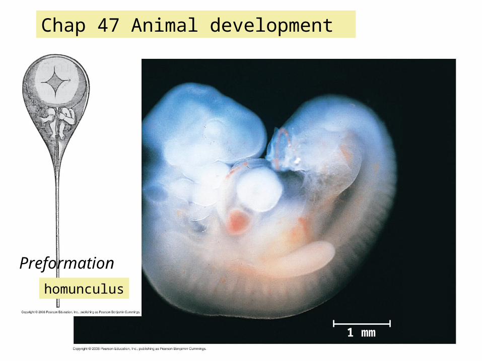
1 mm Chap 47 Animal development Preformation homunculus
-
Upload
magdalena-fajardo -
Category
Documents
-
view
38 -
download
2
description
Chap 47 Animal development. Preformation. homunculus. 1 mm. Development is determined by the zygote ’ s genome and molecules in the egg called cytoplasmic determinants Cell differentiation is the specialization of cells in structure and function - PowerPoint PPT Presentation
Transcript of 1 mm
1 mm
Chap 47 Animal development
Preformation
homunculus

• Development is determined by the zygote’s genome and molecules in the egg called cytoplasmic determinants
• Cell differentiation is the specialization of cells in structure and function
• Morphogenesis is the process by which an animal takes shape
FertilizationCleavageGastrulationOrganogenesis

Fig. 47-3-1
Basal body(centriole)
Spermhead
Sperm-bindingreceptors
Acrosome
Jelly coat Vitelline layer
Egg plasmamembrane
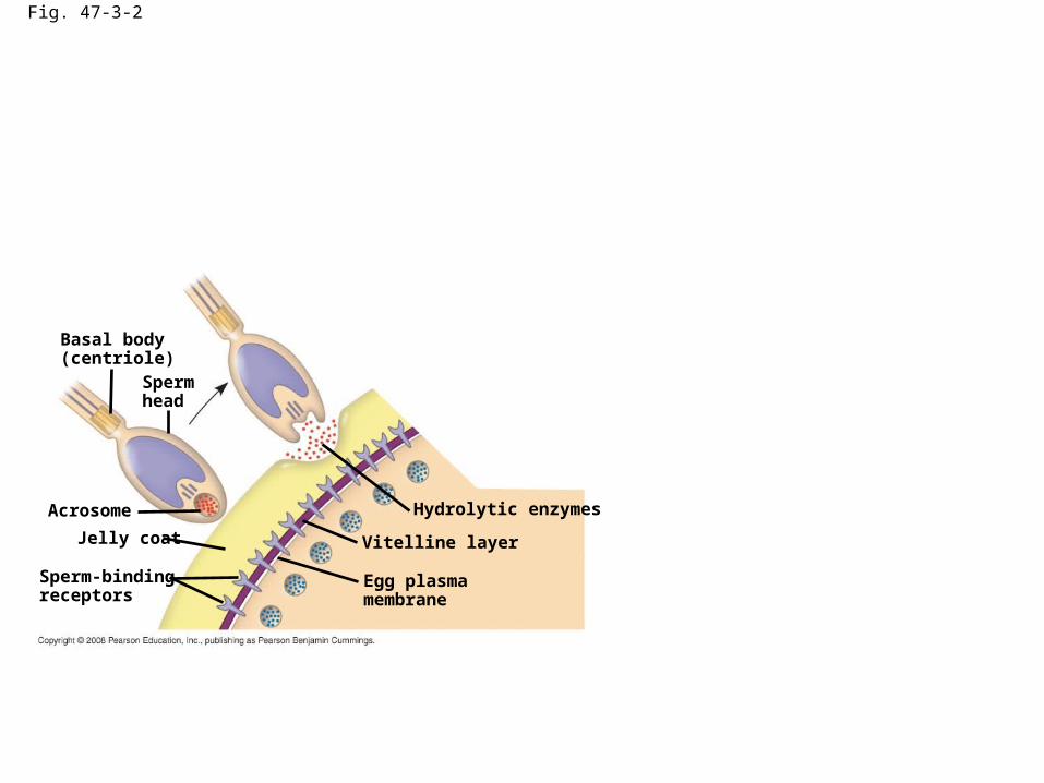
Fig. 47-3-2
Basal body(centriole)
Spermhead
Sperm-bindingreceptors
Acrosome
Jelly coat Vitelline layer
Egg plasmamembrane
Hydrolytic enzymes
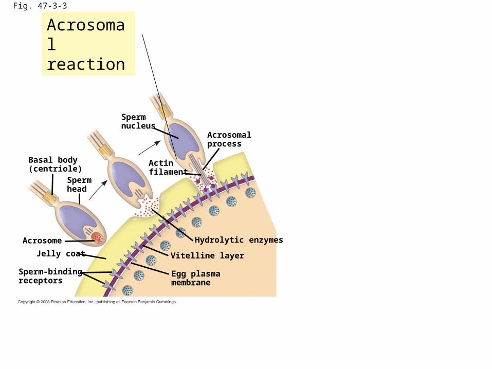
Fig. 47-3-3
Basal body(centriole)
Spermhead
Sperm-bindingreceptors
Acrosome
Jelly coat Vitelline layer
Egg plasmamembrane
Hydrolytic enzymes
Acrosomalprocess
Actinfilament
Spermnucleus
Acrosomal reaction

Fig. 47-3-4
Basal body(centriole)
Spermhead
Sperm-bindingreceptors
Acrosome
Jelly coat Vitelline layer
Egg plasmamembrane
Hydrolytic enzymes
Acrosomalprocess
Actinfilament
Spermnucleus
Sperm plasmamembrane
Fusedplasmamembranes
Acrosomal reaction fast block to polyspermy
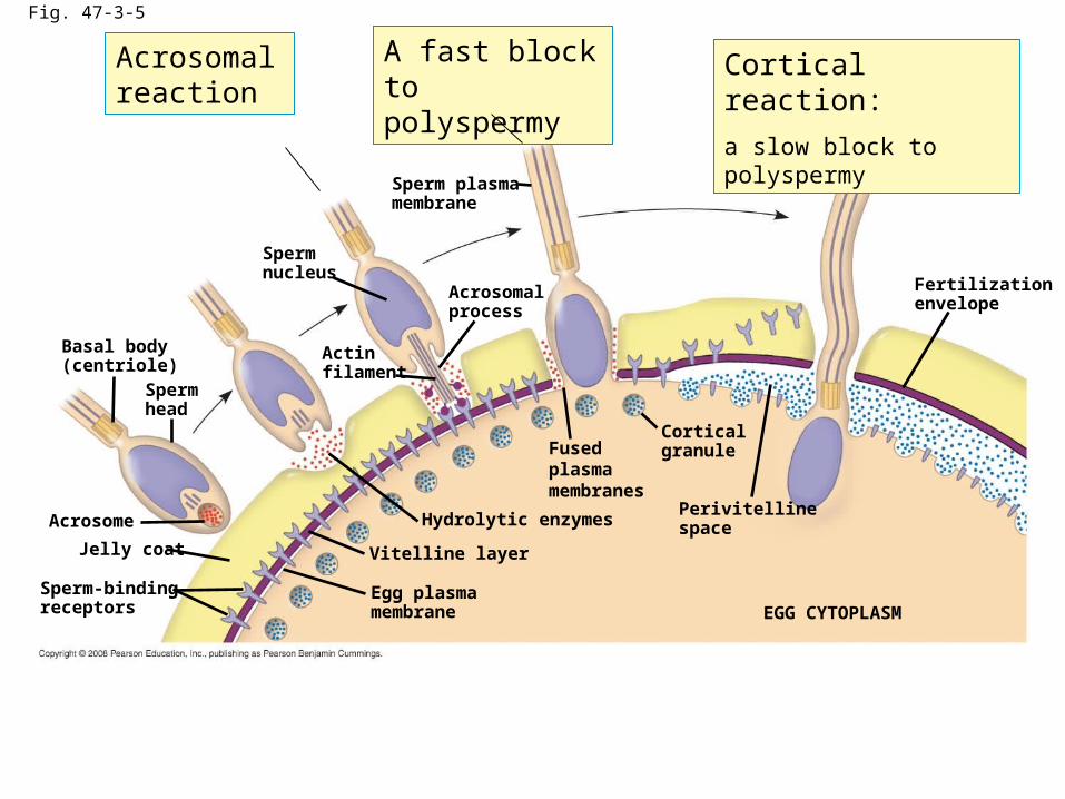
Fig. 47-3-5
Basal body(centriole)
Spermhead
Sperm-bindingreceptors
Acrosome
Jelly coat Vitelline layer
Egg plasmamembrane
Hydrolytic enzymes
Acrosomalprocess
Actinfilament
Spermnucleus
Sperm plasmamembrane
Fusedplasmamembranes
Fertilizationenvelope
Corticalgranule
Perivitellinespace
EGG CYTOPLASM
Acrosomal reaction
A fast block to polyspermy
Cortical reaction:
a slow block to polyspermy

Fig. 47-4 EXPERIMENT
10 sec afterfertilization
1 sec beforefertilization
RESULTS
CONCLUSION
25 sec 35 sec 1 min500 µm
10 sec afterfertilization
20 sec 30 sec500 µm
Point ofspermnucleusentry
Spreadingwave of Ca2+
Fertilizationenvelope
Ca2+
the formation of the fertilization envelope?

Fig. 47-4a
EXPERIMENT
10 sec afterfertilization
25 sec 35 sec 1 min500 µm

Fig. 47-4b
1 sec beforefertilization
RESULTS
10 sec afterfertilization
20 sec 30 sec500 µm

Fig. 47-4c
CONCLUSION
Point ofspermnucleusentry
Spreadingwave of Ca2+
Fertilizationenvelope
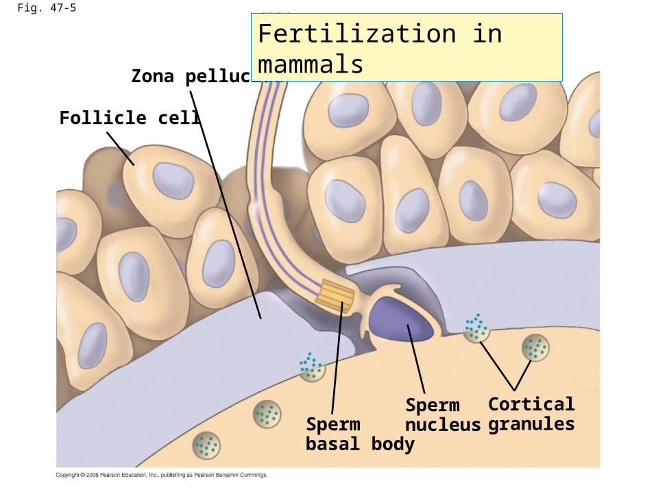
Fig. 47-5
Follicle cell
Zona pellucida
Cortical granules
SpermnucleusSperm
basal body
Fertilization in mammals

Fig. 47-6
(a) Fertilized egg (b) Four-cell stage (c) Early blastula (d) Later blastula
Cleavage Holoblastic cleavage meroblastic cleavage

Fig. 47-7
(a) The three axes of the fully developed embryo
(b) Establishing the axes
Pigmentedcortex
Right
Firstcleavage
Dorsal
Left
Posterior
Ventral
Anterior
Graycrescent
Futuredorsalside
Vegetalhemisphere
Vegetal pole
Animal poleAnimalhemisphere
Point ofspermnucleusentry
(anterior—posterior)(Dorsal– ventral ) (right—left)

Fig. 47-7b-1
(b) Establishing the axes
Vegetalhemisphere
Vegetal pole
Animal poleAnimalhemisphere
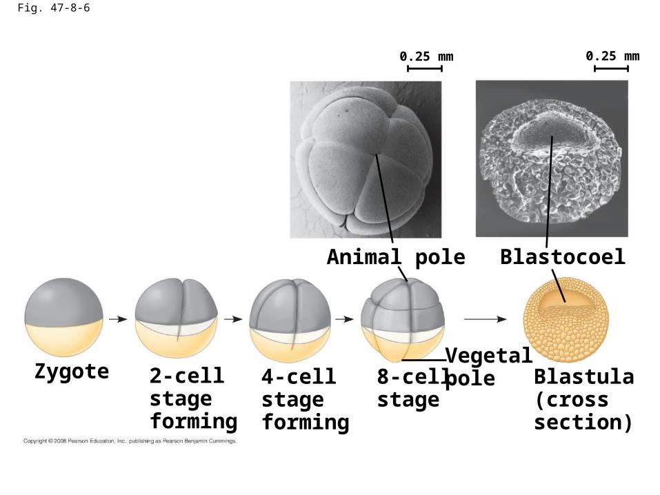
Fig. 47-8-6
Blastula(crosssection)
BlastocoelAnimal pole
4-cellstageforming
2-cellstageforming
Zygote 8-cellstage
Vegetalpole
0.25 mm 0.25 mm

Fig. 47-9-1
Animalpole
Blastocoel
Vegetalpole
Vegetalplate
Mesenchymecells
Future ectoderm
Future mesoderm
Future endoderm
Gastrulation I (sea urchin)
Invagination

Fig. 47-9-2
Future ectoderm
Future mesoderm
Future endoderm
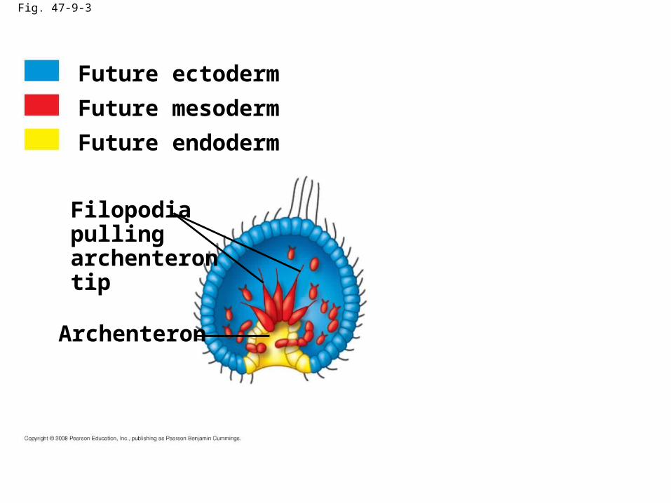
Fig. 47-9-3
Archenteron
Filopodiapullingarchenterontip
Future ectoderm
Future mesoderm
Future endoderm
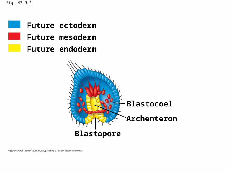
Fig. 47-9-4
Archenteron
Blastopore
Blastocoel
Future ectoderm
Future mesoderm
Future endoderm

Fig. 47-9-5
Digestive tube (endoderm)
Mouth
Ectoderm
Mesenchyme(mesodermforms futureskeleton)
Anus (from blastopore)
Future ectoderm
Future mesoderm
Future endoderm

Fig. 47-9-6
Future ectoderm
Key
Future endoderm
Digestive tube (endoderm)
Mouth
Ectoderm
Mesenchyme(mesodermforms futureskeleton)
Anus (from blastopore)
Future mesoderm
Blastocoel
Archenteron
Blastopore
Blastopore
Mesenchymecells
Blastocoel
Blastocoel
Mesenchymecells
Archenteron
Vegetalplate
Vegetalpole
Animalpole
Filopodiapullingarchenterontip
50 µm
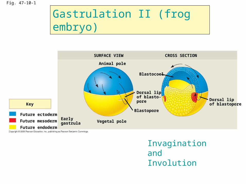
Fig. 47-10-1
Future ectoderm
Key
Future endoderm
Future mesoderm
SURFACE VIEW
Animal pole
Vegetal poleEarlygastrula
Blastopore
Blastocoel
Dorsal lipof blasto-pore
CROSS SECTION
Dorsal lipof blastopore
Invagination and Involution
Gastrulation II (frog embryo)
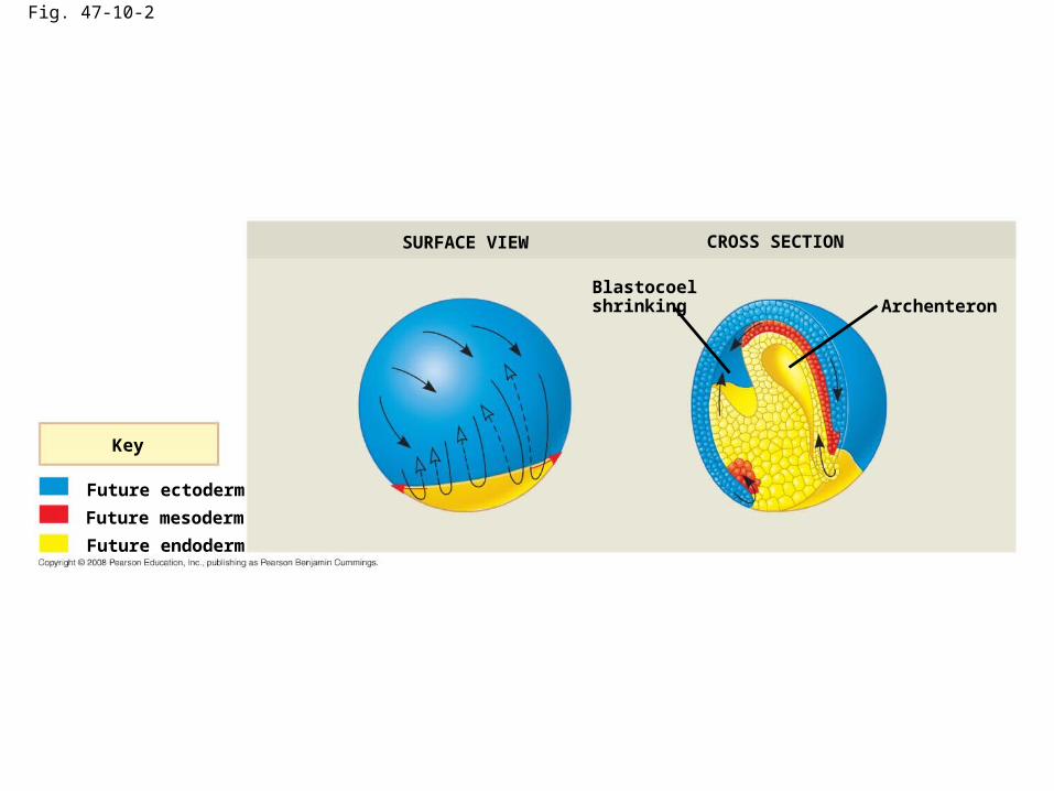
Fig. 47-10-2
Future ectoderm
Key
Future endoderm
Future mesoderm
SURFACE VIEW
Blastocoelshrinking
CROSS SECTION
Archenteron

Fig. 47-10-3
SURFACE VIEW
BlastoporeLategastrula
Blastopore
Blastocoelremnant
Yolk plug
CROSS SECTION
Ectoderm
Mesoderm
Endoderm
Archenteron
Future ectoderm
Key
Future endoderm
Future mesoderm
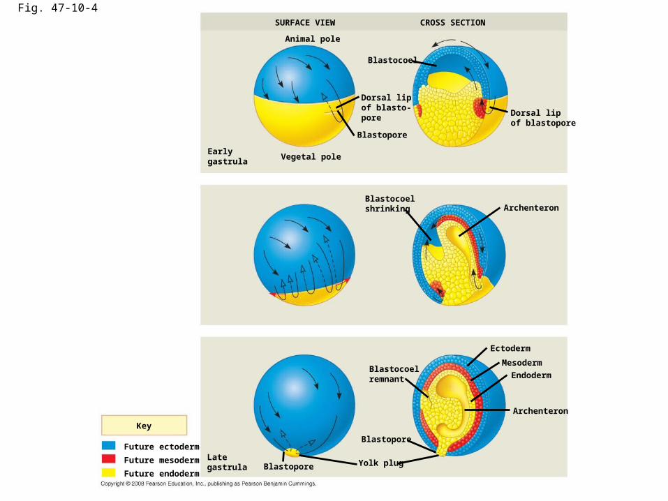
Fig. 47-10-4
Future ectoderm
Key
Future endoderm
Future mesoderm
SURFACE VIEW
Animal pole
Vegetal poleEarlygastrula
Blastopore
Blastocoel
Dorsal lipof blasto-pore
CROSS SECTION
Dorsal lipof blastopore
Lategastrula
Blastocoelshrinking Archenteron
Blastocoelremnant
Archenteron
Blastopore
Blastopore Yolk plug
Ectoderm
Mesoderm
Endoderm
Gastrulation III (a chick embryo)

Fig. 47-11
Endoderm
Futureectoderm
Migratingcells(mesoderm)
Hypoblast
Dorsal Fertilized egg
Blastocoel
YOLK
Anterior
Right
Ventral
Posterior
Left
Epiblast
Primitive streak
Embryo
Yolk
Primitive streak

Fig. 47-12
Neural folds Tail bud
Neural tube
(b) Neural tube formation
Neuralfold
Neural plate
Neuralfold
Neural plate
Neural crestcells
Neural crestcells
Outer layerof ectoderm
Mesoderm
Notochord
Archenteron
Ectoderm
Endoderm
(a) Neural plate formation
(c) Somites
Neural tube
Coelom
Notochord
1 mm
1 mmSEM
Somite
Neural crestcells
Archenteron(digestivecavity)
SomitesEye
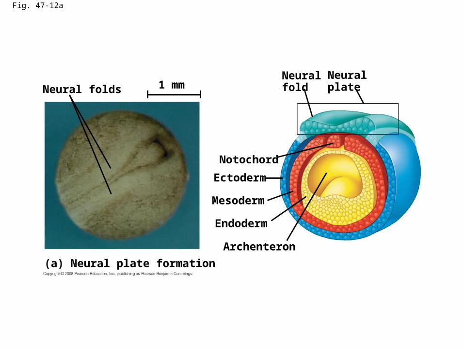
Fig. 47-12a
Neural foldsNeuralfold
Neural plate
Mesoderm
Notochord
Archenteron
Ectoderm
Endoderm
(a) Neural plate formation
1 mm

Fig. 47-12b-1
(b) Neural tube formation
Neuralfold
Neural plate

Fig. 47-12b-2
(b) Neural tube formation

Fig. 47-12b-3
Neural crestcells
(b) Neural tube formation

Fig. 47-12b-4
Neural tube
Neural crestcells
Outer layerof ectoderm
(b) Neural tube formation

Fig. 47-12c
Tail bud
(c) Somites
Neural tube
Coelom
Notochord
1 mmSEM
Somite
Neural crestcells
Archenteron(digestivecavity)
SomitesEye
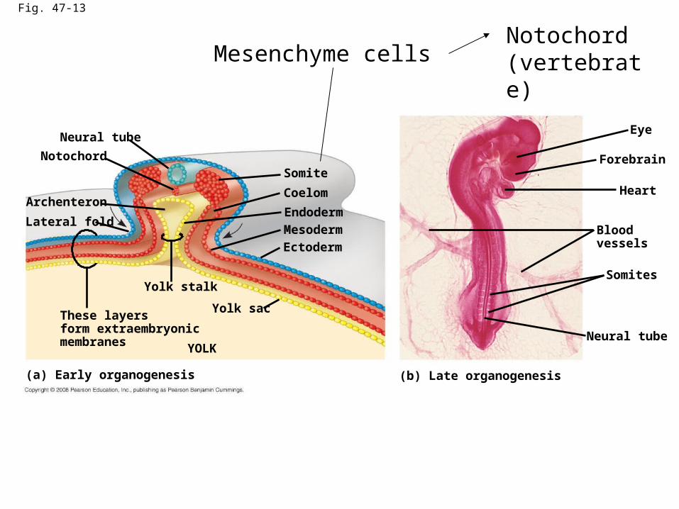
Fig. 47-13
Endoderm
(a) Early organogenesis
Neural tube
Coelom
Notochord
These layersform extraembryonicmembranes YOLK
Heart
Eye
Neural tube
Somite
Archenteron
Mesoderm
Ectoderm
Lateral fold
Yolk stalk
Yolk sac
(b) Late organogenesis
Somites
Forebrain
Bloodvessels
Mesenchyme cellsNotochord (vertebrate)

Fig. 47-14
ECTODERM MESODERM ENDODERM
Epidermis of skin and itsderivatives (including sweatglands, hair follicles)Epithelial lining of mouthand anusCornea and lens of eyeNervous systemSensory receptors inepidermisAdrenal medullaTooth enamelEpithelium of pineal andpituitary glands
NotochordSkeletal systemMuscular systemMuscular layer ofstomach and intestineExcretory systemCirculatory and lymphaticsystemsReproductive system(except germ cells)Dermis of skinLining of body cavityAdrenal cortex
Epithelial lining ofdigestive tractEpithelial lining ofrespiratory systemLining of urethra, urinarybladder, and reproductivesystemLiverPancreasThymusThyroid and parathyroidglands
organogenesis

Fig. 47-15
Embryo
Amnion
Amnioticcavitywithamnioticfluid
Shell
Chorion
Yolk sac
Yolk (nutrients)
Allantois
Albumen
Extraembryonic membrane

Fig. 47-16-5
Yolk sac
Mesoderm
Amnion
Chorion
Ectoderm
Extraembryonicmesoderm
Trophoblast
Endoderm
Hypoblast
Expandingregion oftrophoblast
Epiblast
Maternalbloodvessel
Allantois
Trophoblast
Hypoblast
Endometrialepithelium(uterine lining)
Inner cell mass
Blastocoel
Uterus
Epiblast
Amnioticcavity
Expandingregion oftrophoblast
Yolk sac (fromhypoblast)
Chorion (fromtrophoblast)
Extraembryonicmesoderm cells(from epiblast)
Mammalian development

Fig. 47-17-6
Neural tube
Actin filaments
Microtubules
Ectoderm
Neuralplate
Morphogenesis:
Cell type, position, and adhesion
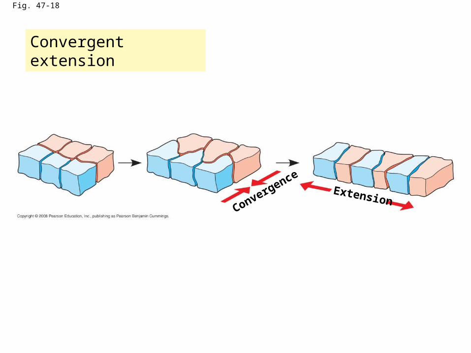
Fig. 47-18
ConvergenceExtension
Convergent extension

Fig. 47-19
Control embryo Embryo without EP cadherin
0.25 mm 0.25 mmRESULTS
CAMs(cell adhesion molecules): Cadherin
EP cadherin is required for proper cell organization in the blastula

Fig. 47-20
Experiment 1
Matrix blocked
RESULTS
Experiment 2
Control
Matrix blockedControl
An organized fibronectin matrix is required for convergent extension.
Injection the molecule to block the interaction of fibronectin and its receptor
Tightly packed in a column

Fig. 47-21
Epidermis
(b) Cell lineage analysis in a tunicate(a) Fate map of a frog embryo
Epidermis
Blastula Neural tube stage(transverse section)
Centralnervoussystem
Notochord
Mesoderm
Endoderm
64-cell embryos
Larvae
Blastomeresinjected with dye
The developmental fate of cells depends on their history and on inductive signals
Fate map

Fig. 47-22
Mouth
Zygote
Intestine
Nervoussystem,outer skin,muscula-ture
Intestine
Hatching
Eggs Vulva
Anus
1.2 mmANTERIOR POSTERIOR
Muscula-ture, gonads
10
0First cell division
Germ line(futuregametes)
Musculature
Outer skin,nervous system
Tim
e a
fter
fe
rtil
iza
tio
n (
ho
urs
)

Fig. 47-23a
Thread
Graycrescent
Experimental egg(side view)
Graycrescent
Control egg(dorsal view)
EXPERIMENT
Gray cresent affects the developmental potential of the first two daught cells

Fig. 47-23b
Thread
Graycrescent
Experimental egg(side view)
Graycrescent
Control egg(dorsal view)
EXPERIMENT
NormalBelly pieceNormal
RESULTS

Fig. 47-24
Primary structures:Neural tube
Dorsal lip ofblastopore
Secondary(induced) embryo
Notochord
Pigmented gastrula(donor embryo)
EXPERIMENT
Primary embryo
RESULTS
Nonpigmented gastrula(recipient embryo)
Secondary structures:Notochord (pigmented cells)
Neural tube (mostly nonpigmented cells)
The Organizer of Spemann and Mangold

Fig. 47-24a
Dorsal lip ofblastopore
Pigmented gastrula(donor embryo)
EXPERIMENT
Nonpigmented gastrula(recipient embryo)

Fig. 47-24b
Primary structures:Neural tube
Secondary(induced) embryo
Notochord
Primary embryo
RESULTS
Secondary structures:Notochord (pigmented cells)Neural tube (mostly nonpigmented cells)
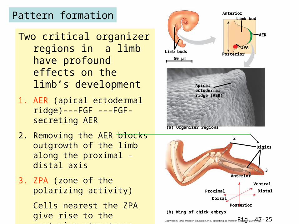
Fig. 47-25
(a) Organizer regions
Apicalectodermalridge (AER)
Digits
Limb buds
(b) Wing of chick embryo
Posterior
AnteriorLimb bud
AER
ZPA
50 µm
Anterior
2
34
Posterior
Ventral
Distal
Dorsal
Proximal
Two critical organizer regions in a limb have profound effects on the limb’s development
1. AER (apical ectodermal ridge)---FGF ---FGF-secreting AER
2. Removing the AER blocks outgrowth of the limb along the proximal –distal axis
3. ZPA (zone of the polarizing activity)
Cells nearest the ZPA give rise to the posterior structures (three digit)
Pattern formation
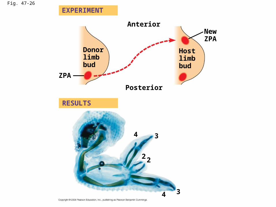
Fig. 47-26
Host limb bud
Posterior
2
34
AnteriorNewZPA
EXPERIMENT
RESULTS
ZPA
Donor limb bud
2
34
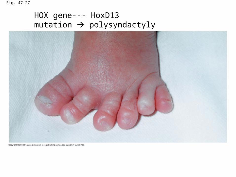
Fig. 47-27
HOX gene--- HoxD13 mutation polysyndactyly

Fig. 47-UN1
Sperm-egg fusion and depolarizationof egg membrane (fast block topolyspermy)
Cortical granule release(cortical reaction)
Formation of fertilization envelope(slow block to polyspermy)
Fertilization

Fig. 47-UN2
Blastocoel
Animal pole
2-cellstageforming
8-cellstage
Blastula
Vegetal pole
Cleavage

Fig. 47-UN3
Gastrulation

Fig. 47-UN4
Neural tube
Coelom
Notochord
Coelom
Notochord
Neural tube
Organogenesis


![INDEX [] · escala real 1:1 real scale 1:1 3,5 mm 10,5 mm 12 mm 5,6 mm. benefits ventajas vorteile avantages ligereza geringes gewicht lÉgÈret ...](https://static.fdocuments.us/doc/165x107/5bbf06ce09d3f280238cf1c7/index-escala-real-11-real-scale-11-35-mm-105-mm-12-mm-56-mm-benefits.jpg)
![alfabeto dei neuroni Trento.ppt [modalità compatibilità]...K+ 5 mM 100 mM 1:20 -80 mV Na+ 150 mM 15 mM 10:1 62 mV Ca2 + 2 mM 0,0002 mM 10.000: 1 123 mV Cl- 150 mM 13 mM 11,5:1 -65](https://static.fdocuments.us/doc/165x107/5faf855e0c275721d342a9f8/alfabeto-dei-neuroni-modalit-compatibilit-k-5-mm-100-mm-120-80-mv-na.jpg)


