1. Introduction - u-szeged.hu
55
1. Introduction 1.1. Properties of titanium Titanium (Ti) the ninth most abundant element in the Earth's crust, occurs in a number of minerals and clays. It is always bonded to other elements in nature. It was discovered in Cornwall, United Kingdom, in 1791 by an amateur geologist and pastor, William Gregor (Boase, 2008). Ti readily reacts with oxygen, when exposed to air. Its alloys are very strong and lightweight, and are widely used in industrial processes, e.g. desalination plants, the aerospace and automotive industries and agriculture, and the element is utilized in medical prostheses, orthopedic implants, dental implants and dental and endodontic instruments (Wang and Fenton, 1996), and also in sporting goods, jewelry and mobile phones. The most important properties of this metal are its corrosion resistance and high strength-to-weight ratio. In an unalloyed condition, Ti is as strong as some steels, but 45% lighter. A passive and protective oxide coating that forms on its surface protects it from further oxidation and other reactions. Nevertheless, it does corrode slowly (Schutz and Thomas, 1987). A surface oxide layer is formed within nanoseconds when Ti is exposed to air (Hanawa, 1998). Passivation treatment provides a controlled and uniformly oxidized surface state. Passivation decreases ionrelease, and leads to a dense and stable oxide film which improves the resistance to corrosion. The treatment with nitric acid eliminates metallic contamination from the surface, but has practically no influence on the overall topography of the Ti surface. The result is a passive layer of TiO 2 (titanium dioxide) with an initial thickness of 2-6 nm (Puippe, 2003), but it slowly grows until it attains a thickness of 25 nm. Oxidative agents are well known to exert a corrosive effect on the alloys used in dentistry, with the exceptions of Ti and some other bioinert materials. Indeed, oxidative processes can thicken and condense the TiO 2 layer on the surface, improving the corrosion stability of the underlying Ti. On the other hand, reducting agents, such as fluoride (F – ), may have the opposite effect and attack this layer (Strietzel et al., 1998). Ti ion release is enhanced in the presence of F – , and this effect is accelerated at low pH. High F – concentrations and an acidic pH are known to impair the corrosion resistance of Ti (Toumelin-Chemla et al., 1996) and consequently crevice formation and pitting corrosion occur (Reclaru and Meyer, 1998; Schiff et al., 2002). Ti is a relatively cheap metal, and commonly used to make surgical and dental implants. However Ti crowns, bridges and the framework of partial removable dentures PDF created with pdfFactory Pro trial version www.pdffactory.com
Transcript of 1. Introduction - u-szeged.hu

1. Introduction
1.1. Properties of titanium
Titanium (Ti) the ninth most abundant element in the Earth's crust, occurs in a number
of minerals and clays. It is always bonded to other elements in nature. It was discovered in
Cornwall, United Kingdom, in 1791 by an amateur geologist and pastor, William Gregor
(Boase, 2008). Ti readily reacts with oxygen, when exposed to air. Its alloys are very strong
and lightweight, and are widely used in industrial processes, e.g. desalination plants, the
aerospace and automotive industries and agriculture, and the element is utilized in medical
prostheses, orthopedic implants, dental implants and dental and endodontic instruments
(Wang and Fenton, 1996), and also in sporting goods, jewelry and mobile phones. The most
important properties of this metal are its corrosion resistance and high strength-to-weight ratio.
In an unalloyed condition, Ti is as strong as some steels, but 45% lighter. A passive and
protective oxide coating that forms on its surface protects it from further oxidation and other
reactions. Nevertheless, it does corrode slowly (Schutz and Thomas, 1987). A surface oxide
layer is formed within nanoseconds when Ti is exposed to air (Hanawa, 1998).
Passivation treatment provides a controlled and uniformly oxidized surface state.
Passivation decreases ionrelease, and leads to a dense and stable oxide film which improves
the resistance to corrosion. The treatment with nitric acid eliminates metallic contamination
from the surface, but has practically no influence on the overall topography of the Ti surface.
The result is a passive layer of TiO2 (titanium dioxide) with an initial thickness of 2-6 nm
(Puippe, 2003), but it slowly grows until it attains a thickness of 25 nm. Oxidative agents are
well known to exert a corrosive effect on the alloys used in dentistry, with the exceptions of
Ti and some other bioinert materials. Indeed, oxidative processes can thicken and condense
the TiO2 layer on the surface, improving the corrosion stability of the underlying Ti. On the
other hand, reducting agents, such as fluoride (F–), may have the opposite effect and attack
this layer (Strietzel et al., 1998). Ti ion release is enhanced in the presence of F–, and this
effect is accelerated at low pH. High F– concentrations and an acidic pH are known to impair
the corrosion resistance of Ti (Toumelin-Chemla et al., 1996) and consequently crevice
formation and pitting corrosion occur (Reclaru and Meyer, 1998; Schiff et al., 2002).
Ti is a relatively cheap metal, and commonly used to make surgical and dental
implants. However Ti crowns, bridges and the framework of partial removable dentures
PDF created with pdfFactory Pro trial version www.pdffactory.com

2
(Huget, 2002; Wang and Fenton, 1996) are not common in prosthetic dentistry because the
technology of the operation process is very expensive. The material used to produce dental
implants is generally “commercially pure” Ti (CP Ti) or the most common Ti alloy, Ti-6Al-
4V (Mändl et al., 2005; Park and Kim, 2000). For the production of Ti brackets for
orthodontic dentistry (Harzer et al., 2001), another Ti alloy with special features is used.
Dental arch wires and orthodontic braces are usually made from the TiNi shape memory alloy.
Large numbers of patients with dental implants also have natural teeth in the oral
cavity. Furthermore, orthodontic patients are at highrisk of the development caries and in need
of F– protection. To maintain good oral hygiene and prevent caries, patients use toothpaste,
gels or mouthwash, almost all of which contain F–. The hydroxyapatite [Ca5(PO4)3OH] of the
enamel is attacked by F– to form fluorapatite, Ca5(PO4)3F. Fluorapatite makes the enamel
more resistant to caries (Triller, 1998). However, as F–-containing mouthwashes have been
found to cause galvanic corrosion between orthodontic wires and brackets, it has been
suggested that a new type of mouthwash for use during orthodontic therapy could be an
interesting development in this field (Schiff et al., 2006).
The effects of the use of ordinary F–-containing toothpastes to clean the teeth of
patients with brackets have also been reported (Harzer et al., 2001). The surface roughness
and biocompatibility of Ti facilitate plaque adherence. The surface of the Ti brackets became
very rough due to the use of toothpaste for 5.5-17 months, and in a few cases corrosion
occurred in the brackets. It was concluded that such toothpaste enhanced plaque accumulation
and discoloration of the bracket surface, but the level of corrosion was not significant.
Nakagawa et al. (2001) investigated the Ti alloys Ti-6Al-4V, Ti-6Al-7Nb and Ti-0.2
Pd form the aspect of their corrosion behavior and established that even a low F–
concentration caused corrosion in an acidic environment. However if the Ti alloy contained at
least 0.2% Pd, corrosion did not take place. The high corrosion resistance of this alloy is due
to the enrichment of Pd in the surface, which promotes the repassivation of Ti.
1.2. Types of dental implants
During the history of dental implantology since 1952, when the first dental implants
were introduced many different types have been employed, but endosteal implants are
currently most common. On the next figures some forms of implants (subperiosteal,
transosteal, endodontic, endosteal) are shown.
PDF created with pdfFactory Pro trial version www.pdffactory.com

3
a. Intramucosal implants (Evasic, 1983).
b. Subperiosteal implants are illustrated in Fig. 1 (Zwerger et al., 2007).
Figure 1. Subperiosteal implants
c. Typical transosteal implants are shown in Fig. 2 (Knapp and Small, 1990).
Figure 2. Transosteal implants
PDF created with pdfFactory Pro trial version www.pdffactory.com

4
d. An endodontic implant is to be seen in Fig. 3 (Frank, 1967).
Figure 3. Endodontic implant
e. Endosteal implants
Endosteal implants (Fig. 4) are the most widely used type of implants in bone. The
various types include screws, cylinders or blades placed surgically into the jawbone. A typical
implant consists of a Ti screw (resembling a tooth root) with a roughened or smooth surface.
Roughened surfaces may be modified by plasma spraying, anodizing, etching or sandblasting
to increase the surface area and the integration potential of the implant. The endosteal
implants used almost exclusively nowadays comprise a root portion, which is placed in the
jawbone, a cervix (transmucosal part), located at the level of the gums, and a head, which
supports the crowns, bridges and in general any structure. The transmucosal part between the
root portion and the head is assorted with the gingival margin (Fig. 4).
Figure 4. Endosteal implants
PDF created with pdfFactory Pro trial version www.pdffactory.com

5
The majority of dental implants are made of CP Ti, which is available in 4 grades
depending on the carbon and iron contents (Park and Kim, 2000, Table 1). For reasons of
economy and mechanical quality, some implant systems are fabricated from Ti-6Al-4V alloy
(Parr et al., 1985).
Table 1. Chemical compositions of various grades of Ti and one of its alloys
[Park and Kim, 2000 (ASTM, 1992)]
Element Grade 1 Grade 2 Grade 3 Grade 4 Ti-6Al-4Va
Nitrogen 0.03 0.03 0.05 0.05 0.05
Carbon 0.10 0.10 0.10 0.10 0.08
Hydrogen 0.015 0.015 0.015 0.015 0.0125
Iron 0.20 0.30 0.30 0.50 0.25
Oxygen 0.18 0.25 0.35 0.40 0.13
Titanium Balance aNominally: aluminum 6.00% (5.50 ~ 6.50), vanadium 4.00% (3.50 ~ 4.50), and other elements: 0.1% maximum individually or 0.4% total. All data are maximum allowable weight percentages.
1.3. Indications of dental implants
Dental implants are used to replace one or more or all missing teeth, and can serve as
an abutment for various prosthetic appliances. Their use allows dentists to avoid the
preparation of teeth for fixed partial dentures, or a more stable and more esthetic treatment
option can be provided for the patients. A well-functioning dental implant can prevent further
tooth loss and preserve the alveolar bone.
The main indications for prosthetic implant treatment are:
(a) One or more missing teeth. Single tooth implants for cases with a bounded saddle: without
preparation of the adjacent teeth for a bridge, the crown is retained only on the implant.
(b) A posterior edentulous ridge: without implants, only removable partial dentures can be
made; with implants, the patient may wear crowns or bridges with great comfort.
(c) A completely edentulous jaw: an implant-retained overdenture, a removable or fixed
bridge, or crowns can be provided for the patient, who will achieve a higher level of
quality of life.
In the first two groups, Ti implants and natural teeth are present together in the oral
cavity. For caries prevention, dentists often suggest various alternative prevention solutions,
mainly mouthwashes.
PDF created with pdfFactory Pro trial version www.pdffactory.com

6
1.4. Macroscopic features of the gingiva
The success and long-term prognosis of endosseous implants depend predominantly
mainly on three factors: osseointegration (anchorage in the host bone), gingival attachment
and the appropriate transmission of masticatory force or load transfer capacity (Fig. 5). This
section focuses on the macroscopic features of the gingiva, which is important for a clear
understanding of the purpose of my studies.
Figure 5. The success and long-term prognosis of endosseous implants depend mainly on
three factors: osseointegration (anchorage in the host bone), gingival attachment and the
appropriate transmission of masticatory force (load transfer capacity).
The gingiva is divided anatomically into free gingiva, and attached and interdental
papillae. The marginal gingiva is the terminal edge of the gingiva surrounding the teeth in a
collar. In around half of the population, it is demarcated from the adjacent, attached gingiva
by a shallow linear depression, the free gingival groove. It forms the soft tissue wall of the
gingival sulcus, which is usually about 1 mm deep. The marginal gingiva is supported and
stabilized by the gingival fibers. The attached gingiva is continuous with the marginal gingiva.
It is firm, resilient, and tightly bound to the underlying periosteum of the alveolar bone. The
facial aspect of the attached gingiva extends to the relatively loose and movable alveolar
mucosa, from which it is demarcated by the mucogingival junction. It is resistant to
masticatory forces and always keratinized. The peri-implant mucosa is the soft tissue that
surrounds dental implants. The interface between the implant and the mucosa consists of an
epithelial and a connective tissue component. The presence of the attached mucosa around
dental implants is important because it is bound very tightly to the underlying alveolar bone
and provides protection for the mucosa during functional use of the structures of the oral
PDF created with pdfFactory Pro trial version www.pdffactory.com
7
cavity, such as chewing. The gingival and the peri-implant mucosa are covered by a
keratinized oral epithelium. The connective tissue barrier and the junctional epithelium
around the tooth or the implant are about 2 mm wide (Fig. 6). The width of the supraalveolar
connective tissue around the tooth is 1 mm, but in the case of a Ti implant it is 1.5 mm
(Berglundh et al., 1991); it is separated from the alveolar bone by collagen-rich connective
tissue. This biological barrier which has a thickness of 3 to 4 mm, has been called the
“biological width”; it protects the zone of osseointegration from factors caused by plaque and
bacteria in the oral cavity (Berglundh and Lindhe, 1996).
Figure 6. Details of the epithelial attachment on a Ti implant surface
Both epithelium types develop a hemidesmosomal connection between the tooth and
the implant surface (Gould et al., 1984; Swope and James, 1981). Pending around the natural
tooth, the main filaments which constitute the connective tissue radiate from the cementum, in
the event of a Ti implant, the main filaments start from the periosteum of the bone and run
parallel to the bone surface. Without attached gingiva, the freely movable alveolar mucosa
(which is more fragile) would suffer injury during eating and cleaning activities, such as
brushing of the teeth. The interdental gingiva occupies the gingival embrasure, which is the
interproximal space beneath the approximal contact area of the teeth. It can be pyramidal or
have a col shape, like a pass in a mountain range (Schroeder and Listgarten, 2000).
Ti implant
Sulcular epithelium
Junctional epithelium
Connective tissue (outer zone)
Bone
PDF created with pdfFactory Pro trial version www.pdffactory.com
8
It has been reported that a basal lamina and hemidesmosomes develop 2 weeks after
implant placement. In vitro and in vivo studies have demonstrated a chemical attachment
between the Ti implant surface oxide layer and the epithelium. There is a similar attachment
between the epithelium and natural tooth surfaces, mediated by a glycoprotein. Connective
tissue fibers adjacent to Ti implant surfaces may bring the tissue into tight apposition to the
implant without the formation of an absolute biological attachment between the implant and
the connective tissue. Modification of the Ti surface morphology may selectively intensify the
attachment of either epithelial cells or fibroblasts. The mechanisms of attachment and of the
factors which enhance the integrity of the biological seal between the implant and the soft
tissues are very important for the prognosis of Ti implants. (Donley and Gilette, 1991).
A very important factor in the implant survival is the state of health of the peri-implant
soft tissue. Any damage to the junctional epithelium results in it being irregular in texture, and
in the formation of "pocket" epithelium, which is a primary symptom of gum disease. The
long-term success of a dental implant depends to a large extent on the gingival attachment to
the neck of the implant. This mucosal seal ensures protection against bacterial attack and
other injurious effects exerted by the oral environment. The epithelial attachment may be
anchored onto a rough or a smooth surface by hemidesmosomes through a preformed
glycoprotein layer. A rough surface is more favorable for plaque accumulation, which is an
undesired effect in this very sensitive region of the implant. Accordingly, in order to avoid
pathogenic plaque accumulation, the neck of an implant abutment must be polished (Fig. 5)
(Bollen et al., 1996).
1.5. Causes of dental implants failure
The most common causes of implant failures are:
1. overloading
2. bacterial infection
3. secondary complications in the incomplete bone tissue
This section will deal only with bacterial infections as these are related to the studies
and aims of the thesis.
The plaque formed on the surface of a dental implant may be indicative of a reaction
in the tissue which causes inflammation primarily in the soft tissue, and the bone too may
soon be is involved. Peri-implantitis is an inflammatory reaction which, without treatment,
sooner or later results in boneloss around the implant (Albrektsson and Isidor, 1994). Peri-
PDF created with pdfFactory Pro trial version www.pdffactory.com

9
implant diseases are caused by inflammatory conditions that affect the soft and hard tissues
around the implant fixtures. The extent and location of peri-implant inflammation differ
greatly from inflammation around the teeth in similar environments (Schou et al., 1993). Peri-
implantitis progresses deep into the bone, which is in contrast with periodontitis, when the
inflammation initially developes only in the connective tissue. Gotfredsen et al. (2002)
reported that the resistivity of peri-implant tissues was less than that of the tissues around the
teeth. Peri-implantitis may involve bone resorption, and the treatment outcome following
nonsurgical management is less predictable. Adjunctive decontaminating treatment includes
the use of antimicrobials, and resistant cases may sometimes be managed with a surgical
approach (Heasman, 2010). The proportion of failed dental implants is higher amount patients
who display poor oral hygiene. Histories of periodontitis and cigarette smoking have been
found to be risk indicators for peri-implant disease (Heitz-Mayfield, 2008).
Fibroblasts play an important role in establishing and maintaining the mucosal seal,
this fibroblast-rich barrier exhibiting a high cell turnover next to the Ti surface (Berglundh et
al., 1994). The microbial biofilm adheres to the surface of the transmucosal abutment of
osseointegrated dental implants. The peri-implant mucosa which covers the alveolar bone is
closely adapted to the implant, in a similar way as the gingival crevice around the natural
tooth (Figure 7.).
Figure 7. On a biomaterial such as a dental implant there is always a competition for the
surface between the tissue cells and the bacteria (Anderson et al., 1996)
1.6. Periodontal pathology diseases and peri-implant infections
Periodontal pathology diseases have been divided into two main types: gingival
diseases and periodontal diseases. The gingival diseases can be subdivided two groups,
depending on the origin: dental plaque-induced or non-plaque-induced (Table 2).
PDF created with pdfFactory Pro trial version www.pdffactory.com

10
Table 2. Classification of the gingival diseases (Armitage, 1999)
A. Dental plaque-induced gingival diseases B. Non-plaque-induced gingival lesions
Gingivitis associated only with dental plaque Gingival diseases of specific bacterial origin
Gingival diseases modified by systemic factors Gingival diseases of viral origin
Gingival diseases modified by medication Gingival diseases of fungal origin
Gingival diseases modified by malnutrition Gingival lesions of genetic origin
Gingival manifestations of a systemic condition
Traumatic lesions
Foreign body reactions
Not otherwise specified
Periodontal diseases can be grouped as shown in Table 3 (Armitage, 1999).
Table 3. Types of periodontal diseases
Chronic periodontitis
Aggressive periodontitis
Periodontitis as a manifestation of a systemic disease
Necrotizing periodontal diseases
Abscesses of the periodontium
Periodontitis associated with endodontic lesions
Developmental or acquired deformities and conditions
In peri-implant tissues, microbial colonization and consequent inflammatory reactions
may occur in a similar manner to the pathogenesis of periodontitis. In partially edentulous
patients, the microbiota developing around an implant closely resembles the microflora of the
crevicular sulcus around natural teeth. A history of periodontitis and the presence of putative
periodontal pathogens are factors that can influence the condition of peri-implant tissues in
partially edentulous subjects (Leonhardt et al., 1993; Mombelli et al., 1995). The subgingival
PDF created with pdfFactory Pro trial version www.pdffactory.com

11
microflora around implants and the gingival sulcus around the teeth in the same jaw have a
similar bacterial morphotype, because the teeth may serve as a reservoir for bacterial
colonization (Quirynen and Listgarten, 1990).
The pockets around the teeth can serve as a reservoir for putative periodontal
pathogens, which cause the early colonization of the peri-implant sulcus in partially
edentulous subjects, when the oral hygiene is not meticulous. In several studies on partially
edentulous patients, the microbiologic flora was found to be analogous in the sulcus around
teeth and implants (van Winkelhoff et al., 2000). The complex peri-implant microbiota
resembling that in adult periodontitis can cause infection and implant failures. Aggregati-
bacter actinomycetemcomitans (previously: Actinobacillus actinomycetemcomitans) and
Porphyromonas gingivalis are not as frequently associated with peri-implant infection in
edentulous individuals as in dentate subjects (Mombelli et al., 1987).
The oral cavity is a favorable environment for a great variety of bacteria. Oral bacteria
include streptococci, lactobacilli, staphylococci and corynebacteria, with a large number of
anaerobes, especially Gram-positive rods. Bacterial cells account for 60-70% of the volume of
the dental plaque. The other components are: salivary polymers and bacterial extracellular
products. The plaque is regarded as a naturally-constructed biofilm, in which the consortia of
bacteria may reach a thickness of 300-500 cells on the surfaces of the teeth or dental implants.
High concentrations of bacterial metabolites accumulated on the teeth and gingival tissues can
cause dental diseases. The dominant bacterial factors in the dental plaque are Streptococcus
species (Streptococcus sanguis and Streptococcus mutans), both of which are considered to be
major factors responsible for the formation of plaque.
The development of a dental biofilm (dental plaque) on a surface is determined by the
physical properties of the surface, the environmental humidity and the free energy of the
bacterial surface. Salivary glycoproteins form an acquired pellicle on a hard surface, which
contains a small amount of immunoglobulins too (Almsthäl et al., 2001). This pellicle can
interconnect with the hard surfaces via van der Waals and electrostatic forces (de Jong et al.,
1984; Quirinen et al., 1989) and its width does not exceed 1 µm. At first, the pellicle is
transparent. The formation of plaque is initiated by a loose attachment of Gram-positive cells;
this connection is reversible. Later, a strong attachment develops, when the extracellular
glycocalyx, a sticky polymer produced by bacterial cells, promotes the connections on the
surface. More and more bacterial cells appear among these polysaccharides and can modify
their phenotype, which results in rapid growth of the bacteria and produces large amounts of
extracellular polymeric substances. The bacterial cells synthesize the glycoprotein from
PDF created with pdfFactory Pro trial version www.pdffactory.com
12
dietary sugars (principally sucrose). The extracellular polymeric substances help the emerging
biofilm community to develop a complex, three-dimensional structure. Biofilm communities
can be formed within some hours, the coccoid bacteria develop several layers, and the form is
usually cubic. After a few days, the Gram-positive rods and filaments (mainly Actinomyces
sp.) outnumber the cocci. A reticulation of water channels serve as the pathways for nutrients
and help with the efficient removal of the waste products for use by the bacteria on the surface.
The oxygen concentration within the biofilm changes. It is increasingly difficult for oxygen to
reach the deeper layers and mainly anaerobes can proliferate near the hard surface until
virtually only aerobes or facultative anaerobes can be found on the surface of the biofilm. The
many interactions between the different bacterial species include a number of synergistic and
antagonistic biochemical interactions. For example, when obligate anaerobes and aerobes are
involved in co-adhesion, they interact to ensure the survival of the anaerobic bacteria in the
oxygen-rich oral cavity (Marsh 2004, 2006). As the biofilm thickens and becomes more
mature, it can consist of 100-300 cell layers. In time, the growth of the plaque slows down
and dead bacterial cells can be seen deep inside the plaque. Around the pellicle Ca-containing
crystals appear in the interbacterial matrix; this is the first sign of calculus. On the surface, the
bacterial colonies demonstrate an active life, reflected in a typical corn-cob formation. The
center is this occupied by long filament- shaped bacteria, connected to a number of cocci and
short rods on the surface (Listgarten, 1976).
The initial dental biofilm bacteria are: Streptococcus species, Veillonella sp., Gram-
negative anaerobic cocci, Gram-positive rods (including Actinomyces sp.) and Gram-negative
rods (Capnocytophaga sp.) Specific anaerobic species subsequently appear: Fusobacteria and
Prevotella intermedia. The late colony bacteria are P. gingivalis, mobile Gram-negative rods
and the spirochetes. Figure 8 illustrates of the biofilm formation and maturation, on the
surface (Rickard et al., 2003).
PDF created with pdfFactory Pro trial version www.pdffactory.com

13
Figure 8. (a) Free-floating or planktonic bacteria come into contact with a coated surface
and within minutes can become attached. (b) The bacteria begin to produce slimy extra-
cellular polymeric substances (EPS). (c) The biofilm can spread through detachment of
small or large clumps of cells, or by "seeding dispersal" that releases individual cells.
(d) Either type of detachment allows bacteria to attach to the surface or the down-
stream biofilm of the original community. Maturation and the formation of clonal
mosaics within the multi-species biofilms can be seen (Marsh 2004, 2006).
PDF created with pdfFactory Pro trial version www.pdffactory.com

14
2. Aims of the study
The physical and chemical changes that may take place on a Ti surfaces as a result of
the use of prophylactic agents such as those which are widespread in everyday oral hygiene
regimens may be important factors as concerns the accumulation of plaque on that surface. In
view of the evidence of the causative effect of plaque accumulation in the development of
peri-implantitis, it is very important to identify the effects of F– on Ti surfaces and the
consequences relating to bacterial colonization.
The aims of my study were as follows:
• To apply atomic force microscopy (AFM) to investigate whether a commonly used F–-
containing mouthwash, NaF solution and a prophylactic gel can modify the surface
roughness of CP Ti.
• To identify the chemical changes caused on a Ti surface caused by F–-containing
materials (mouthwash, NaF solution and prophylactic gel) through the use X-ray
photoelectron spectroscopy (XPS).
• To evaluate whether the surface changes mentioned above could influence the
accumulation and colonization of different microorganisms.
• To assess the changes in the number and maturation of the bacteria after modification
of a Ti surface by F–-containing materials.
PDF created with pdfFactory Pro trial version www.pdffactory.com
15
3. Materials and methods
3.1. Cleaning of Ti samples
A total of 72 polished CP Ti discs (9 mm in diameter, 2 mm in thickness, CP grade 4,
Camlog, Biotechnologies AG, Switzerland and Protetim, Hungary) were used. This type of Ti
contains only very small amounts of other elements: the oxygen content is less than 0.40%,
the nitrogen not more than 0.05%, and the carbon less than 0.10%. The discs were
mechanically polished until the surface was analogous to the polished surface of the neck of
the dental implant abutment, i.e. the roughness did not exceed 0.2 µm. They were first cleaned
in an ultrasonic bath with acetone for 15 min then in absolute ethanol for 15 min, and they
were finally rinsed with distilled (ultrapure) water 3 times for 10 min. The discs were always
held only with Ti forceps, so as not to contaminate the surfaces (Stájer et al., 2008). Each of
the cleaned discs was used in only one subsequent investigation.
3.2. Fluoride treatment and sterilization
Patients are recommended to use a prophylactic rinse once a day for 30 s and the gel
once a week for 2 min. In our investigations, the duration of trials of cleaned Ti discs with F–
was 1 h. This corresponds to the accumulated effect of the regular usage of the rinse for 4
months or of the gel for 7.5 months. In all cases relating to the determination of numerical
data reported in this thesis, groups of 4 discs were treated for 1 h with one or other of the
following materials:
– a mouthwash (Elmex, GABA International AG, Switzerland) containing 250 ppm F–
(pH 4.4),
– an aqueous 1% solution of NaF, containing 3 800 ppm F–, with the pH set to 4.5 with lactic
acid,
– an aqueous 1% solution of NaF, containing 4 159 ppm F–, with the pH set to 6.5 with lactic
acid (prepared according to the instructions of Nakagawa et al., 1999),
– a gel (Elmex, GABA GmbH, Germany, containing 12 500 ppm F–, 2 500 ppm (0.25%) in
the form of amine fluorides: Olaflur or Dectaflur (hexadecylamine hydrofluoride), with
pH 4.8.
The solutions were always prepared fresh and filtered through a Millipore filter with a
0.22 µm filter cartridge.
PDF created with pdfFactory Pro trial version www.pdffactory.com

16
After the 1 h treatment, the discs were removed from the solution or gel, washed with
ultrapure water and dried in air. They were then subjected sterilization with steam at 160 ºC
for 20 min in order to eliminate bacteria from the surfaces. Subsequent investigations were
always performed within the sterilization validity time, which was 14 days. (Stájer et al.,
2012).
3.3. Bacterial incubation
3.3.1. Microbiological sampling, isolation and identification of the oral flora from the
mouth of a patient with chronic periodontitis
The area to be sampled was isolated with cotton rolls, and the tooth surface was
cleaned with 70% ethanol and dried with sterile cotton swabs. Samples were obtained from
the 3 deepest pockets or most diseased sites with individual sterile paper points, which were
placed in the gingival crevice for 15 s and moved around the tooth, and then sent to the
microbiology laboratory in Portagerm multitransport medium (bioMérieux, S.A., Marcy
l’Etoile, France). Culturing was always commenced within 1 h of sampling (Jousimies-Somer
et al., 2002).
Sample culturing: All three paper points were placed together into 1.0 ml of reduced
BHI (brain heart infusion, pH 7.2) solution and mixed on a Vortex shaker for 30 s. After
gentle dispersion, the suspensions were diluted (10-1
–10-6
) in reduced BHI broth and 100-μl
samples of each dilution and 100-μl samples of the corresponding undiluted suspension were
immediately plated on selective and nonselective media. Columbia agar base (Oxoid,
Basingstoke, United Kingdom) supplemented with 5% (v/v) cattle blood, hemin and vitamin
K1 was used to quantify the total cultivable facultative and anaerobic bacterial flora.
Veillonella spp. were isolated from Veillonella agar (Oxoid, Basingstoke, United Kingdom),
while Rogosa agar (Oxoid, Basingstoke, United Kingdom) was used for the selective isolation
of lactobacilli, CFAT (cadmium, fluoride, acriflavine, tellurite) (Oxoid, Basingstoke, United
Kingdom) agar was used for Actinomyces spp., and chocolate agar was used for determination
of the total aerobic bacterial flora. For the selective growth of streptococci and Entero-
bacteriaceae, Mitis Salivarius (Oxoid, Basingstoke, United Kingdom) and Endo (bioMérieux,
S.A., Marcy l’Etoile, France) agar, respectively, were used. For aerobic bacteria, the plates
were cultured at 37 °C in a 5% CO2-containing environment for 48 h. For the isolation of
anaerobic organisms, cultures were set up and incubated in an atmosphere of 90% N2, 5% H2
and 5% CO2 in an anaerobic chamber (Concept 400) for 5 days at 37 °C. The selective agar
PDF created with pdfFactory Pro trial version www.pdffactory.com

17
media for the isolation of Enterobacteriaceae were incubated at 37 °C for 24 h. Each different
colony type from positive cultures was subcultured for purity and identification. The results of
Gram staining and the atmospheric growth requirements of the different colony types were
utilized to determine the additional biochemical tests required for identification of the isolates.
API 20A and ATB ID 32 ANA (bioMérieux, S.A., Marcy l’Etoile, France) tests were applied
to identify anaerobic bacteria, facultative anaerobic Gram-positive cocci and bacilli
(Jousimies-Somer et al., 2002).
Other conventional tests for different bacteria were used where appropriate. A very
complex aerobic-anaerobic bacterial flora was isolated from the given patient. The total
CFU/sample was 7.5x10⁸. Besides Gram-positive anaerobes (Eubacterium sp., Actinomyces
naeslundii, Bifidobacterium sp, Finegoldia magna, Parviromonas micra and
Propionibacterium sp.), Gram-negative anaerobes were dominant (Prevotella disiens, P.
gingivalis, Tannerrella forsythia, Bacteroides ureolyticus and Fusobacterium nucleatum).
Fresh colonies of these bacteria, which were incubated in an atmosphere of 90% N2, 5% H2
and 5% CO2 in an anaerobic environment (Bactron from Sheldon Manufacturing Inc.,
Cornelius, Oregon, USA) for 2 days at 37 ºC were suspended in reduced BHI broth (Oxoid,
Basingstoke, United Kingdom) and used after gentle dispersion (McFarland 1.0 dilution). 2-
ml aliquots of this mixture were immediately plated onto 24-well sterile microtitre plates (two
were used simultaneously), containing different Ti discs. The Ti discs were removed from the
microbiological environment after 2, 4 or 7 days.
3.3.2. Streptococcus mutans preparation
Fresh colonies of the S. mutans ATCC 25175 control strain, incubated in a 5% CO2
atmosphere for 24 h at 37 ºC, were suspended in reduced BHI broth (Oxoid, Basingstoke,
United Kingdom) and used after gentle dispersion (McFarland 0.5 dilution). 20-ml aliquots of
this mixture were immediately plated onto sterile Petri dishes containing the different Ti discs.
After 5 days of incubation under 5% CO2, the samples were removed from the incubation
(Stájer et al., 2009).
3.3.3. Porphyromonas gingivalis preparation
Columbia agar base (Oxoid, Basingstoke, United Kingdom) supplemented with 5
v/v% cattle blood, hemin and vitamin K1 was used for the culturing of P. gingivalis. Fresh
colonies of P. gingivalis strain ATCC 33277, incubated in an atmosphere of 90% N2, 5% H2
and 5% CO2 in an anaerobic environment (Bactron from Sheldon Manufacturing Inc.,
PDF created with pdfFactory Pro trial version www.pdffactory.com

18
Cornelius, Oregon, USA, for 2 days at 37 ºC), were suspended in reduced BHI broth (Oxoid,
Basingstoke, United Kingdom) and used after gentle dispersion (McFarland 1.0 dilution). 2-
ml aliquots of this mixture were immediately plated onto 24-well sterile microtiter plates
containing different Ti discs. After 5 days of anaerobic incubation (under 90% N2, 5% H2 and
5% CO2), the samples were removed from the bacterial culturing.
3.4. Characterization of the Ti surface
3.4.1. AFM Study of the physical properties of Ti surfaces
The surface structure of the discs was analyzed by AFM. A PSIA XE-100 instrument
(PSIA Inc South Korea) was used to acquire information on the roughness of the sample
surface on the micron to nanometer scale, through a technique that measures the forces on the
tip of the instrument probe as it approaches and retracts from the investigated surface. The
tips were P/N 910M-NSC36 contact silicon cantilevers purchased from MikroMasch Eesti
OU (Estonia). Cantilevers with spring constants of 0.95 and 1.75 N/m were used. The
measurements were performed in contact mode, and the height, deflection and 3D images
with areas of 10x10 µm and 5x5 µm were captured. The surface roughness (Ra) was
determined via the AFM software program (at least 10 independent measurements) as the
arithmetic average of the surface height relative to the mean height. Rpv was also determined,
as the difference between the highest (peak) and deepest (valley) values of the surface. Ra was
depicted graphically following section analysis.
3.4.2. XPS study of the chemical properties of Ti surfaces
The chemical composition of the Ti surfaces was studied by XPS, with photoelectrons
generated by Mg Kα primary radiation (hν = 1253.6 eV) and analysis with a hemispherical
electron energy analyzer (Kratos XSAM 800). The X-ray gun was operated at 210 W (14 kV,
15 mA). The binding energies were normalized with respect to the position of the C (1s) peak
of adventitious carbon, which was taken as 285.1 eV. Changes in the XPS spectra were
measured after 10 min of bombardment (repeated several times) with Ar+ generated with an
ion gun energy of 3 kV, at an incident ion beam current density of 4 µA/cm2. This
bombardment led to the removal of ~10 nm from the surface in 10 min. Wide-range scans and
higher-resolution narrow scans of the characteristic Ti 2p peaks were recorded.
PDF created with pdfFactory Pro trial version www.pdffactory.com

19
3.5. Examination of bacterial proliferation and colonization
3.5.1. Scanning electron microscopy (SEM) studies
After treatment with F– and bacterial incubation, the Ti discs were treated with the
following method for fixation: dehydration of the surface bacteria and bacterial biofilm, first
by rinsing with ethanol solutions with increasing etanol content (30–50–70–100%) then by a
mixture of ethanol–acetone (90–10, 70–30, 50–50, 30–70, 10–90, 100% acetone). Critical
point drying (an SPI 1320 apparatus) was applied, after which the discs were gold-coated by
means of an Edwards sputter coater and subjected to SEM with a Hitachi S 2400 instrument.
3.5.2. Protein assay studies
The quantity of P. gingivalis bacterial protein was determined with a micro Coomassie
(Bradford) protein assay kit (Pierce, Rockford, IL USA) in order to check the survival and the
proliferation of the bacteria on the Ti discs surfaces treated with the materials containing
different amounts of F–. For removal of the adhered bacteria, the samples were washed with a
lysis buffer (20 mM Tris-HCl, pH 7.5, 150 mM NaCl, 1 mM Na2EDTA, 1 mM EGTA, 1%
Triton X-100, 2.5 mM sodium pyrophosphate, 1 mM β-glycerophosphate, 1 mM Na3VO4 and
1 μg/ml leupeptin).
PDF created with pdfFactory Pro trial version www.pdffactory.com
20
4. Results
4.1. AFM measurements
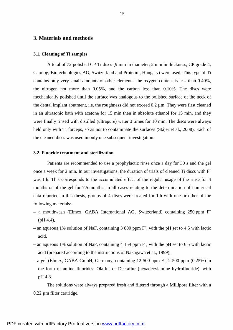
AFM testing of the polished and cleaned Ti samples before F– treatment (Fig. 9)
revealed almost parallel grooves on the surface; these grooves originated from the mechanical
machining of the samples (the grooves appear lighter on proceeding from the depths toward
the surface in the picture). The AFM measurements yielded a value of Ra = 37.0 ± 2 nm for
the control samples.
Figure 9. 3D AFM image of a control Ti surface, revealing the almost parallel
grooves due to the mechanical machining. Image dimensions: 5x5 µm.
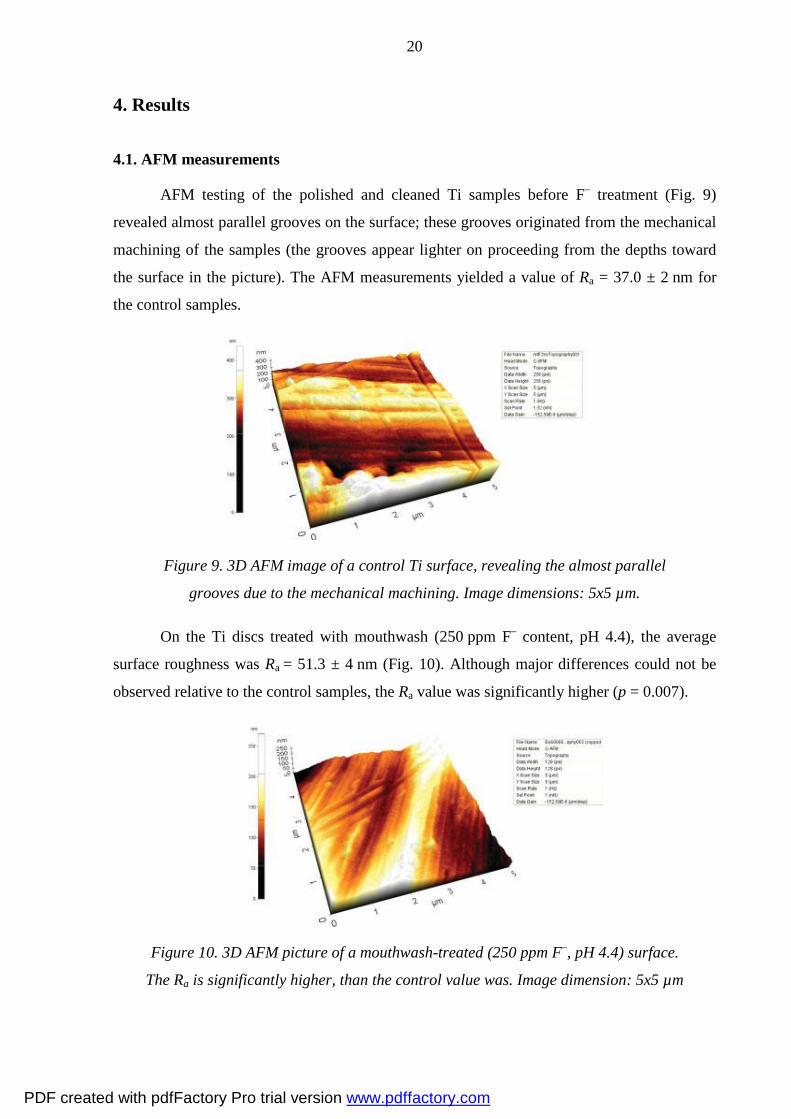
On the Ti discs treated with mouthwash (250 ppm F– content, pH 4.4), the average
surface roughness was Ra = 51.3 ± 4 nm (Fig. 10). Although major differences could not be
observed relative to the control samples, the Ra value was significantly higher (p = 0.007).
Figure 10. 3D AFM picture of a mouthwash-treated (250 ppm F–, pH 4.4) surface.
The Ra is significantly higher, than the control value was. Image dimension: 5x5 µm
PDF created with pdfFactory Pro trial version www.pdffactory.com

21
After treatment with 1% NaF solution (3800 ppm F–, pH = 4.5), the Ti discs displayed
the greatest increase in Ra (Fig. 11): the depth of the grooves was almost 7 times that of the
control : Ra = 254.8 nm ± 20 nm (p < 0.001).
Figure 11. 3D AFM image of a surface treated with 1% Na.
The surface roughness became much higher. Image dimensions: 5x5 µm
For demonstration of the disc surface modification caused by the Elmex gel (12 500
ppm F–), we smeared it with this preventive gel and then rinsed it with distilled water. After
drying, it could be clearly seen that the surface was not as shiny and smooth as previously
(Fig. 12). The AFM picture (Fig. 13) showed deep corrosive regions and granular forms. The
average roughness of the gel-treated surface was significantly increased as compared with the
control: Ra = 48.6 ± 3 nm (p = 0.005).
Figure 12. Macroscopic image of a disc smeared with the gel (12 500 ppm F– and pH 4.8)
revealing the macroscopic surface change caused by the high concentration of F–.
PDF created with pdfFactory Pro trial version www.pdffactory.com

22
Figure 13. 3D AFM picture of a gel-treated Ti discs surface. High differences on the surface
between the higher and deeper parts can be observed. Deep corrosive regions and granular
forms were detected on the surface. Image dimensions: 5x5 µm.
Thus the AFM measurements demonstrated that Ra was increased significantly on all
the treated Ti discs as depicted in Fig. 14: control surface Ra = 37.0 ± 2 nm; mouthwash-
treated discs, Ra = 51.3 ± 4 nm (p = 0.007); 1% NaF solution-treated discs Ra = 254.8 ± 19 nm
(p < 0.001) and gel-treated discs Ra = 48.6 ± 3 nm (p = 0.005). Ra = 254.8 ± 19 nm (p < 0.001)
0
50
100
150
200
250
300
Figure 14. Bar-graph comparison of the surface roughness (Ra)
of the Ti discs after the various treatments.
4.2. XPS measurements
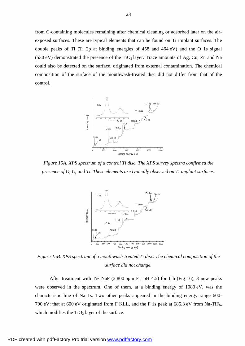
The XPS spectra of control and mouthwash-treated (250 ppm F–, at pH 4.4) Ti discs
are to be seen in Fig. 15A and 15B. In both recordings, the presence of O, C and Ti was
confirmed. The C 1s signal indicates the presence of carbonaceous contamination, resulting
PDF created with pdfFactory Pro trial version www.pdffactory.com
23
from C-containing molecules remaining after chemical cleaning or adsorbed later on the air-
exposed surfaces. These are typical elements that can be found on Ti implant surfaces. The
double peaks of Ti (Ti 2p at binding energies of 458 and 464 eV) and the O 1s signal
(530 eV) demonstrated the presence of the TiO2 layer. Trace amounts of Ag, Cu, Zn and Na
could also be detected on the surface, originated from external contamination. The chemical
composition of the surface of the mouthwash-treated disc did not differ from that of the
control.
0 200 400 600 800 1000 1200
454 456 458 460 462 464 466 468
Cu 2p
Zn 2p
Ti 3p
Inte
nsity
[a.u
.]
Binding energy [eV]
Ti 2p
O 1s
Ag 3d
Ti LMM
Na 1s
Ti 2s
C 1s
Ti 3s
O KLLBinding energy [eV]
Ti 2p
458.7
464.4
Figure 15A. XPS spectrum of a control Ti disc. The XPS survey spectra confirmed the
presence of O, C, and Ti. These elements are typically observed on Ti implant surfaces.
0 100 200 300 400 500 600 700 800 900 1000 1100 1200
454 456 458 460 462 464 466 468
Ag 3d
Zn 2p
Ti 3p
Inte
nsity
[a.u
.]
Binding energy [eV]
Ti 2p
O 1s
Cu 2p
Ti LMM
Na 1s
Ti 2s
C 1s
Ti 3s
O KLLBind ing ene rgy [eV]
Ti 2p
458.6
464. 4
Figure 15B. XPS spectrum of a mouthwash-treated Ti disc. The chemical composition of the
surface did not change.
After treatment with 1% NaF (3 800 ppm F–, pH 4.5) for 1 h (Fig 16), 3 new peaks
were observed in the spectrum. One of them, at a binding energy of 1080 eV, was the
characteristic line of Na 1s. Two other peaks appeared in the binding energy range 600-
700 eV: that at 600 eV originated from F KLL, and the F 1s peak at 685.3 eV from Na2TiF6,
which modifies the TiO2 layer of the surface.
PDF created with pdfFactory Pro trial version www.pdffactory.com

24
Figure 16. XPS spectrum of the surface treated with NaF. The usual peaks (O, C and Ti
on the spectra were found and new F KLL peak and F1s peak appeared,
which proves the modification of the surface composition.
The XPS spectrum of the surface treated with Elmex gel (12 500 ppm F– and pH 4.8)
was similar to that of the NaF-treated surface. Three new peaks were observed: the
characteristic line of Na 1s at a binding energy of 1080 eV resulted from the NaF content, the
line at 600 eV originated from F KLL, and the F 1s peak at 685.3 eV pointed to the presence
of Na2TiF6, modifying the TiO2 layer on the surface (Fig. 17).
Figure 17. XPS spectrum of a Ti discs treated with Elmex gel. The large arrow indicates the
two new peaks: F KLL and F 1s, proving the formation of Na2TiF6.
After Ar+ bombardment for 10, 20, and 30 min, repeated XPS investigation revealed
that ~10 nm was removed from the surface of the material, but the F 1s peak at 684.7 eV
persisted (Fig. 18), proving that the binding between the Ti and F– was very strong.
0 100 200 300 400 500 600 700 800 900 1000 1100 1200
455 460 465 470
Ti 3p
F 1s
Inte
nsity
[a.u
.]
Binding energy [eV]
Ti 2p
O 1s
N 1s
Ti LMM
Na 1s
Ti 2s
C 1s
Ti 3s
O KLLBinding energy [eV]
Ti 2p
458.8
464.5
F KLL
0 200 400 600 800 1000 1200
455 460 465 470
Zn 2p
Ti 3p
F 1s
Inte
nsity
[a.u
.]
Binding energy [eV]
Ti 2p
O 1s
N 1s
Ti LMM
Na 1s
Ti 2s
C 1s
Ti 3s
O KLLBinding energy [eV]
Ti 2p
458.9
464.8
F KLL
PDF created with pdfFactory Pro trial version www.pdffactory.com

25
Figure 18. High-resolution XPS spectrum of the
NaF-treated (3 800 ppm F–, pH 4.5) Ti disc after 10,
20, and 30 min of Ar+ bombardment. After 10 min of
Ar+ bombardment, repeated XPS investigation
revealed that about 10 nm was removed from the
surface of the material, but the F 1s peak at 684.7 eV
persisted, proving that the binding between the Ti
and fluoride was strong.
4.3. SEM results on Ti samples incubated with periodontal pathogenic bacteria
Figures 19A, B and C show typical SEM images of control Ti discs incubated for 2, 4
or 7 days in a suspension of oral bacteria from a patient with chronic periodontitis. The
beginning of biofilm development can readily be observed, and the steps of multiplication and
maturation of the oral bacteria can be clearly followed. The microcolony spreads first in the
surface plane and then upwards, creating palisades of cells. The pioneer species acted as the
substrate for further colonization through a process of coaggregation between different
species.
Figure 19A. SEM image of a control Ti disc incubated for 2 days with pathogenic bacteria
from a patient with chronic periodontitis. Only a few Streptococcus sp. can be seen.
(Magnification: 10 000x)
PDF created with pdfFactory Pro trial version www.pdffactory.com
26
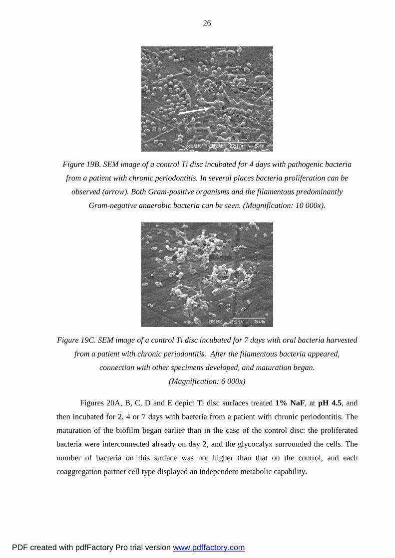
Figure 19B. SEM image of a control Ti disc incubated for 4 days with pathogenic bacteria
from a patient with chronic periodontitis. In several places bacteria proliferation can be
observed (arrow). Both Gram-positive organisms and the filamentous predominantly
Gram-negative anaerobic bacteria can be seen. (Magnification: 10 000x).
Figure 19C. SEM image of a control Ti disc incubated for 7 days with oral bacteria harvested
from a patient with chronic periodontitis. After the filamentous bacteria appeared,
connection with other specimens developed, and maturation began.
(Magnification: 6 000x)
Figures 20A, B, C, D and E depict Ti disc surfaces treated 1% NaF, at pH 4.5, and
then incubated for 2, 4 or 7 days with bacteria from a patient with chronic periodontitis. The
maturation of the biofilm began earlier than in the case of the control disc: the proliferated
bacteria were interconnected already on day 2, and the glycocalyx surrounded the cells. The
number of bacteria on this surface was not higher than that on the control, and each
coaggregation partner cell type displayed an independent metabolic capability.
PDF created with pdfFactory Pro trial version www.pdffactory.com

27
Figure 20A. SEM image of a Ti disc treated with 1% NaF solution (pH 4.5) and then
incubated for 2 days, with oral bacteria from a patient with chronic periodontitis.
The rough, etched surface beneath the bacteria can be observed, with the
interactions between specific bacterial surface molecule. The ordered
arrangement of the bacteria within the biofilm indicate a highly
organized process (Magnification: 4 000x).
Figures 20B and C. SEM images of the surface of Ti discs treated with 1% NaF at pH 4.5 and
then incubated for 4 days with the bacteria from a patient with chronic periodontitis.
A more mature biofilm was visible in a few places. The corn-cob morphological
structure was present in the plaque biofilms. The arrow indicates attached
oral cocci and growing on the surface of filamentous microorganisms.
This formation provided a possibility for cell-cell communication.
(Magnifications: 3 000x and 10 000x).
PDF created with pdfFactory Pro trial version www.pdffactory.com

28
Figures 20D and E. SEM images of the surface of Ti discs after treatment with 1% NaF
solution at pH 4.5, and then incubated for 7 days with mixed bacterial flora.
(Magnifications: 8 000x and 15 000x).
Figures 21A, B, C and D show the surfaces of Ti discs treated with 1 % NaF at
pH 6.5, illustrating the steps of biofilm development. Initially Gram-positive cocci were
situated on the Ti disc surface; secondary colonizers associated with other Gram-positive rods
and filamentous bacteria allowed other bacteria to colonize the Ti implant. Maturation of the
biofilm occurred through the attachment of filamentous, predominantly Gram-negative
aerobic bacteria to the basal biofilm. After 4 days on the surface the corn-cob form appeared
too with longer filament bacteria at the center and a great number of cocci and short rods were
connected with it. After incubation for 7 days there were several layers of bacteria on the
surface. The biofilm was very thick, with all types of cells attached to the surface and
interconnected to each other.
Figure 21A. SEM image of the surface of a
Ti disc after treatment with 1% NaF solution
at pH 6.5, and incubation for 2 days with
bacteria from a patient with chronic
periodontitis. The bacterial proliferation
began earlier than on the control sample as
reproduction was clearly visible and the
filaments appeared on the surface. The
arrow indicates one filamentous bacterial
cell. (Magnification: 6 000x).
PDF created with pdfFactory Pro trial version www.pdffactory.com

29
Figure 21B. SEM image of the surface of a Ti disc treated with 1% NaF solution at pH 6.5
and then incubated for 4 days with bacteria from a patient with chronic periodontitis.
The pioneer cocci cells have begun to interconnect with the rods and filaments.
(Magnification: 6 000x).
Figures 21C and D. SEM images of Ti disc surfaces treated with 1% NaF solution at pH 6.5.
and then incubated for 7 days with bacteria from a patient with chronic periodontitis. Mature
biofilm is seen on the surface, with several layers of bacteria. Cocci, rod-shaped and
filamentous bacteria are also present. Numerous Streptococcus sp. are situated adjacent to
the surface and multiply (see arrow) to cover an ever greater surface area.
(Magnifications: 8 000x and 10 000x).
4.4. SEM results onf Ti samples incubated with Streptococcus mutans
Figure 22 illustrates the adhesion of S. mutans on a control Ti disc surface,
demonstrating the connection between the cells, and the bacterial chain on the surface. A
continuous monolayer is not seen, only separated bacteria cells, in some cases interconnected
(Fig. 22).
PDF created with pdfFactory Pro trial version www.pdffactory.com

30
Figure 22. SEM image of the surface of an untreated (control) Ti disc incubated for 5 days
with S. mutans. There is only one, not continuous bacterium layer on the Ti surface.
(Magnification: 5 000x).
Figure 23 depicts a Ti disc after treatment with mouthwash containing 250 ppm F– at
pH 4.4 and incubated with S. mutans for 5 days. The SEM picture reveals cocci cells covering
the surface in only one layer, and readily observed adhesion in several places. There are a
large number of interconnected multiplied bacteria on the treated Ti surface.
Figure 23. SEM image of a Ti disc treated with mouthwash containing (250 ppm F–) at
pH 4.4 and then incubated with S. mutans for 5 days. The coccus bacteria
are connected to the surface and the arrow indicates the formation
of coccus chains (Magnification: 5 000x).
Figures 24A, B and C relate to the Ti disc treated with 1% NaF solution containing 3
800 ppm F– at pH 4.5 and incubated with S. mutans for 5 days. Figure 24A shows the
macroscopic features of a gold-coated Ti disc prepared for SEM examination.
PDF created with pdfFactory Pro trial version www.pdffactory.com

31
Figure 24A. Macroscopic picture of a Ti disc treated with 1% NaF at pH 4.5 and incubated
with S. mutans for 5 days. The bacterial cells cover almost the whole surface.
Figures 24B and C present SEM images of the surface with the microbial adhesion,
revealing a mature biofilm in several layers, covering the whole surface area. The bacteria
may proliferate inside the polysaccharide matrix produced by the bacteria or some other
biological surface (e.g. the mucosa). Other microorganisms can infiltrate into this formation,
and may cause the growth of a microbial consortium. The glycocalyx provides protection for
the S. mutans and promotes the proliferation of bacterial cells.
Figure 24B. SEM image of a Ti disc treated with the 1% NaF (3 800 ppm F–) at pH 4.5 and
incubated with S. mutans for 5 days. The biofilm can be seen, its form is maturated
which developed in several layers. The bacteria covered the whole area
of this Ti surface (Magnification: 2 000x).
PDF created with pdfFactory Pro trial version www.pdffactory.com
32
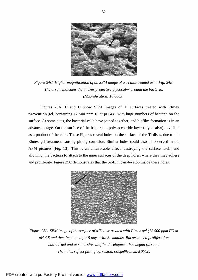
Figure 24C. Higher magnification of an SEM image of a Ti disc treated as in Fig. 24B.
The arrow indicates the thicker protective glycocalyx around the bacteria.
(Magnification: 10 000x).
Figures 25A, B and C show SEM images of Ti surfaces treated with Elmex
prevention gel, containing 12 500 ppm F– at pH 4.8, with huge numbers of bacteria on the
surface. At some sites, the bacterial cells have joined together, and biofilm formation is in an
advanced stage. On the surface of the bacteria, a polysaccharide layer (glycocalyx) is visible
as a product of the cells. These Figures reveal holes on the surface of the Ti discs, due to the
Elmex gel treatment causing pitting corrosion. Similar holes could also be observed in the
AFM pictures (Fig. 13). This is an unfavorable effect, destroying the surface itself, and
allowing, the bacteria to attach to the inner surfaces of the deep holes, where they may adhere
and proliferate. Figure 25C demonstrates that the biofilm can develop inside these holes.
Figure 25A. SEM image of the surface of a Ti disc treated with Elmex gel (12 500 ppm F–) at
pH 4.8 and then incubated for 5 days with S. mutans. Bacterial cell proliferation
has started and at some sites biofilm development has begun (arrow).
The holes reflect pitting corrosion. (Magnification: 8 000x).
PDF created with pdfFactory Pro trial version www.pdffactory.com

33
Figure 25B. Higher magnification of an SEM image of a Ti disc treated as in Fig. 25A. The
holes in the Ti surface were caused by the high fluoride content of the gel. S. mutans
bacteria can attach the inner surface of the deep hole, where they may adhere and
proliferate. The biofilm can develop inside these holes (Magnification: 10 000x).
Figure 25C. Higher magnification of an SEM image of a Ti disc treated as in Fig. 25A,
revealing a typical bacterium biofilm formed in several layers with glycocalyx.
Adhesion to the protective mucous substance of the bacterium promots
reproduction (Magnification: 6 000x).
4.5. Results of incubation with Porphyromonas gingivalis
4.5.1. SEM results
After incubation for 5 days active reproduction of the bacteria was observed on the
control discs (Fig. 26). The bacterial cells did not cover the entire Ti surface. The formation
of a monolayer had not yet started and transverse bacterium chains could be seen as the
Porphyromonas multiplied.
PDF created with pdfFactory Pro trial version www.pdffactory.com
34
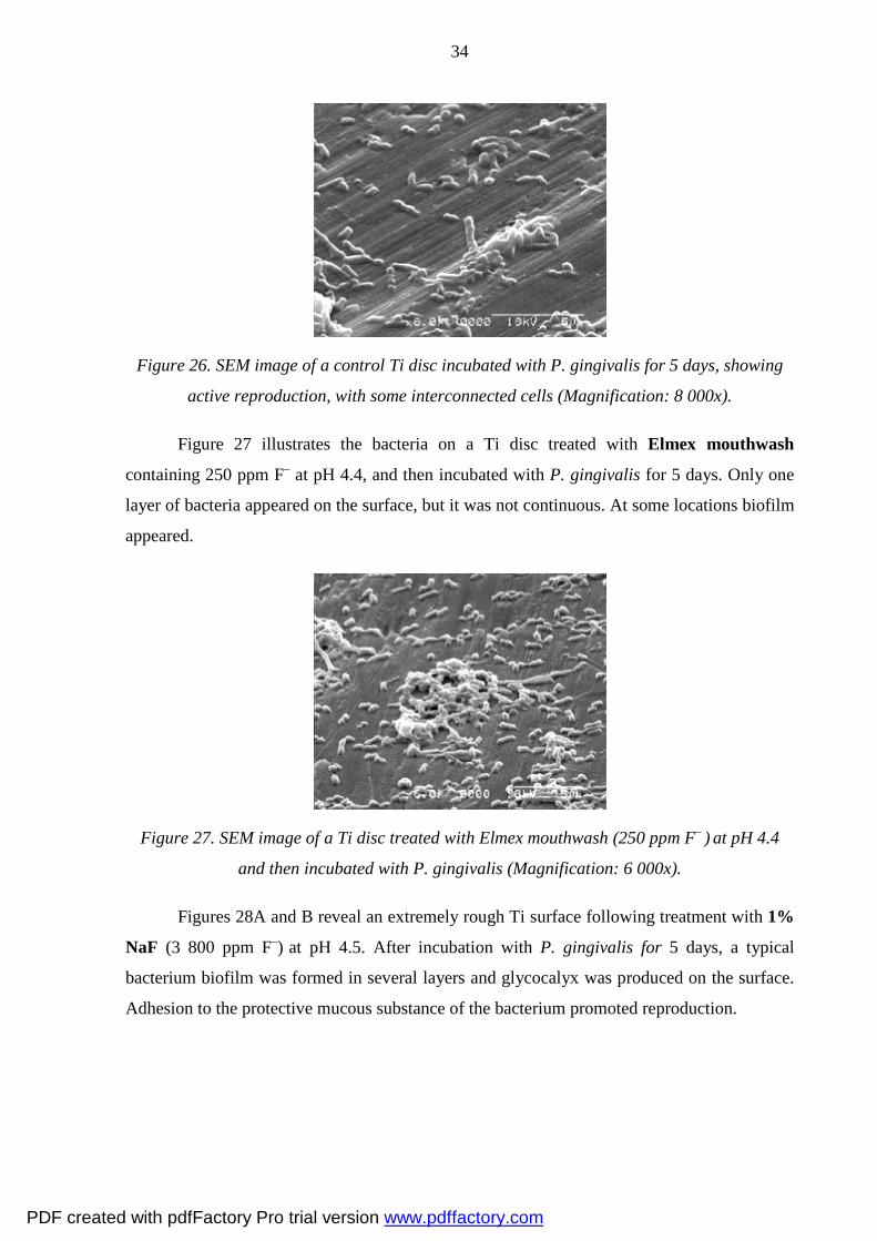
Figure 26. SEM image of a control Ti disc incubated with P. gingivalis for 5 days, showing
active reproduction, with some interconnected cells (Magnification: 8 000x).
Figure 27 illustrates the bacteria on a Ti disc treated with Elmex mouthwash
containing 250 ppm F– at pH 4.4, and then incubated with P. gingivalis for 5 days. Only one
layer of bacteria appeared on the surface, but it was not continuous. At some locations biofilm
appeared.
Figure 27. SEM image of a Ti disc treated with Elmex mouthwash (250 ppm F– ) at pH 4.4
and then incubated with P. gingivalis (Magnification: 6 000x).
Figures 28A and B reveal an extremely rough Ti surface following treatment with 1%
NaF (3 800 ppm F–) at pH 4.5. After incubation with P. gingivalis for 5 days, a typical
bacterium biofilm was formed in several layers and glycocalyx was produced on the surface.
Adhesion to the protective mucous substance of the bacterium promoted reproduction.
PDF created with pdfFactory Pro trial version www.pdffactory.com

35
Figure 28A and B. SEM images of bacteria on Ti discs treated with 1% NaF (3 800 ppm F– )
at pH 4.5 and then incubated with P. gingivalis for 5 days. Biofilm developed
on the surface. (Magnifications: 3 000x (A) and 8 000x (B)).
Figures 29A and B show SEM images of the surfaces of Ti discs after treatment with
Elmex gel (12 500 ppm F–) at pH 4.8 and incubation with P. gingivalis for 5 days. Deep
corrosive regions and holes can be seen. Some of the bacteria are present in multiple layers, in
an interconnected manner, and additionaly a biofilm has been formed. The polysaccharide
coating (glycocalyx) forms a protective layer around the bacterial population.
Figure 29A. SEM image revealing holes and bacteria on the surface of a Ti disc treated with
Elmex gel (12 500 ppm F–) at pH 4.8 and incubated with P.gingivalis for 5 days.
The arrow shows the beginning of corn-cob formation on the surface
(Magnification: 6 000x).
PDF created with pdfFactory Pro trial version www.pdffactory.com

36
Figure 29B. Higher magnification of an SEM image of a Ti disctreated as in Fig. 29A,
demonstrating thick glycocalyx growth (Magnification: 10 000x).
4.5.2. Protein content assays
The quantity of P. gingivalis bacterial protein was determined with a micro Coomassie
protein assay kit in order to check the survival and proliferation of the bacteria on the Ti disc
surfaces after different treatments.
After incubation with P. gingivalis for 5 days, the protein assays indicated that there
were no significant differences in the number of bacteria on the surfaces of Ti discs treated
with Elmex mouthwash (250 ppm F– at pH 4.4), with 1% NaF solution (3 800 ppm F– at
pH 4.5) or with Elmex gel (12 500 ppm F– at pH 4,8) as compared with the control (Fig. 30),
with levels of 96.7 ± 25%, 106.7 ± 24% and 118.6 ± 26% respectively, vs. 100%).
Figure 30. Protein contents on the surfaces of Ti discs incubation with P. gingivalis for 5 days
following treatment with Elmex mouthwash, NaF solutions or Elmex gel, relative to the
amount measured on the control
PDF created with pdfFactory Pro trial version www.pdffactory.com
37
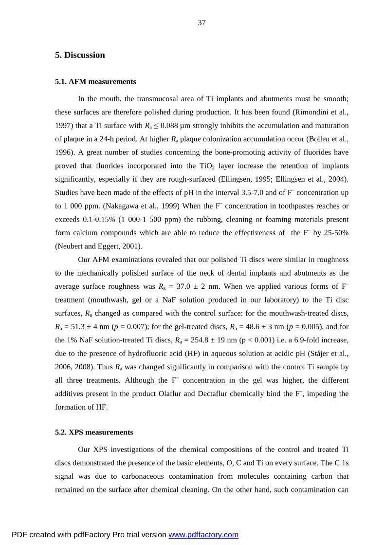
5. Discussion
5.1. AFM measurements
In the mouth, the transmucosal area of Ti implants and abutments must be smooth;
these surfaces are therefore polished during production. It has been found (Rimondini et al.,
1997) that a Ti surface with Ra ≤ 0.088 µm strongly inhibits the accumulation and maturation
of plaque in a 24-h period. At higher Ra plaque colonization accumulation occur (Bollen et al.,
1996). A great number of studies concerning the bone-promoting activity of fluorides have
proved that fluorides incorporated into the TiO2 layer increase the retention of implants
significantly, especially if they are rough-surfaced (Ellingsen, 1995; Ellingsen et al., 2004).
Studies have been made of the effects of pH in the interval 3.5-7.0 and of F– concentration up
to 1 000 ppm. (Nakagawa et al., 1999) When the F– concentration in toothpastes reaches or
exceeds 0.1-0.15% (1 000-1 500 ppm) the rubbing, cleaning or foaming materials present
form calcium compounds which are able to reduce the effectiveness of the F– by 25-50%
(Neubert and Eggert, 2001).
Our AFM examinations revealed that our polished Ti discs were similar in roughness
to the mechanically polished surface of the neck of dental implants and abutments as the
average surface roughness was Ra = 37.0 ± 2 nm. When we applied various forms of F–
treatment (mouthwash, gel or a NaF solution produced in our laboratory) to the Ti disc
surfaces, Ra changed as compared with the control surface: for the mouthwash-treated discs,
Ra = 51.3 ± 4 nm (p = 0.007); for the gel-treated discs, Ra = 48.6 ± 3 nm (p = 0.005), and for
the 1% NaF solution-treated Ti discs, Ra = 254.8 ± 19 nm (p < 0.001) i.e. a 6.9-fold increase,
due to the presence of hydrofluoric acid (HF) in aqueous solution at acidic pH (Stájer et al.,
2006, 2008). Thus Ra was changed significantly in comparison with the control Ti sample by
all three treatments. Although the F– concentration in the gel was higher, the different
additives present in the product Olaflur and Dectaflur chemically bind the F–, impeding the
formation of HF.
5.2. XPS measurements
Our XPS investigations of the chemical compositions of the control and treated Ti
discs demonstrated the presence of the basic elements, O, C and Ti on every surface. The C 1s
signal was due to carbonaceous contamination from molecules containing carbon that
remained on the surface after chemical cleaning. On the other hand, such contamination can
PDF created with pdfFactory Pro trial version www.pdffactory.com

38
be adsorbed later on air-exposed surfaces (NIST XPS Database). These elements are typically
observed on Ti implant surfaces. Slight amounts of Ag, Cu, Zn and Na were also detected on
these samples, which originated from external contamination (Handbook of X-Ray
Photoelectron Spectroscopy. 1979). The presence of the TiO2 layer was confirmed by the
double peaks of Ti (Ti 2p at binding energies of 458 and 464 eV) and the O 1s signal (530
eV) (Ameen et al., 1993, Kilpadi et al., 1998). After NaF solution or gel treatments, 3 new
peaks were seen in the spectra. One of them, at a binding energy of 1 080 eV, was the
characteristic line of Na 1s, which resulted from NaF. Two other peaks appeared in the region
600-700 eV. That at 600 eV originated from F KLL (Huang, 2002) and the F 1s peak at 685.3
eV from Na2TiF6, which modifies the TiO2 layer of the surface (Huang, 2003; NIST XPS
Database). XPS revealed that the high F– concentration and acidic pH of the gel and the 1%
NaF solution resulted in strong corrosion and modification of the composition of the Ti
surface.
This result is in accordance with literature findings (Huang, 2003). Our XPS
investigations further demonstrated that, after 10 min of Ar+ bombardment, ~10 nm was
removed from the surface of the discs, but the F 1s peak at 684.7 eV persisted, proving that
the binding between Ti and F– was very strong. The complex Na2TiF6 modifies the TiO2 layer
on the surface (Stájer et al., 2006, 2008). The smoothness and the chemical stability of the Ti
surface may deteriorate following the application of 1% NaF solution or Elmex gel, favoring
the adhesion of bacterial flora.
5.3. Incubation with bacteria from a patient with chronic periodontitis
On the control discs, the process of biofilm development could be followed. Initially
only a few Streptococcus sp. cells were observed to be adhered to the surface. After 2 days,
they were associated with Gram-positive organisms containing surface proteins that bind to
specific salivary glycoproteins in the pellicle. After 4 days, proliferation had started, and other
types of bacteria also appeared on the Ti discs. After 7 days, the maturation of the biofilm was
in advanced stage, but was not yet completed. Large numbers of different bacteria, fungi and
protozoa can live in the oral cavity. When these organisms adhere to a surface, they form an
organized mass called dental plaque or biofilm.
Pontoriero et al. (1994) allowed plaque to accumulate around implants and teeth for 3
weeks and found a correlation between the levels of plaque accumulation and peri-implant
mucositis and the similar response of the soft tissues around the teeth and implants when
PDF created with pdfFactory Pro trial version www.pdffactory.com
39
these were exposed to plaque. Our understanding of how these organisms arrive in the dental
biofilm community and how they interact with each other and their hosts has been improved
immeasurably by applying the principles of ecology to the study of this complex system. In
addition to their ability to coaggregate into complex structures, a number of other interactions
occur between the microbial species that comprise bacteria. These interactions can be either
inhibitory or stimulatory, depending on the species involved. On the control Ti discs, the steps
of biofilm (dental plaque) formation could be followed clearly. This plaque contains more
than 600 different microorganisms, and is typically the precursor of tooth decay, contributing
to the overall dynamic environment in the oral cavity that frequently undergoes rapid changes,
depending on the pH, nutrient availability and oxygen tension (ten Cate, 2006).
Maturation of the biofilm (after 4 days in our study) occurs via the attachment of
filamentous, predominantly Gram-negative aerobic bacteria to the basal biofilm. This process
also reflects interactions between specific bacterial surface molecules. The ordered
arrangement of bacteria within the biofilm indicates a highly organized process. In a recent
study of microbial colonization on Ti implants, periodontal pathogens were demonstrated to
be present in different proportions on all failed implant surfaces (Shibli et al., 2007). Specific
bacteria within the biofilm community were able to interact with other species to either help
or impair the host, besides to providing positive cooperation between the different species of
the biofilm. After 7 days, clustered groups of bacteria appeared too. Each cluster is created on
the basis of similarities and differences in nutritional and atmospheric environments. Polymer
production results in the development of the extracellular matrix, which consists of soluble
and insoluble glucans, fructans and heteropolymers. This matrix is one of the key structural
aspects of the plaque biofilm, much like that of other biofilms.
We performed SEM analyses of Ti discs treated with 1% NaF solutions (pH 4.5) and
incubated with oral bacteria from the sulcus of a patient with chronic periodontitis for 2, 4 or
7 days. The SEM images clearly showed that the proliferation of the cells had alredy begun
after 2 days, the cells becoming interconnected and producing glycocalyx. The steps of
biofilm formation continued without difficulty. Corn-cob formation and clustered groups of
bacteria could be observed on the treated Ti discs. The bacteria did not cover the surface
everywhere. Initiation was associated with Gram-positive organisms (mostly Streptococcus
sp.) that contain surface proteins that bind to specific surfaces of Ti implants. Negatively
charged adhesions on bacterial cells may bind to negatively charged glycoproteins through
bivalent cations (usually Ca2+ or Mg2+). Secondary colonizers associated with other Gram-
positive rods and filaments (mostly Lactobacillus sp.) allow other bacteria to colonize Ti
PDF created with pdfFactory Pro trial version www.pdffactory.com

40
implants. The maturation of biofilm occurs through the attachment of filamentous
predominantly Gram-negative aerobic bacteria to the basal biofilm. The early composition of
the biofilm reflects the ability of the early colonizing biomass to create a low redox potential
suitable for anaerobes (e.g. P. gingivalis) (Marsh, 2004). Anaerobes survive in the high
oxygen concentrations present in the oral cavity, without having much protection from other
bacteria. Immediately after tooth brushing, such a thin biofilm is almost always present on the
tooth surface (Sbordone and Bortolaia, 2003).
In our study, Ti discs were treated with 1% NaF solution at pH 6.5 and incubated for 2,
4 or 7 days with oral bacteria flora from the patient with periodontal disease, in order to
investigate the changes in the bacterial cells on the Ti surface. The multiplication of the
bacteria began after day 2, and after day 4 the maturity of the biofilm had reached the second
stage. At the end of this investigation (after 7 days), several layers of bacteria cells were
present on the surfaces. While the discs were removed from the incubation medium, many
bacterial cells were lost because the huge amount could not remain on the surface. SEM
revealed that after 7 days more and more Streptococcus sp. strived to adhere to the empty
surface to grow the biofilm. Subramani et al. (2009) established that increases in surface
roughness and surface free energy facilitate biofilm formation on dental implant and abutment
surfaces. The surface chemistry and the design features of the implant-abutment configuration
also play significant roles in biofilm formation. (Subramani et al., 2009). The maturation of
the biofilm seemed to be advanced relative to the previously mentioned samples. The
immersion of the Ti discs in the 1% NaF solution at pH 6.5 apparently led to the surface
changes that generated a very favorable environment for the oral bacteria and the biofilm
developed more easily. These results demonstrate that the changes caused on Ti surfaces by
the above-mentioned treatment can promote favor the development of a biofilm.
5.4. Incubation with Streptococcus mutans
The behavior of S. mutans on the Ti surface was examined because it is considered to
be the primary etiologic agent of dental caries a global health problem that affects 60-90% of
the population. S. mutans has 4 different serotypes. The Gram-positive cocci bacterium is
facultative aerobic; although it is often found in the normal, healthy human oral cavity, it is a
significant contributor to tooth decay. The microbe was first described in 1924 (Clarke, 1924).
Our investigations with S. mutans demonstrated a monolayer of separate bacterial cells, on the
control Ti discs, which did not multiply after 5 days. The primary colonizing bacteria may
PDF created with pdfFactory Pro trial version www.pdffactory.com

41
connect to the surface in two ways: 1. Hydrophobic interactions, when lipophilic adhesions on
the bacterial cell surfaces come into contact with hydrophobic receptors on the epithelial cells.
S. mutans is frequently exposed to toxic compounds from oral healthcare products, food
additives and tobacco. Streptococci account for about 20% of the oral bacteria and determine
the development of the biofilm. Although S. mutans can be antagonized by pioneer
colonizers, when it becomes dominant in oral biofilms, dental caries can develop and thrive
(Biswas and Biswas, 2011). 2. S. mutans prossesses an enzyme, glycosyl transferase, which is
involved in the initial attachment of the bacterial cells to the tooth surface. Through the
transformation of sucrose to dextran polymers (glucans) it facilitates the formation of plaque.
It appears as a regular component of human normal oral flora in relatively large numbers. The
salivary component mucin contains glycoproteins which can easily colonize on the tooth
surfaces and form a thin film called enamel pellicle on the tooth. The adsorbed mucin
probably serves as a molecular receptor for ligands on the bacterial cell surface. The lactic
acid that originates from the utilization of dietary carbohydrate can demineralize the tooth
enamel. The extracellular glucans formed by S. mutans and other organisms can also produce
intracellular polysaccharides from sugars, which are stored in the cells and then metabolized
to lactic acid. S. mutans produces more lactic acid and is more acid-tolerant than most other
streptococci. S. mutans is important in the initiation of dental caries, because its presence
leads to bacterial colonization on the tooth surfaces, plaque formation and the localized
demineralization of tooth enamel. However, it is not the only bacterium which causes dental
decay. After bacteria appear on the enamel, various oral bacteria begin to grow in the interior
regions of the tooth. Lactobacilli, actinomyce, and various proteolytic bacteria are secondary
invaders, which assist the progression of the lesions (Todar’s online textbook of bacteriology).
Prevention is very important in oral health. During orthodontic treatment, dentists
recommend different types of preventive solutions and gels, and prophylactic agents are used
in dental surgery too. Unfortunately, the pH of the mouth rinses and gels used for caries
prevention in dentistry range between pH 3.5 and pH 7.0, and their F– concentration is
between 1 000 and 10 000 ppm (Nakagawa et al., 1999). For dental practitioners, it is
essential to know whether F– attacks Ti surfaces, i.e. whether the prophylactic solutions are
able to modify the corrosion resistance of the surfaces of Ti dental implants, dental prostheses
or wires in the case of orthodontic braces.
When Elmex mouthwash was applied on Ti discs, we observed that the number and
proliferation of bacterial cells did not change on the surface relative to the control ones. The
treatment with Elmex gel resulted in an increase in the number of S. mutans cells which grew
PDF created with pdfFactory Pro trial version www.pdffactory.com

42
on the samples. At some some sites a biofilm appeared, with a glycocalyx wrapping. In the
presence of corrosion holes, the cells adhered inside the deep areas and proliferated there. If
the surface of Ti exhibits crevices and pits, S. mutans can adhere there (Barbour et al., 2007).
SEM analysis of Ti discs treated with NaF solution before incubation showed that
several layers of bacteria cells covered the surface, and at higher magnification the biofilm
covered the whole of the Ti discs. We earlier reported (Stájer et al., 2009), in agreement with
Rimondini et al. (1997), that the amount and rate of maturation of the biofilm correlated with
the roughness of the Ti surface. Both Gram-positive colonies and Gram-negative cocci were
observed to be present as in a previous study (Li et al., 2001). A change in free energy on the
Ti surface influences the adherence of S. (Fujioka-Hirai et al., 1987).
5.5. Incubation with Porphyromonas gingivalis
5.5.1. SEM studies with Porphyromonas gingivalis
We carried out an SEM investigation of the proliferation of P. gingivalis on Ti discs
treated with Elmex mouthwash, 1% NaF or Elmex gel. On the controls the formation of a
monolayer had not yet started after 5 days of incubation; only transverse bacterial chains
could be seen as the Porphyromonas multiplied. The colonization of P. gingivalis on polished
and F–-modified Ti surfaces can play an important role in the development of peri implantitis
inside the mouth. On intact Ti surfaces, P. gingivalis can proliferate in association with other
bacterial strains and together they may develop the biofilm of the surface.
P. gingivalis is a non-motile Gram-negative, rod-shaped, anaerobic pathogenic
bacterium, which produces dark-brown or black porphyrin pigments. Porphyromonas sp.,
commonly found in humans, and especially in the oral cavity, grow in black colonies on
blood agar. They cause gingivitis or other oral inflammatory processes in approximately 70-
90% of the pubescent population, which could be a possible precursor of adult periodontal
diseases (Choi et al., 1990). As in Bacteroides, they form an outer membrane, the
peptidoglycan layer, and a cytoplasmic membrane. The cell surface adhesion molecules on
Porphyromonas interact with other bacteria, when epithelial cells and extracellular matrix
proteins assist them to live in their human host, attaching equally well to both smooth and
grooved coated Ti surfaces (Wu-Yuan et al., 1995). P. gingivalis is present in periodontal
tissues: it receives energy from dissolved sugars and other simple carbohydrates which the
mouth generally contains. The clinical evidence points to the presence of P. gingivalis in
periodontal diseases (Haffajee and Socransky, 1994). It is found in elevated levels in
PDF created with pdfFactory Pro trial version www.pdffactory.com
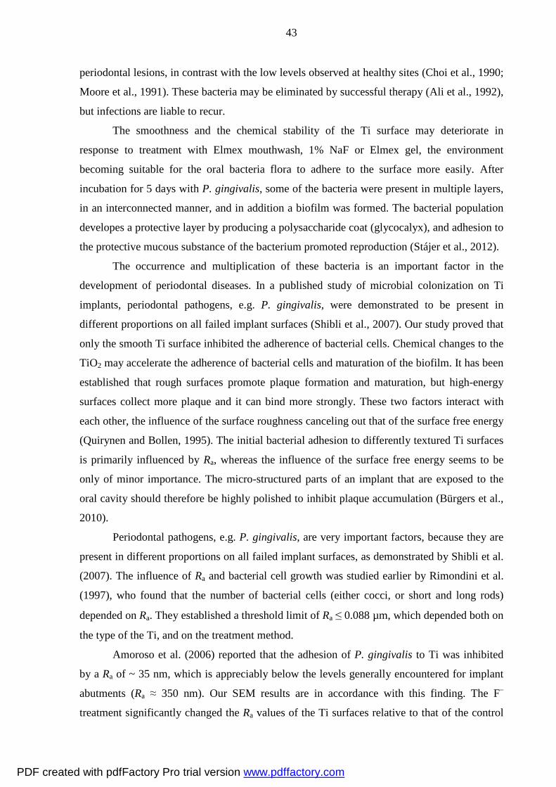
43
periodontal lesions, in contrast with the low levels observed at healthy sites (Choi et al., 1990;
Moore et al., 1991). These bacteria may be eliminated by successful therapy (Ali et al., 1992),
but infections are liable to recur.
The smoothness and the chemical stability of the Ti surface may deteriorate in
response to treatment with Elmex mouthwash, 1% NaF or Elmex gel, the environment
becoming suitable for the oral bacteria flora to adhere to the surface more easily. After
incubation for 5 days with P. gingivalis, some of the bacteria were present in multiple layers,
in an interconnected manner, and in addition a biofilm was formed. The bacterial population
developes a protective layer by producing a polysaccharide coat (glycocalyx), and adhesion to
the protective mucous substance of the bacterium promoted reproduction (Stájer et al., 2012).
The occurrence and multiplication of these bacteria is an important factor in the
development of periodontal diseases. In a published study of microbial colonization on Ti
implants, periodontal pathogens, e.g. P. gingivalis, were demonstrated to be present in
different proportions on all failed implant surfaces (Shibli et al., 2007). Our study proved that
only the smooth Ti surface inhibited the adherence of bacterial cells. Chemical changes to the
TiO2 may accelerate the adherence of bacterial cells and maturation of the biofilm. It has been
established that rough surfaces promote plaque formation and maturation, but high-energy
surfaces collect more plaque and it can bind more strongly. These two factors interact with
each other, the influence of the surface roughness canceling out that of the surface free energy
(Quirynen and Bollen, 1995). The initial bacterial adhesion to differently textured Ti surfaces
is primarily influenced by Ra, whereas the influence of the surface free energy seems to be
only of minor importance. The micro-structured parts of an implant that are exposed to the
oral cavity should therefore be highly polished to inhibit plaque accumulation (Bürgers et al.,
2010).
Periodontal pathogens, e.g. P. gingivalis, are very important factors, because they are
present in different proportions on all failed implant surfaces, as demonstrated by Shibli et al.
(2007). The influence of Ra and bacterial cell growth was studied earlier by Rimondini et al.
(1997), who found that the number of bacterial cells (either cocci, or short and long rods)
depended on Ra. They established a threshold limit of Ra ≤ 0.088 µm, which depended both on
the type of the Ti, and on the treatment method.
Amoroso et al. (2006) reported that the adhesion of P. gingivalis to Ti was inhibited
by a Ra of ~ 35 nm, which is appreciably below the levels generally encountered for implant
abutments (Ra ≈ 350 nm). Our SEM results are in accordance with this finding. The F–
treatment significantly changed the Ra values of the Ti surfaces relative to that of the control
PDF created with pdfFactory Pro trial version www.pdffactory.com

44
(Ra = 37.0 ± 2 nm), on which fewer bacteria were observed. Ra increased to ~ 51 nm (Elmex
mouthwash), 49 nm (Elmex gel) and 255 nm (1% NaF solution), the SEM investigations
revealing a more pronounced bacterial growth on these surfaces.
The XPS studies demonstrated modifications in the chemical composition of the
surfaces of the Ti discs treated with 1% NaF or Elmex gel, due to the concomitant effect of
the high F– concentration (3 800–12 500 ppm) and the acidic pH (4.5-4.8).
The findings of this thesis clearly prove that the surface characteristics induced by F–
treatment affected the growth and maturation of P. gingivalis, with the roughness of the
surface as the predominant factor.
5.5.2. Protein assays
The SEM investigations of the surfaces of Ti discs treated with different F–-
containing substances indicated more extensive growth of the bacterial biofilm and earlier
maturation. Although the protein content assays revealed slight increases relative to the
control in the number of bacteria on the surfaces of Ti discs treated with NaF solution or
Elmex gel, the changes were not significant. These apparently contradictory results can be
explained with regard to the relatively short incubation time (5 days). Mabilleau et al. (2006)
established that Ra was highly increased when Ti discs were immersed in F–, H2O2 and lactic
acid containing artificial saliva. They found that the number of bacteria was significantly
increased on discs incubated with activated J774.2 cells or Streptococcus mitis for 21 days.
PDF created with pdfFactory Pro trial version www.pdffactory.com

45
6. Conclusions
The main conclusions of my Ph.D. work are as follows:
1. AFM measurements demonstrated that Ra was increased significantly on all Ti discs
treated with F–-containing materials. Elmex rinse was tested, because many patients use this
for caries prevention worldwide. Treatment with 1% NaF resulted in a roughness
enhancement of almost 7-fold. After Elmex gel treatment, the AFM picture revealed deep
corrosive regions and granular forms, but the roughness change was not so high. The reason
for this is that, although the F– concentration in Elmex gel is higher than that in 1% NaF
solution, the Olaflur and Dectaflur in the gel chemically bind the F–, impeding the formation
of HF.
For a dental practitioner, it is essential to know whether fluorides attack Ti surfaces,
i.e. whether prophylactic solutions are able to modify the corrosion resistance of the surface
of Ti dental implants, crowns and bridges, partial prostheses, orthodontic brackets and wires.
We were the first to study in detail the changes in roughness of CP Ti surfaces due to these
treatments.
The SEM and AFM measurements revealed that crevice and pitting corrosion can
occur on the surface of Ti implants, and it may therefore be concluded that these destructive
effects can also take place on fixed and removable dentures, orthodontic braces and wires
made of Ti.
2. Dentists often offer preventive mouthwashes (e.g. Elmex rinse) for home use. Our
XPS investigations indicated that these rinses did not change the chemical composition of the
Ti surface. The XPS spectra of 1% NaF solution and Elmex gel-treated surfaces showed that a
high F– concentration and acidic pH can cause significant corrosion and result in the formation
of the complex Na2TiF6. Our results are in accordance with the findings of other studies. We
have shown that this complex binds strongly to the surface and modifies the TiO2 layer. The
prophylactic gels (e.g. Elmex gel) are mainly used in pediatric dentistry to prevent caries
while orthodontic braces and wires are situated in the oral cavity.
We are not aware of previous investigations the effects of preventive mouthwashes
and gels (e.g. Elmex gel) on the physical and chemical properties of CP Ti surfaces.
3. The above-mentioned surface treatments favored the accumulation of S. mutans and
of P. gingivalis on CP Ti discs during incubation with bacteria from a patient with periodontal
disease. The SEM pictures showed that after a few days the total number of bacterial cells was
PDF created with pdfFactory Pro trial version www.pdffactory.com
46
higher and the development of maturation was faster than on the untreated Ti surfaces. The
steps of bacterial plaque development were similar in every case, even when oral bacteria
flora and individual bacteria were used. The acidic environment and the F– concentration of
these materials were the determining factors. Higher F– content caused deeper corrosions,
where the bacteria cells could adhere, and could not be removed by the patients or oral
hygienists.
4. The protein content assay of P. gingivalis did not indicate any significant
differences between the untreated and treated samples, although slight increases in the number
of bacterial cells were displayed by the Elmex gel and NaF-treated discs. The incubation time
was only 5 days, and this short time can probably account for this result, which is in contrast
with the literature data.
PDF created with pdfFactory Pro trial version www.pdffactory.com

47
7. Summary
Similarly to other reductive agents, F– may affect the structure of the oxide layer on a
Ti surface. Prophylactic mouthwashes or gels used for the prevention of caries usually contain
F– and are applied at low pH. Hence, the aim of the present work was to study whether
various concentrations of F– at acidic pH cause changes in the surface structure of polished
dental Ti implants and abutments, and alter the adherence, colonization and proliferation of
bacteria.
Polished CP grade 4 Ti discs were treated with a mouthwash containing 0.025% F–
(pH 4.4), a 1% aqueous solution of NaF (3800 ppm, pH 4.5) or a gel containing 1.25% F–
(pH 4.8). The surfaces were investigated by AFM and XPS, to assess the physical and
chemical changes in the smooth Ti surface.
The AFM measurements revealed that the roughness (Ra) of the treated sample
surfaces was increased significantly: 1.3-fold for the gel, 1.4-fold for the mouthwash, and 6.9-
fold for the 1% NaF solution relative to the untreated control surface. The high F–
concentration and acidic pH characteristic of the 1% NaF solution and the gel caused
significant corrosion and resulted in the formation of the complex Na2TiF6, revealed by XPS
measurements. This binds strongly to the surface and modifies the native TiO2 layer. XPS
revealed that the high F– concentration and acidic pH of the gel and 1% NaF solution resulted
in strong corrosion and modification of the composition of the Ti surface.
In microbiological tests, the bacterial flora was used from the sulcus of a patient with
periodontal disease. We observed the development of a biofilm on the control and later the
treated surfaces; the incubation period was 2, 4 or 7 days. In this case the initial treatment was
with 1% NaF solution at pH 4.5 or pH 6.5.
We next treated Ti disc surfaces with Elmex mouthwash, 1% NaF solution or
prophylaxis gel before incubation for 5 days with S. mutans (the initial bacterium responsible
for the formation of the biofilm) or P. gingivalis (which causes gingivitis or other oral
inflammatory processes). Bacterial colonization was observed by SEM, and the quantity of
the bacterial protein of P. gingivalis was determined by means of protein assay.
The microbiological investigations showed that the steps in the formation of a biofilm
occurred on all of the Ti surfaces. The number of bacterial cells was increased on the treated
surfaces and the proliferation began earlier than on the control discs. When the surface of the
Ti was not sufficiently smooth and chemical changes took place on the surface. The biofilm
PDF created with pdfFactory Pro trial version www.pdffactory.com

48
underwent maturation earlier. The quantity of P. gingivalis protein was slightly (but not
significantly) increased on the surfaces treated with 1% NaF and gel in comparison with the
control surface.
Agents with high F– concentration at acidic pH increased the roughness of the Ti
surface. The SEM images revealed a correlation between the roughness and the chemical
composition of the surface and thickness and maturity of the bacterial biofilm. This study
suggested that a high F– concentration at acidic pH (as in the preventive products) may
promote the adhesion of the different bacteria on the Ti surface. This may therefore affect the
development and maintenance of a healthy transgingival epithelial junction on Ti implants.
The predictability and success rate of oral Ti implants in patients depend mainly on a healthy
periodontal environment.
PDF created with pdfFactory Pro trial version www.pdffactory.com

49
8. Acknowledgments
First and foremost, I would like to extend my thanks to Dr. Márta Radnai and
Dr. Kinga Turzó (Faculty of Dentistry, University of Szeged) for their enormous help and
encouraging support throughout my Ph.D. work. Spezial thanks are due Prof. András Fazekas,
who introduced me to scientific research, and who directed my work from the beginnings of
my clinical career. Thanks regarde Dr. Edit Urbán (Department of Clinical Microbiology,
Faculty of Medicine, University of Szeged) for the preparation of the bacterial incubation and
the valuable discussions, Dr. Albert Oszkó (Department of Physical Chemistry and Materials
Science, Faculty of Science and Informatics, University of Szeged for the XPS measurements,
and Dr. Erzsébet Mihalik (Department of Botany and Botanical Garden, University of
Szeged) for the SEM investigations. Many thanks are also due to Prof. Zoltán Rakonczay and
Dr. István Pelsőczi Kovács (Faculty of Dentistry, University of Szeged) for their help. I am
grateful to the Dean of Faculty of Dentistry, Prof. Katalin Nagy, for her support. Many people
have kindly contributed to this thesis, providing both technical and moral support: friends and
colleagues alike. I would like to take this opportunity to thank all of them. Last, but not least,
I am most grateful to my family for their patience and appreciation of my scientific work.
PDF created with pdfFactory Pro trial version www.pdffactory.com

50
9. REFERENCES
Albrektsson T, Isidor F. Consensus report of session IV. In: Lang NP, Karring T, editors. Proceedings of the 1st European Workshop on Periodontology. London: Quintessence Publishing; 1994. pp. 365–369.
Ali RW, Lie T, Skaug N. Early effects of periodontal therapy on the detection frequency of four putative periodontal pathogens in adults. J. Periodontol 1992;63:540–547.
Almstähl A, Wikström M, Groenink J. Lactoferrin, amylase and mucin MUG5B and their relation to the oral microflora in hyposalivation of different origins. Oral Microbiol Immunol 2001;16:345-352.
Ameen AP, Short RD, Johns R, Schwach G. The surface analysis of implant materials 1. The surface composition of a titanium dental implant material. Clin Oral Implants Res 1993;4:144-150.
Amoroso PF, Adams RJ, Waters MG, Williams DW. Titanium surface modification and its effect on the adherence of Porphyromonas gingivalis: an in vitro study. Clin Oral Implants Res 2006;17: 633–637.
Anderson JM, Gristina AG, Hanson SR, Harker LA, Johnson RJ, Merritt K, Naylor PT & Schoen FJ. (1996). Host Reactions to Biomaterials and Their Evaluation, In: Biomaterials Science: An Introduction to Materials in Medicine, Ratner BD, Hoffman AS, Schoen AJ, Lemons JE (Eds.). 309-312, Academic Press, ISBN 0-12-582461-0, San Diego, California, USA).
Armitage GC: Development of a classification system for periodontal diseases and conditions. Ann Periodontol 1999;4:1-6.
Barbour ME, O’Sullivan DJ, Jenkinson HF, Jagger DC: The effects of polishing methods on surface morphology, roughness and bacterial colonisation of titanium abutments. J Mater Sci Mater Med 2007;18:1439-1447.
Berglundh T, Lindhe J. Dimension of the periimplant mucosa. Biological width revisited. J Clin Periodontol 1996;23:971–973.
Berglundh T, Lindhe J, Ericsson I, Marinello CP, Liljenberg B, Thomsen P. The soft tissue barrier at implants and teeth. Clin Oral Implants Res 1991;2:81-90.
Berglundh T, Lindhe J, Jonsson K, Ericsson I. The topography of the vascular systems in the periodontal and peri-implant tissues in the dog. J Clin Periodontol 1994;21:189–193.
Biswas S, Biswas I. Role of VltAB, an ABC transporter complex, in viologen tolerance in Streptococcus mutans. Antimicrob Agents Chemother 2011;55:1460-1469.
Boase GC, McConnell A (October 2005). "Gregor, William (1761–1817), mineralogist and Church of England clergyman". Oxford Dictionary of National Biography, online edition. Oxford University Press. Retrieved 2008-05-21. http://www.oxforddnb.com/view/article/11451.
PDF created with pdfFactory Pro trial version www.pdffactory.com

51
Bollen CML, Papaioannou W, Van Eldere J, Schepers E, Quirinen M, Van Steenberghe D. The influence of abutment surface roughness on plaque accumulation and peri-implant mucositis. Clin Oral Imp Res 1996;7:201-211.
Bürgers R, Gerlach T, Hahnel S, Schwarz F, Handel G, Gosau M. In vivo and in vitro biofilm formation on two different titanium implant surfaces. Clin Oral Implants Res 2010;21:156-164.
Choi JI, Nakagawa T, Yamada S, Takazoe I, Okuda K. Clinical, Microbiological and immunological studies on recurrent periodontal disease. J. Clin. Periodontol 1990;17:26-434.
Clarke JK. On the bacterial factor in the etiology of dental caries. Brit J Exp Pathol 1924;5:141–147.
de Jong HP, de Boer P, Busscher HJ, Van Pelt AWJ, Arends J. Surface free energy changes of human enamel during pellicle formation. An in vivo study. Caries Res 1984;18:408-415.
Donley TG, Gillette WB. Titanium endosseous implant-soft tissue interface: a literature review. J Periodontol 1991;62:153-160.
Ellingsen JE, Johansson CB, Wennerberg A, Holmén A. Improved retention and bone-to-implant contact with fluoride-modified titanium implants. Int J Oral Maxillofac Implants 2004;19:659-666.
Ellingsen JE. Pre-treatment of titanium implants with fluoride improves their retention in bone. J Mat Sci Mat Med 1995;6:749–753.
Evasic RW. Intramucosal implants: a review of concepts and techniques-single inserts and tandem denserts. J Prosthet Dent 1983;49:695-701.
Frank AL. Improvement of the crown-root ratio by endodontic endosseous implants. J Am Dent Assoc 1967;74:451-462.
Fujioka-Hirai Y, Akagawa Y, Minagi S, Tsuru H, Miyage Y, Suginaka H. Adherence of Streptococcus mutans to implant materials. J Biomed Mater Res 1987;21:913-920.
Gotfredsen K, Berglundh T, Lindhe J. Bone reactions at implants subjected to experimental peri-implantitis and static load. A study in the dog. J Clin Periodontol 2002;29:144-151.
Gould TRL, Westbury L, Brunette DM. Ultrastructural study of the attachment of human gingiva to titanium in vivo. J Prosthet Dent 1984;52:418-420.
Haffajee AD, Socransky SS. Microbiological etiological agents of destructive periodontal diseases. Periodontol 2000 1994;5:78-111.
Hanawa T. Behavior of titanium in biological systems. Bulletin of Kanagawa Dental College. 1998;26:120-127.
Handbook of X-Ray Photoelectron Spectroscopy. Edited by G. E. Muilenberg (Perkin-Elmer Corporation), Eden Prairie, Minnesota, 1979.
PDF created with pdfFactory Pro trial version www.pdffactory.com

52
Harzer W, Schröter A, Gedrange T, Muschter F. Sensitivity of titanium brackets to the corrosive influence of fluoride-containing toothpaste and tea. Angle Orthod 2001;71:318-323.
Heasman P, Esmail Z, Barclay C. Peri-implant diseases. Dent Update 2010;37:511-2, 514-516.
Heitz-Mayfield LJ. Peri-implant diseases: diagnosis and risk indicators. J Clin Periodontol 2008;35:292-304.
http://www.nycdentist.com/dental-information/56/Intramucosal-Dental-Implants
Huang H. Effect of fluoride and albumin concentration on the corrosion behavior of Ti-6Al-4V alloy. Biomaterials 2003;24:275–282.
Huang H. Effects of fluoride concentration and elastic tensile strain on the corrosion resistance of commercially pure titanium. Biomaterials 2002;23:59–63.
Huget EF. Base Metal Casting Alloys in: Dental Materials and Their Selection. Ed: O’Brien WJ, Chicago: Quintessence Publishing Co, Inc, Chicago; 2002.p.233.
Jousimies-Somer HR, Summanen P, Baron J, Citron DM, Strong CA, Wexler HM, Finegold SM. Wadsworth anaerobic bacteriology manual. 6th ed. Star Publishing Co., Belmont, Calif. 2002.
Kilpadi DV, Raikar GN, Liu J, Lemons JE, Vohra Y, Gregory JC. Effect of surface treatment on unalloyed titanium implants: spectroscopic analyses. J Biomed Mater Res 1998;40:646-659.
Knapp JG, Small IA. Fixed mandibular completed denture prostheses supported by mandibular staple bone plate implant. J Prosthet Dent 1990;63:73-76.
Leonhardt A, Adolfsson B, Lekholm U, Wiksröm M, Dahlén G. A longitudinal microbiological study on osseointegrated titanium implants in partially edentulous patients. Clin Oral Implants Res 1993;4:113–120.
Li X, Guo T, Zhou Z: Influence of surface roughness of pure titanium on accumulation of bacterium. Zhongua Kou Qiang Yi Xue Za Zhi 2001;36:289-291.
Listgarten MA. Structure of the microbiological flora associated with periodontal health and disease in man. A light and electron microscopic study. J Periodontol 1976;47:1-18.
Mabilleau G, Bourdon S, Joly-Guillou ML, Filmon R, Baslé MF, Chappard D. Influence of fluoride, hydrogen peroxide and lactic acid on the corrosion resistance of commercially pure titanium. Acta Biomater 2006;2:121-129.
Mändl S, Gerlach JW, Rauscenbach B. Surface modification of NiTi for orthopaedic braces by plasma immersion ion implantation. Surf Coat Technol 2005;196:293-297.
Marsh PD. Dental plaque as a biofilm and a microbial community – implications for health and disease. BMC Oral Health 2006; 15. Suppl 1:S14
Marsh, PD. Dental plaque as a microbial biofilm. Caries Res 2004;38:204-211.
PDF created with pdfFactory Pro trial version www.pdffactory.com

53
Mombelli A, Marxer M, Gaberthüel T, Grunder U, Lang NP. The microbiota of osseointegrated implants in patients with a history of periodontal disease. J Clin Periodontol 1995;22:124–130.
Mombelli A, van Oosten MA, Schurch E Jr, Land NP. The microbiota associated with successful or failing osseointegrated titanium implants. Oral Microbiol Immunol 1987; 2:145–151.
Moore WE, Moore LH, Ranney RR, Smibert RM, Burmeister JA, Schenkein HA. The microflora of periodontal sites showing active destructive progression. J Clin Periodontol 1991;18:729–739.
Nakagawa M, Matsuya S, Udoh K. Corrosion behavior of pure titanium and titanium alloys in fluoride-containing solutions. Dent Mater J 2001;20:305–314.
Nakagawa M, Matsuya S, Shiraishi T, Ohta M. Effect of fluoride concentration and pH on corrosion behavior of titanium for dental use. J Dent Res 1999;78:1568–1572.
Neubert R, Eggert F. Fluoridhaltige Zahnpasten. Deut Apoth Ztg 2001;141: 42-45.
NIST XPS Database. Principal Photoelectron Lines Result. 2000. Available at: http:/srdata.nist.gov/xps ;)
Park JB, Kim YK. Metallic biomaterials, 2nd ed. In: Bronzino JD, editor. The Biomedical Engineering Handbook. Boca Raton: CRC Press and IEEE Press, Second Edition; Vol. 1, 2000. p. 37-5–37-11.
Parr GR, Gardner LK, Toth RW. Titanium: The mystery metal of implant dentistry. Dental materials aspects. J Prosthet Dent 1985;54:410-414.
Pontoriero P, Tonelli MP, Carnevale G, Mombelli A, Nyman SR, Lang NP. Experimentally induced peri-implant mucositis: a clinical study in humans. Clin Oral Implants Res 1994;5:254-259.
Puippe JC. Surface treatments of titanium implants. European Cells and Materials 2003;5:32-33.
Quirynen M, Bollen CM. The influence of surface roughness and surface-free energy on supra- and subgingival plaque formation in man. A review of the literature. J Clin Periodontol 1995;22:1-14.
Quirynen M, Listgarten M. Distribution of bacterial morphotypes around natural teeth and titanium implants ad modum Brånemark. Clin Oral Implants Res 1990; 1(1):8–13.
Quirynen M, Marechal M, Busscher HJ, Weerkamp AH, Arends J, Darius PL, van Steenberghe D. The influence of surface free energy on planimetric plaque growth in man. J Dent Res 1989;68:696-699.
Reclaru L, Meyer JM. Effects of fluorides on titanium and other dental alloys in dentistry. Biomaterials 1998;19:85-92.
Rickard AH, Gilbert P, High NJ, Kolenbrander PE, Handley PS. Bacterial coaggregation: an integral process in the development of multi-species biofilms. Trends Microbiol 2003;11:94-100.
PDF created with pdfFactory Pro trial version www.pdffactory.com
54
Rimondini L, Faré S, Brambilla E, Felloni A, Consonni C, Brossa F, Carrassi A. The effect of surface roughness on early in vivo plaque colonization on titanium, J Periodontol 1997;68:556–562.
Sbordone L, Bortolaia C. Oral microbial biofilms and plaque-related diseases: microbial communities and their role in the shift from oral health to disease. Clin Oral Investig 2003;7:181-188.
Schiff N, Boinet M, Morgon L, Lissac M, Dalard F, Grosgogeat B. Galvanic corrosion between orthodontic wires and brackets in fluoride mouthwashes. Eur J Orthod 2006;28:298-304.
Schiff N, Grosgogeat B, Lissac M, Dalard F. Influence of fluoride content and pH on the corrosion resistance of titanium and its alloys. Biomaterials 2002;23:1995-2002.
Schou S, Holmstrup P, Stoltze K, Hjørting-Hansen E, Kornman KS. Ligature induced marginal inflammation around osseointegrated implants and ankylosed teeth. Clin Oral Implants Res 1993;4:12-22.
Schroeder HE, Listgarten MA. The gingival tissues: the architecture of periodontal protection. Periodontol 2000 1997;13:91-120.
Schutz RW, Thomas DE. Corrosion of titanium and titanium alloys. ASM Handbook. Vol. 13. Corrosion. ASM International, Member/Customer Service Center, Materials Park, OH 44073-0002, USA, 1987. pp. 669-706.
Shibli JA, Vitussi TR, Garcia RV, Zenóbio EG, OtaTsuzuki C, Cassoni A, Piattelli A, d’Avila S. Implant surface analysis and microbiologic evaluation of failed implants retrieved from smokers. J Oral Implantol 2007;33:232–238.
Stájer A, Radnai M, Pelsőczi KI, Turzó K, Oszkó A, Fazekas A. Fluoridok hatása titán implantátumok felületi szerkezetére. Fogorv Szemle 2006;99:53-59.
Stájer A, Ungvári K, Pelsőczi KI, Polyánka H, Oszkó A, Mihalik E, Rakonczay Z, Radnai M, Kemény L, Fazekas A, Turzó K. Corrosive effects of fluoride on titanium: Investigation by X-ray photoelectron spectroscopy, atomic force microscopy, and human epithelial cell culturing. J Biomed Mater Res A. 2008;87:450-458.
Stájer A, Urbán E, Mihalik E, Rakonczay Z, Nagy E, Fazekas A, Turzó K, Radnai M, Nagy K. A Streptococcus mutans kolonizációja különböző fluoridot tartalmazó prevenciós oldatokkal kezelt titánfelszínen. Fogorv Szemle 2009;102:117-122.
Stájer A, Urbán E, Pelsőczi KI, Mihalik E, Rakonczay Z, Nagy K, Turzó K, Radnai M. Effect of caries preventive products on the growth of bacterial biofilm on titanium surface. Acta Microbiol Immunol Hung 2012;59:51-61.
Strietzel R, Hösch A, Kalbfleish H, Buch D. In vitro corrosion of titanium. Biomaterials 1998;19:1495–1499.
Subramani K, Jung RE, Molenberg A, Hammerle CH: Biofilm on dental implants: a review of the literature. Int J Oral Maxillofac Implants 2009;24:616-626.
Swope EM, James RA. A longitudinal study on hemidesmosome formation at the dental implant-tissue overflow. J Oral Implantol 1981;9:412-422.
PDF created with pdfFactory Pro trial version www.pdffactory.com

55
ten Cate JM. Biofilms, a new approach to the microbiology of dental plaque. Odontology 2006;94:1-9.
Todar’s online textbook of bacteriology, p. 5, Kenneth Todar; Available at: http://www.textbookofbacteriology.net/normalflora_5.html
Toumelin-Chemla F, Rouelle F, Burdairon G. Corrosive properties of fluoride-containing odontologic gels against titanium. J Dent 1996;24:109–115.
Triller M: Fluoride, a preventive agent of caries: mechanisms, sources, risks. Arch Pediatr 1998;5:1149-1152.
van Winkelhoff AJ, Goené RJ,Bencshop C, Folmer T. Early colonization of dental implants by putative periodontal pathogens in partially edentulous patients. Clin Oral Implants Res 2000;11:511–520.
Wang RR, Fenton A. Titanium for prosthodontic applications: A review of the literature. Quintessence Int 1996;27:401-408.
Wu-Yuan CD, Eganhouse KJ, Keller JC, Walters KS. Oral bacterial attachment to titanium surfaces: a scanning electron microscopy study. J Oral Implantol 1995;21:207-213.
Zwerger S, Abu-Id MH, Kreusch T. Long-term results of fitting subperiosteal implants: report of twelve patient cases. Mund Kiefer Gesichtschir 2007;11:359-362.
10. LIST OF ABBREVIATIONS
AFM: Atomic force microscopy
ATCC: American Type Culture Collection
BHI: brain heart infusion
CFAT: cadmium, fluoride, acriflavine, tellurite
CFU: colony-forming unit
NIST: National Institute of Standards and Technology
SEM: Scanning electron microscopy
XPS: X-ray photoelectron microscopy
PDF created with pdfFactory Pro trial version www.pdffactory.com