1 Environmental Microbiology Talaro Chapter 26. 2 Environmental Microbiology –Study of microbes in...
36
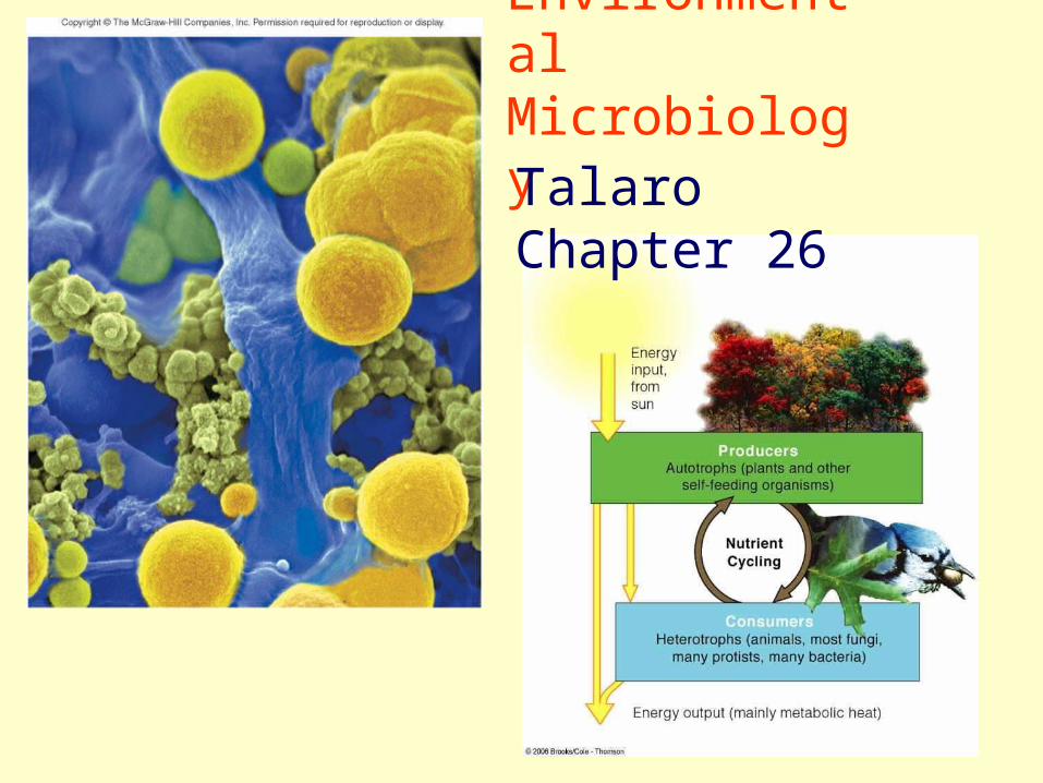
1 Environment al Microbiolog y Talaro Chapter 26
-
Upload
marvin-morgan -
Category
Documents
-
view
246 -
download
0
Transcript of 1 Environmental Microbiology Talaro Chapter 26. 2 Environmental Microbiology –Study of microbes in...
1
Environmental MicrobiologyTalaro Chapter 26
2
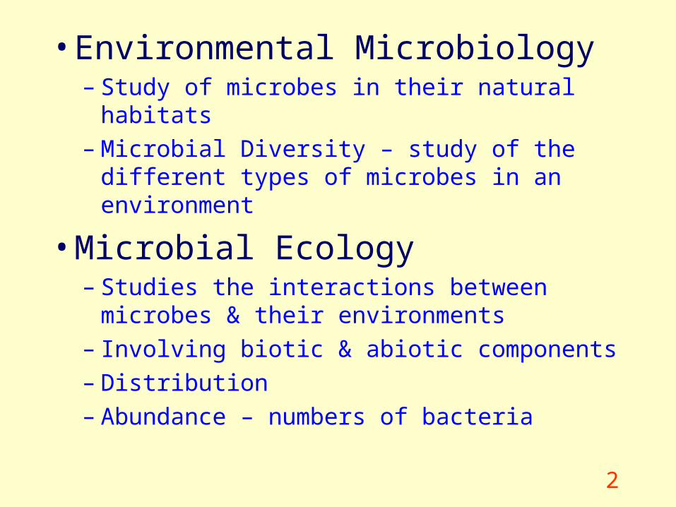
• Environmental Microbiology – Study of microbes in their natural habitats– Microbial Diversity – study of the different types
of microbes in an environment
• Microbial Ecology– Studies the interactions between microbes & their
environments– Involving biotic & abiotic components– Distribution– Abundance – numbers of bacteria
3
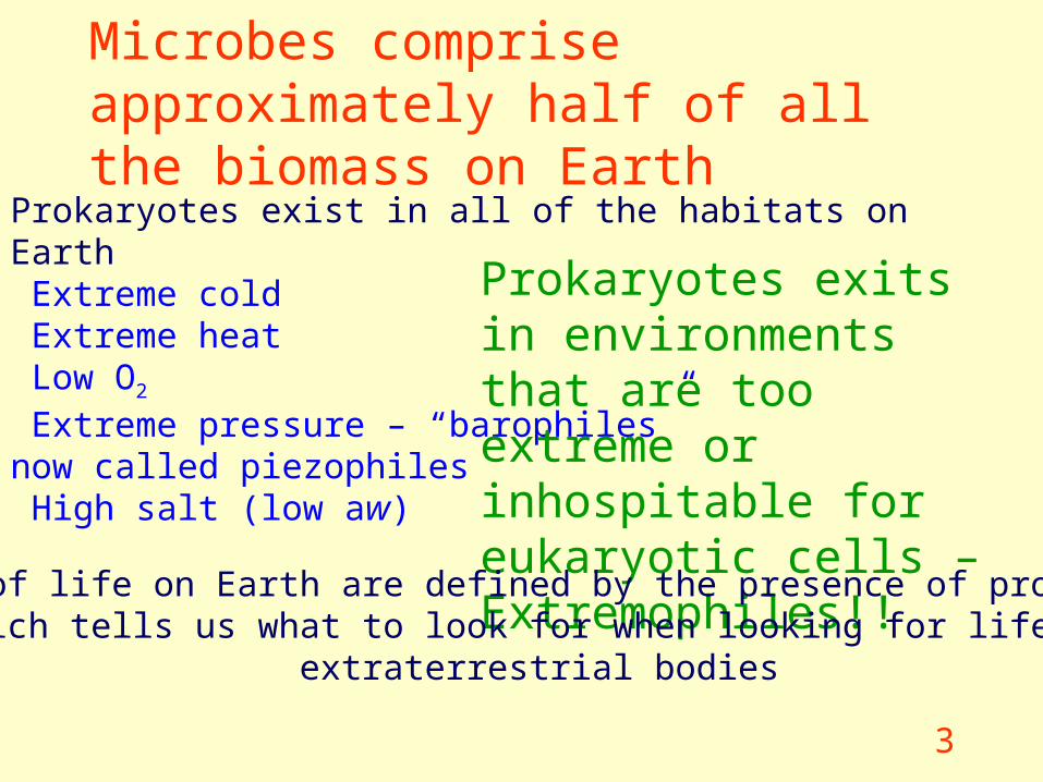
Microbes comprise approximately half of all the biomass on Earth
Prokaryotes exist in all of the habitats on Earth Extreme cold Extreme heat Low O2
Extreme pressure – “barophiles” now called piezophiles High salt (low aw)
Prokaryotes exits in environments that are too extreme or inhospitable for eukaryotic cells – Extremophiles!!
Limits of life on Earth are defined by the presence of prokaryotes which tells us what to look for when looking for life on
extraterrestrial bodies
4
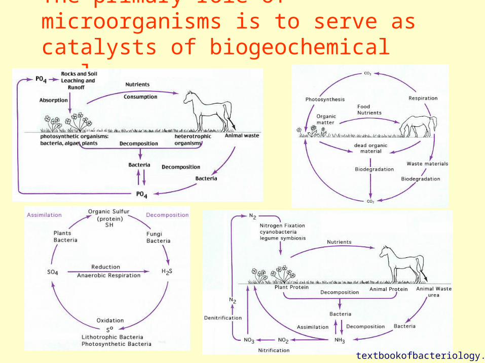
The primary role of microorganisms is to serve as catalysts of biogeochemical cycles
textbookofbacteriology.net
5
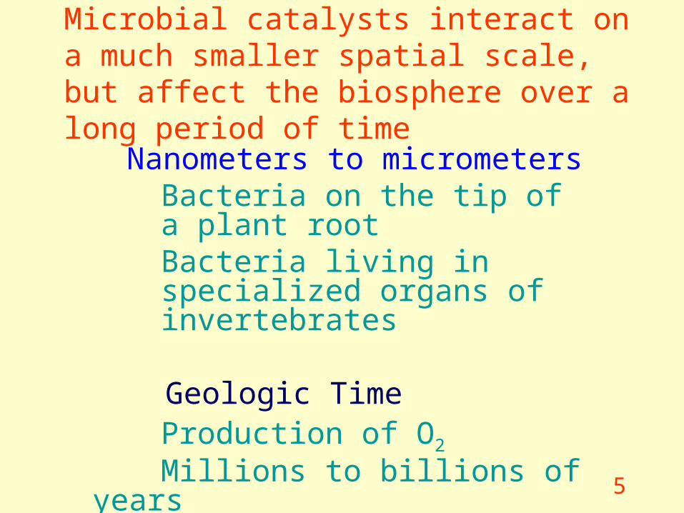
Microbial catalysts interact on a much smaller spatial scale, but affect the biosphere over a long period of time
Nanometers to micrometersBacteria on the tip of a plant rootBacteria living in specialized organs of invertebrates
Geologic TimeProduction of O2
Millions to billions of years
6
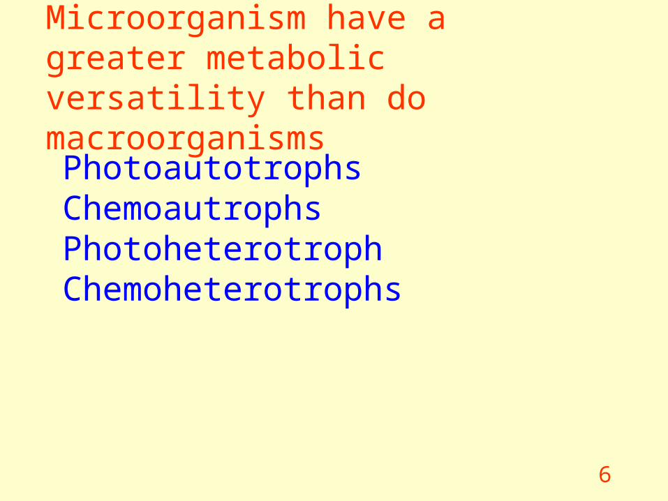
Microorganism have a greater metabolic versatility than do macroorganisms
PhotoautotrophsChemoautrophsPhotoheterotrophChemoheterotrophs
7
Only a small number of bacteria are pathogenic!
Plant and animals are dependent upon the actions of prokaryotes
Archaea and Bacteria participate in mutualistic relationships that benefit both organisms
And there are bacteria that are pathogens of animals and plants
Prokaryotes do not Exist in Isolation
8
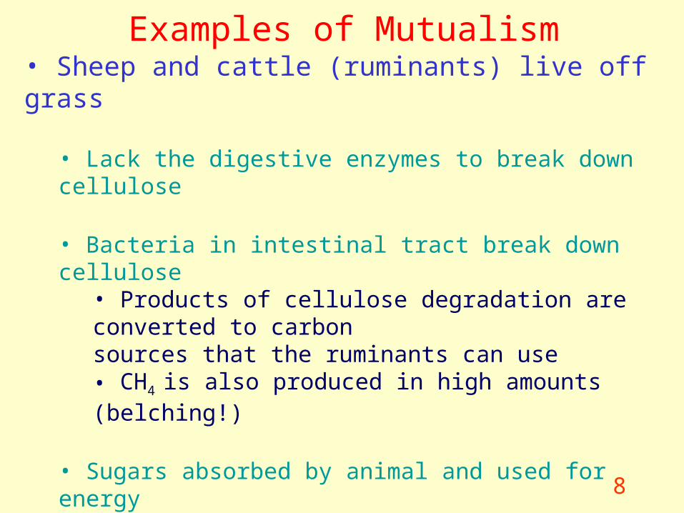
Examples of Mutualism• Sheep and cattle (ruminants) live off grass
• Lack the digestive enzymes to break down cellulose
• Bacteria in intestinal tract break down cellulose• Products of cellulose degradation are converted to carbonsources that the ruminants can use • CH4 is also produced in high amounts (belching!)
• Sugars absorbed by animal and used for energy
• Plants unable to fix atmospheric N2
• Symbiotic bacteria infect roots
• Plant requires nitrogen for proteins
9
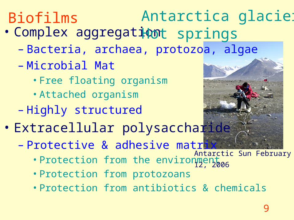
Biofilms• Complex aggregation
– Bacteria, archaea, protozoa, algae– Microbial Mat
• Free floating organism
• Attached organism
– Highly structured
• Extracellular polysaccharide– Protective & adhesive matrix
• Protection from the environment
• Protection from protozoans
• Protection from antibiotics & chemicals
Antarctica glaciersHot springs
Antarctic Sun February 12, 2006
10
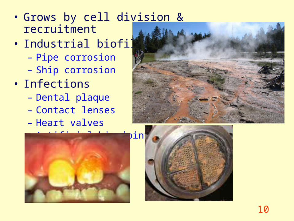
• Grows by cell division & recruitment• Industrial biofilms
– Pipe corrosion– Ship corrosion
• Infections– Dental plaque– Contact lenses– Heart valves– Artificial hip joints
11
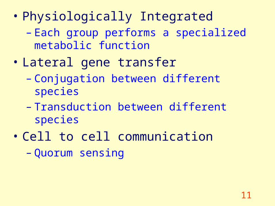
• Physiologically Integrated– Each group performs a specialized metabolic
function
• Lateral gene transfer– Conjugation between different species– Transduction between different species
• Cell to cell communication– Quorum sensing
12
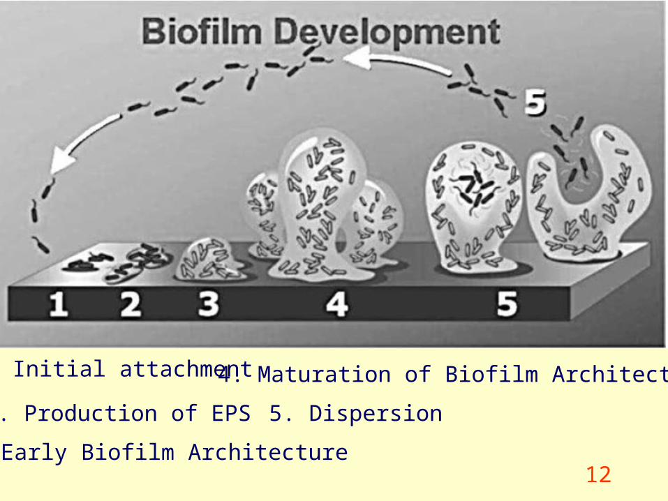
1. Initial attachment
2. Production of EPS 5. Dispersion
4. Maturation of Biofilm Architecture
3. Early Biofilm Architecture
13www.microbes.org/labs.asp
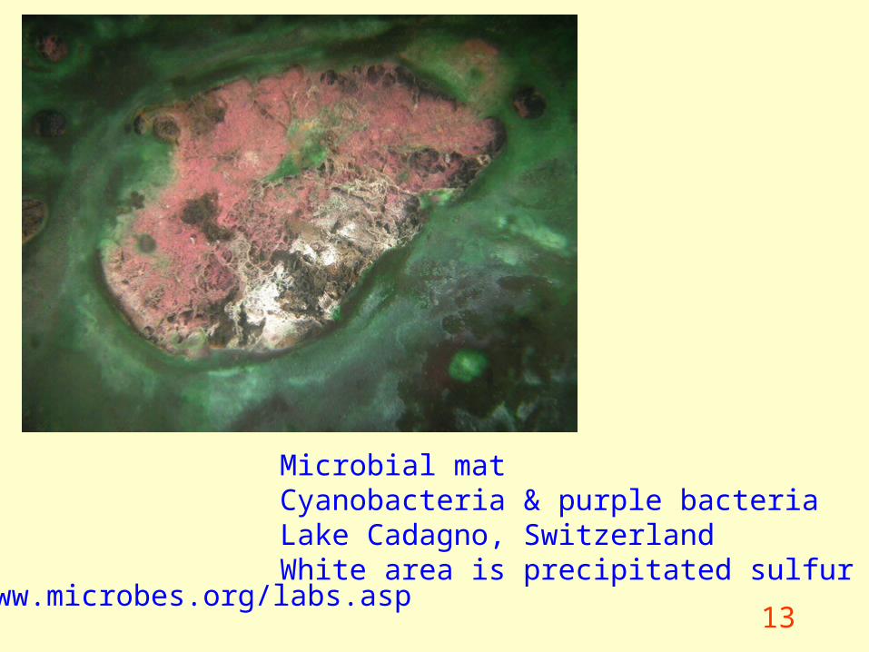
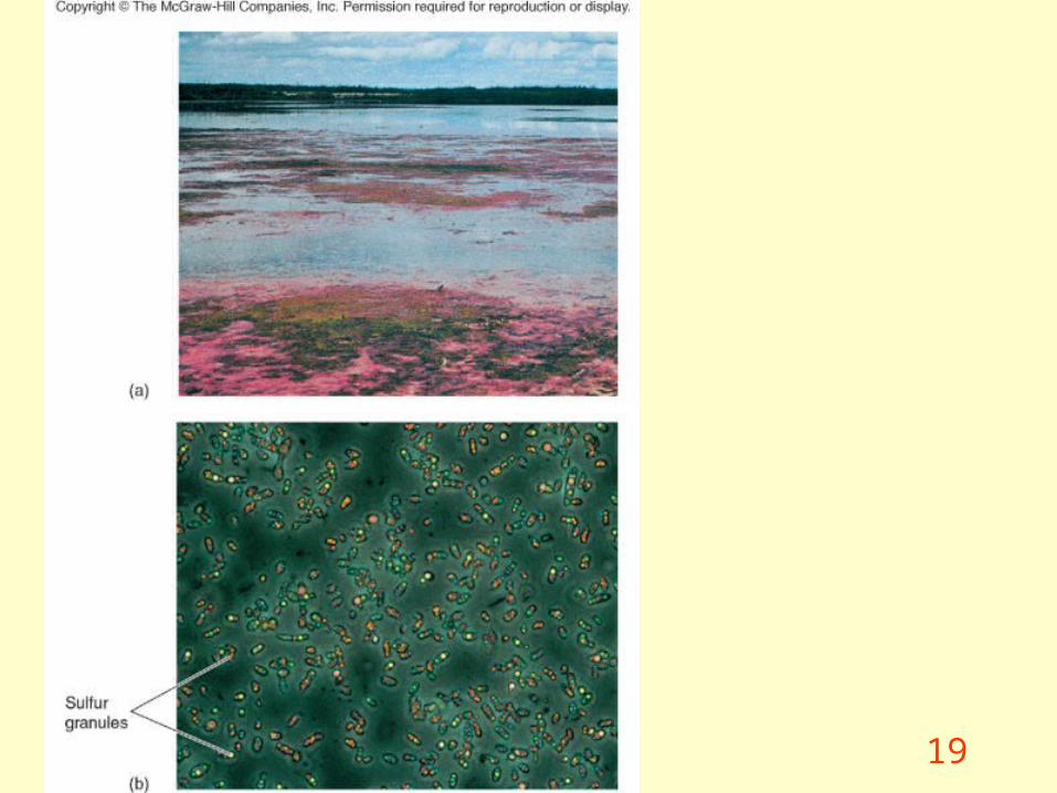
Microbial matCyanobacteria & purple bacteriaLake Cadagno, SwitzerlandWhite area is precipitated sulfur
14
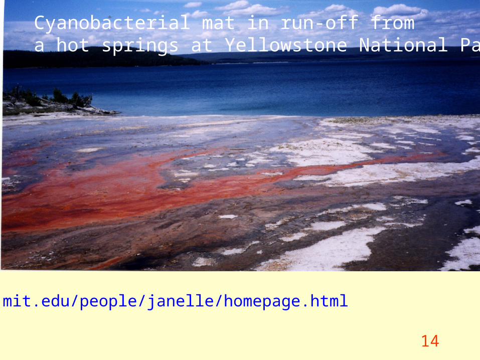
Cyanobacterial mat in run-off froma hot springs at Yellowstone National Park
www.mit.edu/people/janelle/homepage.html
15
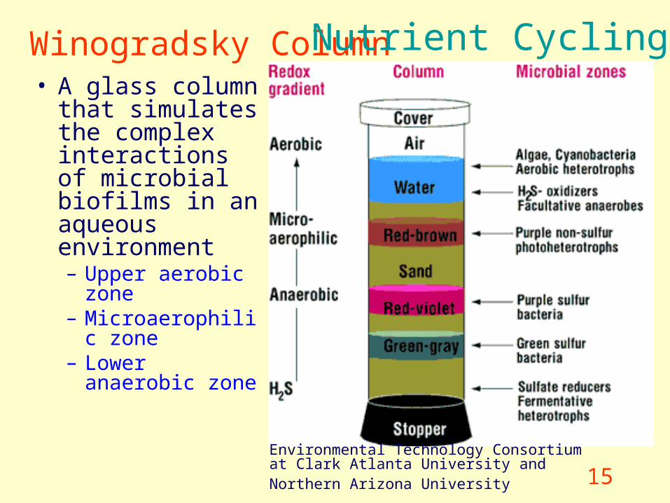
Winogradsky Column • A glass column
that simulates the complex interactions of microbial biofilms in an aqueous environment– Upper aerobic
zone– Microaerophilic
zone– Lower anaerobic
zone
Environmental Technology Consortium at Clark Atlanta University and Northern Arizona University
Nutrient Cycling
16
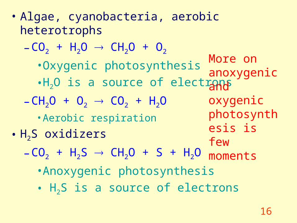
• Algae, cyanobacteria, aerobic heterotrophs
– CO2 + H2O CH2O + O2
• Oxygenic photosynthesis
• H2O is a source of electrons
– CH2O + O2 CO2 + H2O
• Aerobic respiration
• H2S oxidizers
– CO2 + H2S CH2O + S + H2O
• Anoxygenic photosynthesis
• H2S is a source of electrons
More on anoxygenic and oxygenic photosynthesis is few moments
17
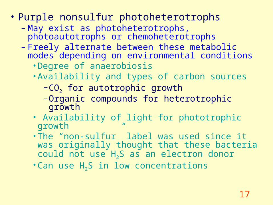
• Purple nonsulfur photoheterotrophs– May exist as photoheterotrophs, photoautotrophs or
chemoheterotrophs – Freely alternate between these metabolic modes
depending on environmental conditions • Degree of anaerobiosis• Availability and types of carbon sources
– CO2 for autotrophic growth – Organic compounds for heterotrophic growth
• Availability of light for phototrophic growth• The “non-sulfur” label was used since it was
originally thought that these bacteria could not use H2S as an electron donor
• Can use H2S in low concentrations
18
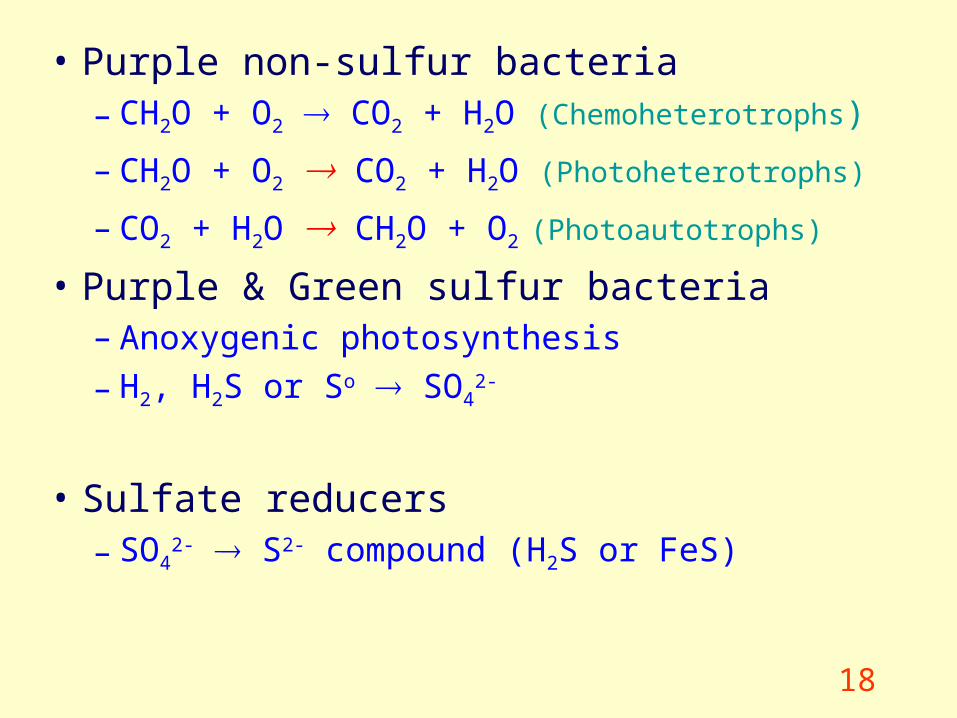
• Purple non-sulfur bacteria– CH2O + O2 CO2 + H2O (Chemoheterotrophs)
– CH2O + O2 CO2 + H2O (Photoheterotrophs)
– CO2 + H2O CH2O + O2 (Photoautotrophs)
• Purple & Green sulfur bacteria– Anoxygenic photosynthesis
– H2, H2S or So SO42-
• Sulfate reducers– SO4
2- S2- compound (H2S or FeS)
19
20
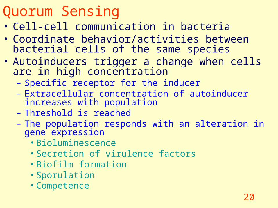
Quorum Sensing• Cell-cell communication in bacteria • Coordinate behavior/activities between bacterial cells of the
same species• Autoinducers trigger a change when cells are in high
concentration– Specific receptor for the inducer– Extracellular concentration of autoinducer increases with
population – Threshold is reached– The population responds with an alteration in gene expression
• Bioluminescence• Secretion of virulence factors• Biofilm formation• Sporulation• Competence
21
Energy & Nutrient Flow
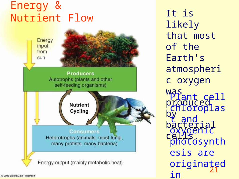
It is likely that most of the Earth's atmospheric oxygen was produced by bacterial cells.
Plant cell chloroplast and oxygenic photosynthesis are originated in prokaryotes.
22
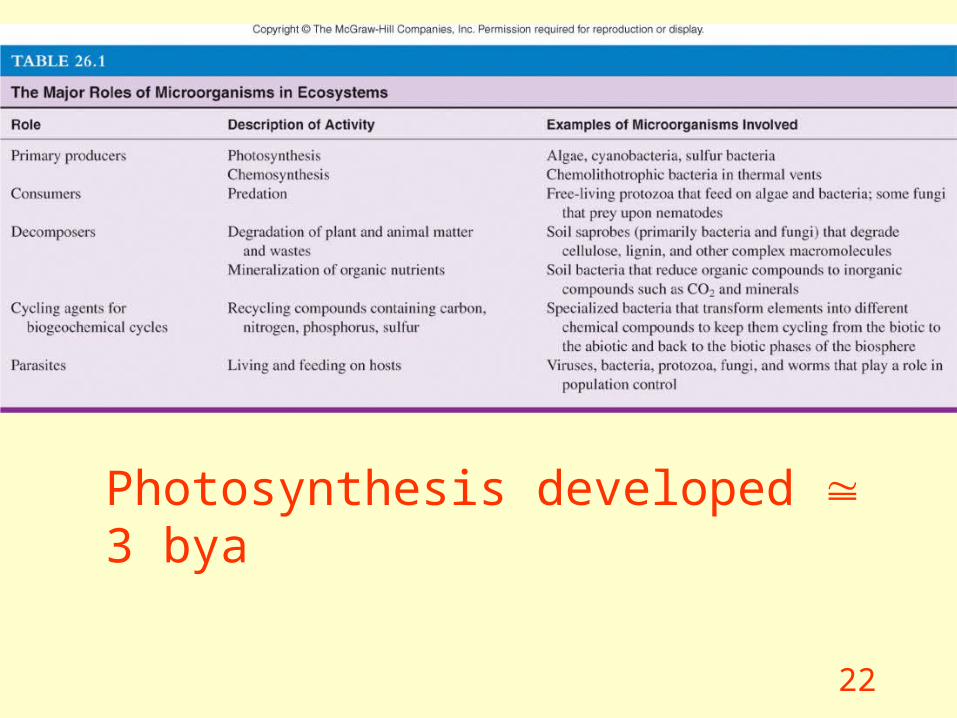
Photosynthesis developed 3 bya
23
Anoxygenic Photosynthesis – Anaerobic bacterial photosynthesis that does not produce O2
– CO2 + H2S (CH2O)n + S + H2O• H2, H2S or So or organic compounds serves as a source of electrons
– Need electrons to make fix C and make ATP
– Purple and green photosynthetic sulfur bacteria• Aquatic & anaerobic• Pigments that absorb different • Bacteriochlorophyll (800 - 1000 nm [far red]) • Carotenoids (400 - 550 nm)
– Phycobilins are not present • Only 1 photosystem
– Rhodobacter • Oxidize succinate or butyrate during CO2 fixation • Hypothesized to be have become an endosymbiont of eucaryotes • Mitochondrion 16S rRNA sequences
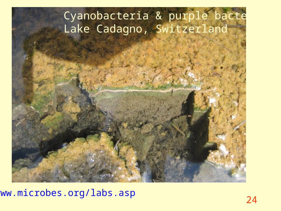
24www.microbes.org/labs.asp
Cyanobacteria & purple bacteriaLake Cadagno, Switzerland

25
• Start here next time
26
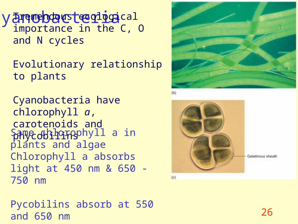
CyanobacteriaTremendous ecological importance in the C, O and N cycles
Evolutionary relationship to plants
Cyanobacteria have chlorophyll a, carotenoids and phycobilins
Same chlorophyll a in plants and algaeChlorophyll a absorbs light at 450 nm & 650 - 750 nm
Pycobilins absorb at 550 and 650 nm
27
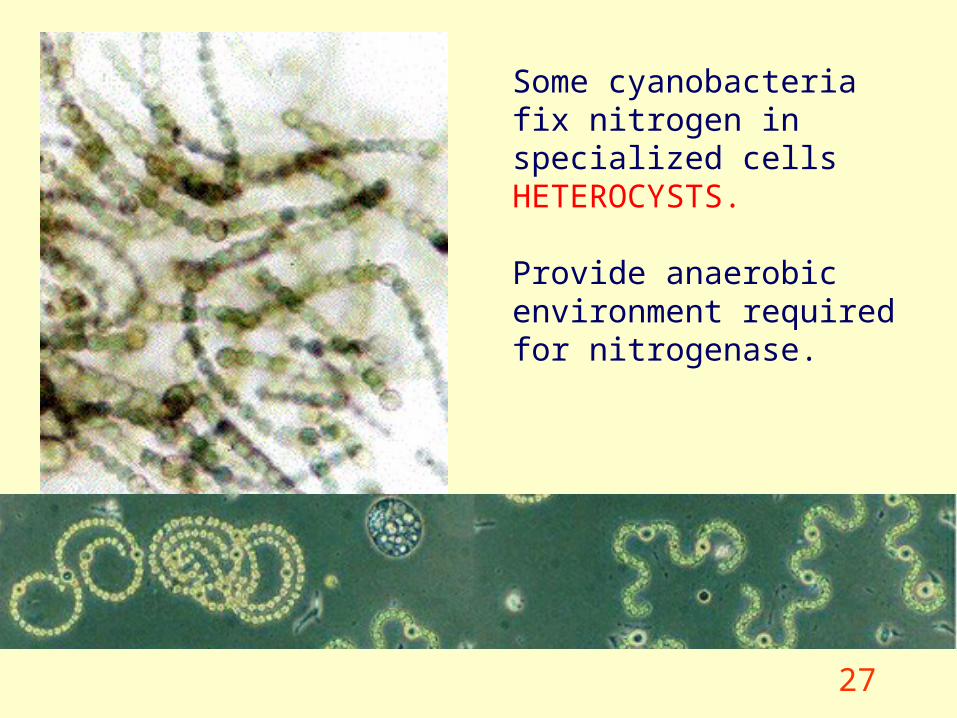
Some cyanobacteria fix nitrogen in specialized cells HETEROCYSTS.
Provide anaerobic environment required for nitrogenase.
28
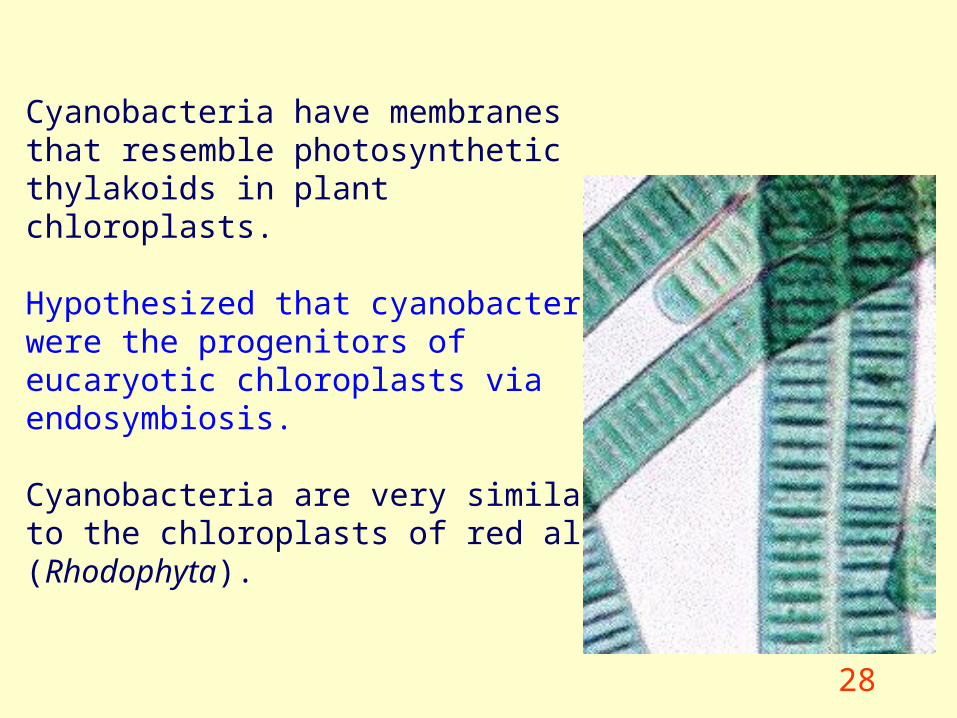
Cyanobacteria have membranes that resemble photosynthetic thylakoids in plant chloroplasts.
Hypothesized that cyanobacteria were the progenitors of eucaryotic chloroplasts via endosymbiosis.
Cyanobacteria are very similar to the chloroplasts of red algae (Rhodophyta).
29
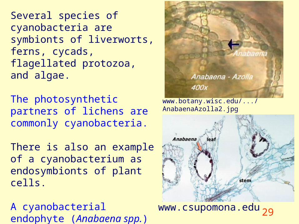
Several species of cyanobacteria are symbionts of liverworts, ferns, cycads, flagellated protozoa, and algae.
The photosynthetic partners of lichens are commonly cyanobacteria.
There is also an example of a cyanobacterium as endosymbionts of plant cells.
A cyanobacterial endophyte (Anabaena spp.) fixes nitrogen that becomes available to the water fern, Azolla.
www.csupomona.edu
www.botany.wisc.edu/.../AnabaenaAzolla2.jpg
30
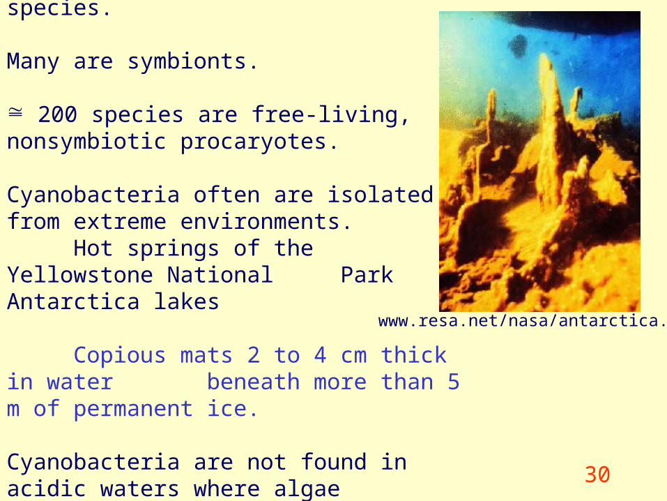
Several thousand cyanobacteria species. Many are symbionts.
200 species are free-living, nonsymbiotic procaryotes.
Cyanobacteria often are isolated from extreme environments.
Hot springs of the Yellowstone National Park Antarctica lakes
Copious mats 2 to 4 cm thick in water beneath more than 5 m of permanent ice.
Cyanobacteria are not found in acidic waters where algae (euckaryotic) predominate.
www.resa.net/nasa/antarctica.htm
31
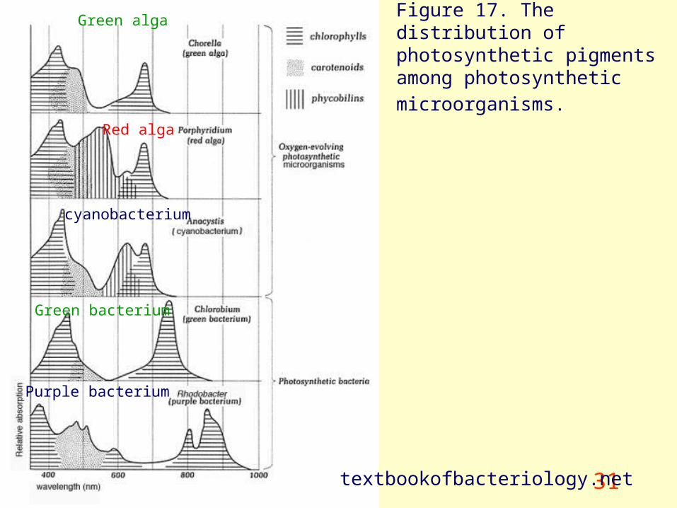
Figure 17. The distribution of photosynthetic pigments among
photosynthetic microorganisms.
textbookofbacteriology.net
Green alga
Red alga
cyanobacterium
Green bacterium
Purple bacterium
32
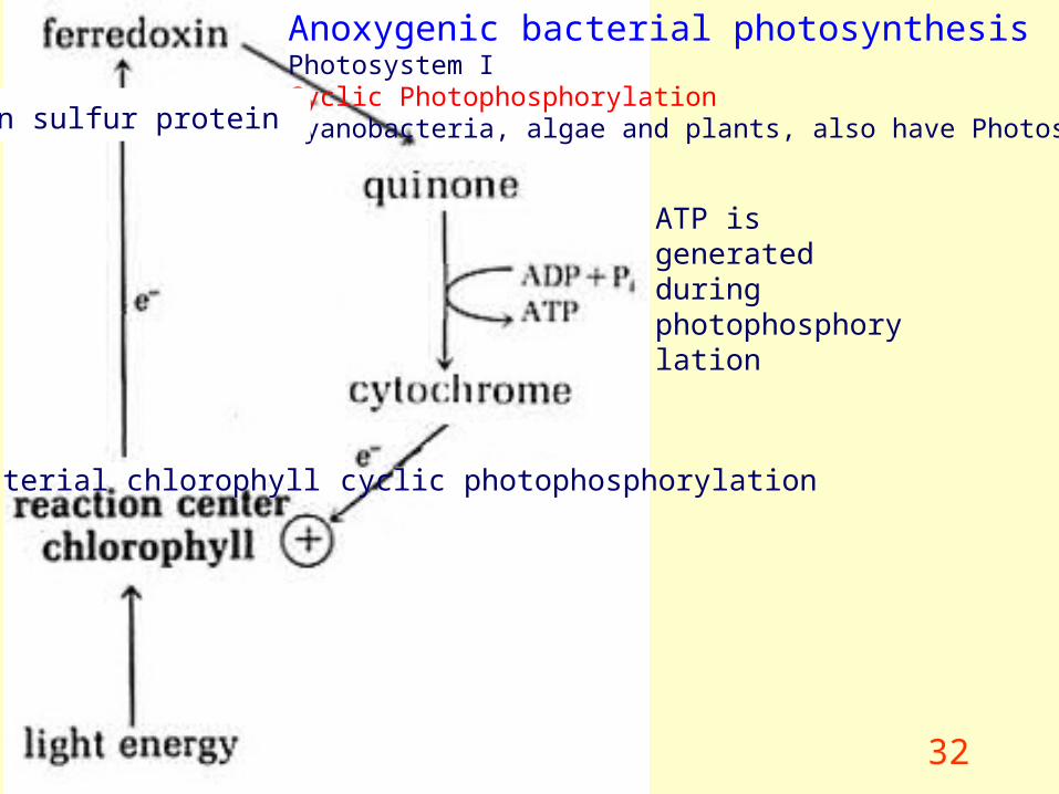
Anoxygenic bacterial photosynthesis Photosystem ICyclic PhotophosphorylationCyanobacteria, algae and plants, also have Photosystem II
bacterial chlorophyll
iron sulfur protein
ATP is generated during photophosphorylation
cyclic photophosphorylation
33textbookofbacteriology.net
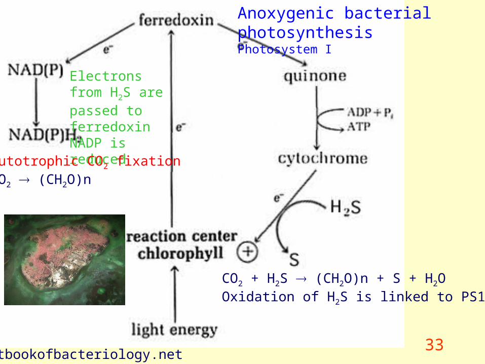
CO2 + H2S (CH2O)n + S + H2OOxidation of H2S is linked to PS1
Anoxygenic bacterial photosynthesis Photosystem I
Electrons from H2S are passed to ferredoxinNADP is reduced
Autotrophic CO2 fixationCO2 (CH2O)n

34
Anoxygenic photosynthesis
Limitations on the amount of C that can be fixed
Need more electrons to fix more C
35textbookofbacteriology.net
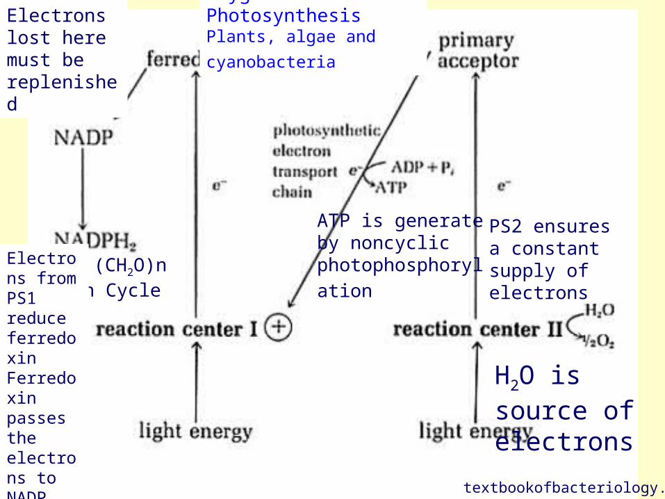
Oxygenic Photosynthesis
Plants, algae and cyanobacteria
PS2 ensures a constant supply of electrons
Electrons lost here must be replenished
H2O is source of electrons
CO2 (CH2O)nCalvin Cycle
ATP is generate by noncyclic
photophosphorylation Electrons from PS1 reduce ferredoxin Ferredoxin passes the electrons to NADP
36
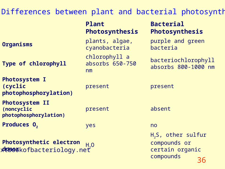
Table 6. Differences between plant and bacterial photosynthesis
Plant Photosynthesis Bacterial Photosynthesis
Organismsplants, algae, cyanobacteria
purple and green bacteria
Type of chlorophyllchlorophyll a absorbs 650-750 nm
bacteriochlorophyll absorbs 800-1000 nm
Photosystem I (cyclic photophosphorylation)
present present
Photosystem II(noncyclic photophosphorylation)
present absent
Produces O2 yes no
Photosynthetic electron donor H2OH2S, other sulfur compounds
or certain organic compounds
textbookofbacteriology.net