1. Clinical Impact of Glycated Albumin as Another Glycemic Control Marker
12
Endocrine Journal 2010, 57 (9), 751-762 Clinical impact of glycated albumin as another glycemic control marker Masafumi Koga 1) and Soji Kasayama 2) 1) Department of Internal Medicine, Kinki Central Hospital, Itami 664-8533, Japan 2) Department of Medicine, Nissay Hospital, Osaka 550-0012, Japan Abstract. It is known that glycation among various proteins is increased in diabetic patients compared with non-diabetic subjects. Currently, among these glycated proteins, glycated hemoglobin (HbA 1C ) is used as the gold standard index of glycemic control in clinical practice for diabetes treatment. However, HbA 1C does not accurately reflect the actual status of glycemic control in some conditions where plasma glucose changes during short term, and in patients who have diseases such as anemia and variant hemoglobin. In comparison, another index of glycemic control, glycated albumin (GA), more accurately reflects changes in plasma glucose during short term and also postprandial plasma glucose. Although GA is not influenced by disorders of hemoglobin metabolism, it is affected by disorders of albumin metabolism. This review summarizes diseases and pathological conditions where GA measurement is useful. These include the status of glycemic control changes during short term, diseases which cause postprandial hyperglycemia, iron deficiency anemia, pregnancy, chronic liver disease (liver cirrhosis), chronic renal failure (diabetic nephropathy), and variant hemoglobin. Key words: Glycated albumin, Glycated hemoglobin (HbA 1C ), Postprandial hyperglycemia, Iron deficiency anemia, Chronic liver disease IT IS KNOWN that glycation among various pro- teins is increased in diabetic patients compared with non-diabetic subjects (Table 1) [1]. Some of these glycated proteins are suggested to be involved in the development and progression of chronic diabetic com- plications [2]. Among these glycated proteins, gly- cated hemoglobin (HbA 1C ) is commonly used as the gold standard index of glycemic control in the clini- cal setting [3, 4]. Based on the Diabetes Control and Complications Trial (DCTT), it is recommended to bring HbA 1C lower than 7.0% in order to prevent the development and progression of chronic diabetic com- plications [5]. Since lifespan of erythrocytes is ap- proximately 120 days, HbA 1C reflects plasma glucose levels for the past few months [6]. Therefore, in cases where glycemic control status improves and worsens during short term, HbA 1C is not an appropriate index of glycemic control status at the time of measure- ment. Furthermore, HbA 1C measurements are affected by variant hemoglobin and some diseases that shorten the lifespan of erythrocytes, such as hemolytic anemia and renal anemia. Thus, HbA 1C does not properly rep- resent glycemic control status in such conditions [7-9]. Fructosamine was previously introduced as an in- dex of glycemic control since it is not influenced by disorders with abnormal hemoglobin metabolism. Fructosamine represents all of serum glycated proteins that have become stable ketoamines, and is measured by reduction coloring reaction with nitro blue tetrazo- lium as the substrate by means of its potent reducing power in alkaline solution. Fructosamine is not influ- enced by anemia or variant hemoglobin. Moreover, the metabolic turnover of serum albumin, which ac- counts for a large proportion of serum protein, is faster than that of hemoglobin, and thus fructosamine is su- perior to HbA 1C for reflecting glycemic control status during short term [10]. However, fructosamine levels are strongly influenced by the concentration of serum proteins and low-molecular-weight substances coex- isting in the plasma (e.g. bilirubin, hemoglobin, uric acid etc.) [10]. Glycated albumin (GA) was developed to solve Received May 6, 2010; Accepted Jul. 6, 2010 as K10E-138 Released online in J-STAGE as advance publication Aug. 17, 2010 Correspondence to: Masafumi Koga, M.D., Ph.D., Department of Internal Medicine, Kinki Central Hospital, Kuruma-zuka 3-1, Itami, Hyogo 664-8533, Japan. E-mail: [email protected] Disclosure statement: The authors have nothing to disclose. REVIEW ©The Japan Endocrine Society
-
Upload
arjunapamungkas -
Category
Documents
-
view
15 -
download
3
description
CIGA
Transcript of 1. Clinical Impact of Glycated Albumin as Another Glycemic Control Marker

Endocrine Journal 2010, 57 (9), 751-762
Clinical impact of glycated albumin as another glycemic control markerMasafumi Koga1) and Soji Kasayama2)
1) Department of Internal Medicine, Kinki Central Hospital, Itami 664-8533, Japan2) Department of Medicine, Nissay Hospital, Osaka 550-0012, Japan
Abstract. It is known that glycation among various proteins is increased in diabetic patients compared with non-diabetic subjects. Currently, among these glycated proteins, glycated hemoglobin (HbA1C) is used as the gold standard index of glycemic control in clinical practice for diabetes treatment. However, HbA1C does not accurately reflect the actual status of glycemic control in some conditions where plasma glucose changes during short term, and in patients who have diseases such as anemia and variant hemoglobin. In comparison, another index of glycemic control, glycated albumin (GA), more accurately reflects changes in plasma glucose during short term and also postprandial plasma glucose. Although GA is not influenced by disorders of hemoglobin metabolism, it is affected by disorders of albumin metabolism. This review summarizes diseases and pathological conditions where GA measurement is useful. These include the status of glycemic control changes during short term, diseases which cause postprandial hyperglycemia, iron deficiency anemia, pregnancy, chronic liver disease (liver cirrhosis), chronic renal failure (diabetic nephropathy), and variant hemoglobin.
Key words: Glycated albumin, Glycated hemoglobin (HbA1C), Postprandial hyperglycemia, Iron deficiency anemia, Chronic liver disease
It Is known that glycation among various pro-teins is increased in diabetic patients compared with non-diabetic subjects (Table 1) [1]. Some of these glycated proteins are suggested to be involved in the development and progression of chronic diabetic com-plications [2]. Among these glycated proteins, gly-cated hemoglobin (HbA1C) is commonly used as the gold standard index of glycemic control in the clini-cal setting [3, 4]. Based on the Diabetes Control and Complications Trial (DCTT), it is recommended to bring HbA1C lower than 7.0% in order to prevent the development and progression of chronic diabetic com-plications [5]. Since lifespan of erythrocytes is ap-proximately 120 days, HbA1C reflects plasma glucose levels for the past few months [6]. Therefore, in cases where glycemic control status improves and worsens during short term, HbA1C is not an appropriate index of glycemic control status at the time of measure-
ment. Furthermore, HbA1C measurements are affected by variant hemoglobin and some diseases that shorten the lifespan of erythrocytes, such as hemolytic anemia and renal anemia. Thus, HbA1C does not properly rep-resent glycemic control status in such conditions [7-9].
Fructosamine was previously introduced as an in-dex of glycemic control since it is not influenced by disorders with abnormal hemoglobin metabolism. Fructosamine represents all of serum glycated proteins that have become stable ketoamines, and is measured by reduction coloring reaction with nitro blue tetrazo-lium as the substrate by means of its potent reducing power in alkaline solution. Fructosamine is not influ-enced by anemia or variant hemoglobin. Moreover, the metabolic turnover of serum albumin, which ac-counts for a large proportion of serum protein, is faster than that of hemoglobin, and thus fructosamine is su-perior to HbA1C for reflecting glycemic control status during short term [10]. However, fructosamine levels are strongly influenced by the concentration of serum proteins and low-molecular-weight substances coex-isting in the plasma (e.g. bilirubin, hemoglobin, uric acid etc.) [10].
Glycated albumin (GA) was developed to solve
Received May 6, 2010; Accepted Jul. 6, 2010 as K10E-138 Released online in J-STAGE as advance publication Aug. 17, 2010Correspondence to: Masafumi Koga, M.D., Ph.D., Department of Internal Medicine, Kinki Central Hospital, Kuruma-zuka 3-1, Itami, Hyogo 664-8533, Japan. E-mail: [email protected] statement: The authors have nothing to disclose.
Review
©The Japan Endocrine Society

752 Koga et al.
as an index of glycemic control.
1. Change in short-term Glycemic Control status
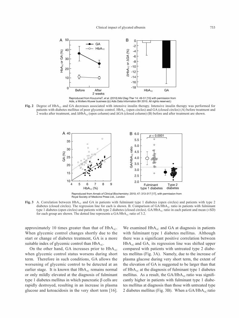
Since the half-life of serum albumin is shorter than that of erythrocytes, GA changes rapidly in cas-es where the status of glycemic control changes dur-ing short term [6, 14]. Intensive insulin therapy was performed as the initial treatment in 8 patients with type 2 diabetes mellitus with poor glycemic control (Fig. 2) [15]. There was only a mild decrease in av-eraged HbA1C from 10.9% to 10.0%, while averaged GA decreased markedly from 35.6% to 25.0%. The changes of HbA1C and GA during 2 weeks were -0.9% and -10.6%, respectively, and the decrease of GA was
above weak points of fructosamine [11]. GA is a ke-toamine which is formed by binding of albumin and glucose by nonenzymatic oxidation reaction (Fig. 1). Similar to fructosamine, GA is an index of glyce-mic control which is not affected by disorders of he-moglobin metabolism. Additionally, it reflects the short-term status of glycemic control compared with HbA1C. Furthermore, GA is not influenced by serum albumin concentration because it calculates the ra-tio of total serum albumin. Although GA used to be measured by the high-performance liquid chromatog-raphy (HPLC) method, it became rapidly and easily determined by an enzyme method for automated gen-eral biochemical analyzers recently developed [12, 13]. Here, we summarize the recent observations by focusing on the clinical usefulness of measuring GA
table 1 Glycated proteins within the body* Matrix proteins Enzymes Plasma proteins
・Collagen ・Cathepsin B ・Albumin
・Myelin ・Lysozyme ・Immunoglobulin
・Fibronectin ・Pancreatic ribose ・Apo A-I, II
・Fibrin ・Copper/zinc SOD ・Apo B
Membrane proteins ・Carbonate dehydratase ・Apo C-I
・Red cell Glu transport protein ・β-N-acetyl hexominase ・Apo E
・Red cell spectrin ・Alcohol dehydrogenase ・Haptoglobin
・Red cell membrane protein ・Aldose reductase ・Ferritin
・Endothelial plasma membrane protein ・Aldehyde reductase ・Transferrin
Intracellular proteins ・Sorbitol dehydrogenase ・α1-antitrypsin
・Hemoglobin ・Na+/K+-ATPase ・Plasminogen
・Crystallin Hormones ・Plasminogen activator
・Tubulin ・Thyroid hormone ・Fibrinogen
・Calmodulin ・Insulin ・Fibrin
・Antithrombin III
・β2-microglobulin
・Ceruloplasmin
*Reproduced from Taniguchi N (1997) Significance of protein glycation reaction in a living body; 2-8 [1], with permission from Igaku-Shoin Ltd., Japan
Fig. 1 Albumin structure and glycated positions.
753Clinical impact of glycated albumin
approximately 10 times greater than that of HbA1C. When glycemic control changes shortly due to the start or change of diabetes treatment, GA is a more suitable index of glycemic control than HbA1C.
On the other hand, GA increases prior to HbA1C when glycemic control status worsens during short term. Therefore in such conditions, GA allows the worsening of glycemic control to be detected at an earlier stage. It is known that HbA1C remains normal or only mildly elevated at the diagnosis of fulminant type 1 diabetes mellitus in which pancreatic β cells are rapidly destroyed, resulting in an increase in plasma glucose and ketoacidosis in the very short term [16].
We examined HbA1C and GA at diagnosis in patients with fulminant type 1 diabetes mellitus. Although there was a significant positive correlation between HbA1C and GA, its regression line was shifted upper compared with patients with untreated type 2 diabe-tes mellitus (Fig. 3A). Namely, due to the increase of plasma glucose during very short term, the extent of the elevation of GA is suggested to be larger than that of HbA1C at the diagnosis of fulminant type 1 diabetes mellitus. As a result, the GA/HbA1C ratio was signifi-cantly higher in patients with fulminant type 1 diabe-tes mellitus at diagnosis than those with untreated type 2 diabetes mellitus (Fig. 3B). When a GA/HbA1C ratio
0
10
20
30
40
50
Before After2 weeks
Hb
A1C
or
GA
(%) HbA1C
GABA
-18
-16
-14
-12
-10
-8
-6
-4
-2
0
HbA1C GA
∆H
bA
1C o
r ∆
GA
(%)
Reproduced from KouzumaT, et al. (2010) Mol Diag Ther 14: 49-51 [15] with permission from Adis, a Wolters Kluwer business ((c) Adis Data Information BV 2010. All rights reserved.)
BA
10
15
20
25
30
35
40
4 5 6 7 8 9HbA1C (%)
GA
(%)
2.0
2.5
3.0
3.5
4.0
4.5
5.0
5.5
6.0G
A/H
bA
1C r
atio
p < 0.0001
Fulminanttype 1 diabetes
Type 2diabetes
Reproduced from Annals of Clinical Biochemistry; 2010; 47: 313-317 [17], with permission from Royal Society of Medicine Press Ltd., London
Fig. 3 A. Correlation between HbA1C and GA in patients with fulminant type 1 diabetes (open circles) and patients with type 2 diabetes (closed circles). The regression line for each is shown. B. Comparison of GA/HbA1C ratio in patients with fulminant type 1 diabetes (open circles) and patients with type 2 diabetes (closed circles). GA/HbA1C ratio in each patient and mean (±SD) for each group are shown. The dotted line represents a GA/HbA1C ratio of 3.2.
Fig. 2 Degree of HbA1C and GA decreases associated with intensive insulin therapy. Intensive insulin therapy was performed for patients with diabetes mellitus of poor glycemic control. HbA1C (open circles) and GA (closed circles) (A) before treatment and 2 weeks after treatment, and ΔHbA1C (open column) and ΔGA (closed column) (B) before and after treatment are shown.

754 Koga et al.
reflecting decreased endogenous insulin secretion is involved in increased value of the GA/HbA1C ratio. Combining the results that maximum plasma glucose is involved in the GA/HbA1C ratio in patients with type 1 diabetes mellitus [22], it is suggested that a de-crease in the insulin secretion increases the glycemic excursion, causing increase of the GA/HbA1C ratio.
Recently, plasma glucose levels throughout the day can be measured by means of the continuous glucose monitor (CGM) system. A study on the relationship between the CGM system data and the index of gly-cemic control was reported. Among the patients with diabetes mellitus showing poor glycemic control, GA indicated a more potent relationship with differences of plasma glucose levels and plasma glucose fluctua-tion index than HbA1C and 1,5-anhydroglucitol (1,5-AG) [24].
In gastrectomized subjects, oral glucose tolerance test often shows marked hyperglycemia (oxyhypergly-cemia) 30 to 60 min after glucose loading. We showed that in non-diabetic subjects who underwent gastrec-tomy, GA and HbA1C were both higher than in controls but the GA/HbA1C ratio was also significantly higher. Namely, the extent of increase of GA was greater than that of HbA1C in the gastrectomized subjects, thus bet-ter reflecting postprandial hyperglycemia [25].
The reasons why serum GA reflects postprandi-al hyperglycemia better than HbA1C are unknown. The shortened lifespan of erythrocytes in diabetic pa-tients with poor glucose control [26], lagging GLUT1-
≥3.2 is regarded as a cutoff value, the sensitivity and specificity of differentiating fulminant type 1 diabetes mellitus at diagnosis from untreated type 2 diabetes mellitus were 97% and 98%, respectively. Therefore, we suggested that a high GA/HbA1C ratio is helpful for diagnosing fulminant type 1 diabetes mellitus [17].
2. Postprandial Hyperglycemia
A number of epidemiological studies have shown that postload hyperglycemia becomes a risk factor for cardiovascular diseases. The DECODE study [18] or Funagata study [19] revealed that postload plasma glucose in the glucose tolerance test is a more potent risk factor for cardiovascular events than fasting plas-ma glucose. Furthermore, it has been reported that administration of the α glucosidase inhibitor acarbose to patients with impaired glucose tolerance or diabetes mellitus was associated with cardiovascular risk re-duction (STOP-NIDDM trial) [20, 21].
HbA1C is considered primarily to be an index which reflects the mean plasma glucose levels. On the other hand, there have been recently several reports suggest-ing that GA is an index which more strongly reflects postprandial plasma glucose rather than mean plasma glucose. The GA/HbA1C ratio in patients with type 1 diabetes mellitus was shown to be significantly higher than in patients with type 2 diabetes mellitus [22]. In general, plasma glucose fluctuates over a greater range in patients with type 1 diabetes mellitus compared with patients with type 2 diabetes mellitus. In view of this phenomenon, in patients with type 1 diabetes mel-litus and those with type 2 diabetes mellitus who show no difference in HbA1C value, the GA value is signifi-cantly higher in the former. The authors speculated that GA may more strongly reflect postprandial plas-ma glucose and range of plasma glucose fluctuations than HbA1C.
When relationship between HbA1C and GA was ex-amined in patients with type 2 diabetes mellitus, GA/HbA1C ratio was found to be significantly higher in pa-tients receiving insulin treatment than in patients re-ceiving diet therapy or oral hypoglycemic drugs [23]. Insulin secretion determined by the homeostasis mod-el assessment pancreatic β–cell function (HOMA-%β) was significantly lower in the patients receiving insu-lin treatment than that in the patients not receiving in-sulin treatment. There was a significant inverse cor-relation in GA/HbA1C ratio and HOMA-%β (Fig. 4),
R = 0.315p < 0.0001
2.0
2.5
3.0
3.5
4.0
4.5
0HOMA-% β (%)
GA
/Hb
A1C
rat
io
Copyright 2010 American Diabetes Association From Diabetes Care, Vol 33, 2010; 509-511 [23] Reprinted with permission from the Reproduced from the American Diabetes Association
50 100 150
Fig. 4 Correlation between HOMA-%β and GA/HbA1C ratio in patients with type 2 diabetes.

755Clinical impact of glycated albumin
Phelps et al. [40] showed biphasic changes in HbA1C levels during pregnancy, with HbA1C levels lowest at gestational week 24. A longitudinal study also demonstrated similar biphasic changes in HbA1C levels [41]. One of the reasons why HbA1C decreases from the first trimester to the second trimester of preg-nancy is considered to be the decrease in plasma glu-cose levels, although the reason why HbA1C increases again from the second trimester to the third trimester is unknown. Sanaka, et al. [42] reported the phenom-enon that HbA1C increases from the second trimes-ter to the third trimester of pregnancy in non-diabet-ic cases, whereas GA does not change much during this period. Based on this result, it was suggested that HbA1C show high values independent of plasma glu-cose levels from the second trimester to the third tri-mester during pregnancy. It is known that iron de-mand is increased and iron deficiency often occurs in the third trimester of pregnancy. Our investigations in non-diabetic pregnant women demonstrated that HbA1C increased from the second trimester to the third trimester of pregnancy while GA did not show any significant change. Mean corpuscular hemoglobin (MCH), transferring saturation, and serum ferritin de-creased from the second trimester to the third trimes-ter of pregnancy, and thus most women became iron
mediated glucose uptake by erythrocyte resulting in a relatively lower degree of rise in HbA1C [27], different glycation rates between albumin and hemoglobin [28] and a direct effect of insulin and oral hypoglycemic agents on serum albumin metabolism [29] may be in-volved. These mechanisms need to be clarified in fu-ture studies.
If GA presents relatively higher values than HbA1C in patients associated with postprandial hyperglycemia, it may be more appropriate to measure GA as an index of glycemic control in patients with diabetes mellitus. Recently, it has been revealed that GA is an index that predicts the development of coronary artery disease (CAD) as well as its severity [30, 31]. The relationship between GA and diabetic vascular complications will be further investigated.
3. Anemia (Hemolytic Anemia, Iron Deficiency Anemia, Iron Deficiency Status,
treatment by Iron Preparations)
It is well known that HbA1C presents lower values in relation to glycemia in patients with hemolytic anemia, because lifespan of erythrocytes is shortened in these patients [7]. Meanwhile, patients with iron deficiency anemia conversely presents higher HbA1C values rela-tive to plasma glucose levels [32, 33]. We found that HbA1C also shows higher levels in relation to glycemia even in iron deficient state without anemia [34]. Iron deficiency anemia is the most frequently seen anemia. Since approximately one half of premenopausal wom-en are in iron deficient status, a great number of pre-menopausal women present high HbA1C values relative to plasma glucose levels [33, 35]. On the other hand, when patients with iron deficiency anemia are treat-ed with iron supplements, HbA1C transiently decreas-es because lifespan of erythrocytes shortens [36, 37]. In contrast, GA is not influenced by these conditions, and thus GA is a preferable index of glycemic control in premenopausal women who frequently suffer from iron deficiency anemia (Fig. 5) [37].
4. Pregnancy
In pregnant women displaying diabetes mellitus and those with gestational diabetes, intensive glyce-mic control during pregnancy is needed to lower the risk of intrauterine fetal death, fetal growth disorders and maternal complications [38, 39].
*** **
8
9
10
11
12
13
14
15
Hem
oglo
bin
(g/d
L)
**5
6
7
8
9
Hb
A1C
(%)
HbA1C
15
18
21
24
27
GA
(%)
GA
Before 4w 8w 12w
Reproduced from Journal of the Japan Diabetes Society; Koga M, et al. 2009; 52: 341-345 [37], with permission from the Japan Diabetes Society
Fig. 5 Effects of iron treatment on hemoglobin, HbA1C and GA in diabetic patients with iron deficiency anemia.
*: p<0.05, **: p<0.01 ***: p<0.001 vs. before treatment.

756 Koga et al.
5. Chronic Liver Diseases (Liver Cirrhosis)
Since liver is a pivotal organ regulating plasma glu-cose levels, glucose metabolic abnormalities occur fre-quently in patients with chronic liver diseases (CLD), such as chronic hepatitis and liver cirrhosis. In pa-tients with CLD, about 70-90% are diagnosed as im-paired glucose tolerance and 30-60% of them as dia-betes mellitus [47]. It is important to maintain a good glycemic control status because CLD patients with poor glycemic control have been shown to offer poor prognosis [48].
HbA1C has been shown to be apparently lower in re-lation to glycemia due to shortened half-life of eryth-rocytes [49] originating from hypersplenism in CLD patients. On the contrary, GA and fructosamine levels are apparently higher in relation to glycemia in these patients due to prolonged half-life of serum albumin originating from reduced capacities of albumin syn-thesis in vivo [50, 51]. Similarly, it has been reported that serum 1,5-AG levels do not reflect glycemic con-trol state accurately in CLD patients [52]. Taken to-gether, it was difficult to monitor glycemic control sta-tus accurately in CLD patients, because none of the
deficient in the third trimester. Since HbA1C showed significant inverse correlations with these values, it was concluded that the increase of HbA1C in the third trimester of pregnancy was caused by iron deficiency [43]. In our further investigations of pregnant wom-en with diabetes mellitus, HbA1C increased and trans-ferrin saturation and serum ferritin decreased from the second trimester to the third trimester of pregnan-cy, while GA showed no significant change (Fig. 6). Since HbA1C again showed a significant inverse corre-lation with transferrin saturation, it was concluded that HbA1C increases due to iron deficiency in the third tri-mester also in pregnant women with diabetes mellitus [44]. These findings suggest that HbA1C is not an ap-propriate index of glycemic control during pregnancy. Meanwhile, GA is not affected by iron deficiency and has the advantage of reflecting the short-term status of glycemic control, and is thus considered to be a pref-erable index of glycemic control during pregnancy.
Since the threshold of reabsorption of glucose de-creases in renal tubules resulting in renal glycouria during pregnancy, serum 1,5-AG indicates a low value [45, 46]. Therefore, serum 1,5-AG is inadequate as an index of glycemic control during pregnancy.
BA
C D
*## #
4.0
5.0
6.0
7.0
Hb
A1C
(%)
10
15
20
25
GA
(%)
** ******# #
0I II III IV I II III IV
I II III IV I II III IV
10
20
30
40
Ser
um t
rasf
errin
satu
ratio
n (%
)
*** ***## ***#
0.0
0.5
1.0
1.5
2.0
2.5
Ferr
itin(
log-
ng/m
L)
Copyright 2010 American Diabetes Association From Diabetes Care, Vol 33, 2010; 270-272 [44] Reprinted with permission from the Reproduced from the American Diabetes Association
Fig. 6 Changes in HbA1C (A), GA (B), serum transferrin saturation (C), and serum ferritin (D) during pregnancy in diabetic patients. Term I, 20-23 weeks; Term II, 24-27 weeks; Term III, 28-31 weeks; Term IV, 32-35 weeks. * p<0.05, ** p<0.01, and *** p<0.001 vs. Term I; # p<0.05, and ## p<0.01 vs. Term II.

757Clinical impact of glycated albumin
values relative to glycemia in CLD patients, the GA/HbA1C ratio is set higher in these patients. We re-vealed that although there was a significant positive correlation between HbA1C and GA in CLD patients, its regression line was shifted higher in comparison with that in patients with type 2 diabetes mellitus (Fig. 7A). There was no correlation between GA/HbA1C ra-tio with mean plasma glucose, while there were sig-nificantly inverse correlations with hepaplastin test (HPT), cholinesterase and platelet count, which are indices of hepatic function (Fig. 7B). These results showed that GA/HbA1C ratio reflects the hepatic func-tion independently of plasma glucose levels [55]. Therefore, simultaneous measurement of HbA1C and GA can give the glycemic control index CLD-HbA1C as well as the hepatic function index GA/HbA1C ratio in CLD patients (Fig. 8).
known markers reflects it precisely. We investigated the relationship between plasma
glucose levels and glycemic control markers in CLD patients [51]. In comparison with HbA1C estimated from the mean plasma glucose levels [53], measured HbA1C showed lower values and GA/3 (GA indicates approximately 3 times the value of HbA1C, thus the HbA1C value can be predicted from GA/3) showed higher values in CLD patients. The discrepancy be-tween HbA1C or GA/3 and estimated HbA1C increased when hepatic function decreased. Instead, CLD-HbA1C calculated as the mean of HbA1C and GA/3 was found closely matched with HbA1C estimated from the mean plasma glucose levels. Therefore, CLD-HbA1C was shown to be useful as an index of glycemic con-trol in CLD patients [54].
Since HbA1C shows lower and GA shows higher
R = -0.696
p < 0.0001
2
4
6
8
10
0 50 100 150Hepaplastin test (%)
GA
/Hb
A1C
rat
io
BA
y = 2.6 x + 1.6
y = 2.5 x + 8.7
10
20
30
40
HbA1C (%)
GA
(%)
Reproduced from Annals of Clinical Biochemistry; 2009; 46: 368-372 [55], with permission from Royal Society of Medicine Press Ltd., London
2 4 6 8 10 12
Fig. 7 A. Correlation between HbA1C and GA in patients with chronic liver disease (CLD) (open circles) and patients with type 2 diabetes (closed circles). B. Correlation of hepaplastin test with GA/HbA1C ratio in patients with CLD.
Simultaneous measurement of HbA1C and GA
Calculation of CLD-HbA1C
(Mean of HbA1C and GA/3)Glycemic control marker
independent of hepatic function
Evaluation of glycemic control status
Calculation of GA/HbA1C ratioHepatic function marker
independent of plasma glucose levels
Evaluation of hepatic function
Fig. 8 Proposal of diabetes-related tests for patients with chronic liver diseases (CLD) complicated with diabetes
758 Koga et al.
HbA1C measurements through various mechanisms, the glycemic control status should be determined by using GA in patients with variant hemoglobin [61].
8. Disorders of Albumin Metabolism
Although GA is not influenced by anemia and vari-ant hemoglobin, it is influenced in patients with dis-orders of albumin metabolism [62]. GA shows lower values in relation to glycemia in patients with nephrot-ic syndrome, hyperthyroidism [63], and glucocorticoid administration in which albumin metabolism increas-es. Meanwhile, GA presents higher values relative to plasma glucose levels in patients with liver cirrhosis [51, 54] and hypothyroidism [63] in which albumin metabolism decreases.
Moreover, in obese subjects GA values were found to set lower in relation to glycemia [64-66]. It is known that chronic micro-inflammation is evoked by inflammatory cytokines (adipocytokine) secreted from adipocytes in obese subjects [67], and there was a sig-nificant positive correlation between BMI and high-sensitive C-reactive protein as well. Furthermore, we found a significant inverse correlation between high-sensitive C-reactive protein and GA [66]. Based on these results, we propose the theory that chronic micro-inflammation increases albumin catabolism in obese subjects [68], and as a result of the shortened half life of albumin, GA decreases relative to plasma glucose levels [66]. Indeed, it has been shown that GA was set lower in relation to plasma glucose levels in smokers, hyperuricemic patients, hypertriglyceridemia and men with nonalcoholic fatty liver disease (NAFLD) with high alanine aminotransferase (ALT) levels in whom chronic inflammation is evoked [69-72].
9. Use of HbA1C and GA Measurement to Diagnose Diabetes Mellitus
Measurements of HbA1C have not been world-wide-ly standardized. By the measurement standardized by Japan Diabetes Society (JDS), HbA1C levels are shown to set approximately 0.4% lower compared with those standardized by National Glycated Hemoglobin Standard Program (NGSP) [73]. Thus, GA/HbA1C ra-tio based upon the measurement standardized by JDS is approximately 3.0, whereas GA/HbA1C ratio based upon the measurement standardized by NGSP is ap-proximately 2.8.
6. Chronic Renal Failure (Diabetic nephropathy)
In patients with chronic renal failure, HbA1C shows lower values in relation to glycemia due to renal ane-mia. Furthermore, when erythropoietin is adminis-tered to patients with renal anemia, HbA1C shows even lower values because lifespan of erythrocytes is short-ened [56, 57]. Meanwhile, it has been reported that GA is a useful index of glycemic control in hemo-dialysis patients with diabetes because GA is not af-fected by renal anemia [56-58]. Additionally, in the examination of patients with diabetes mellitus receiv-ing peritoneal dialysis, it was shown that GA reflects properly the status of glycemic control whereas HbA1C does not [59]. In contrast, in patients with diabetic nephropathy (stage III or IV) presenting marked pro-teinuria, it should be noted that GA shows a lower val-ue relative to plasma glucose levels as a result of the increased turnover of albumin metabolism.
Observations of 98 hemodialysis patients with dia-betes mellitus for 11 years indicated that the progno-sis in the group with a GA value ≥29.0% at the start of hemodialysis was significantly poorer than that in the group with a GA value <29.0% [60]. These results show that glycemic control status in hemodialysis pa-tients with diabetes is also involved in their prognosis, and that glycemic control status should be judged by GA, not by HbA1C.
7. Variant Hemoglobin
Conventionally, abnormal hemoglobin has been found through other symptoms such as anemia and cy-anosis. In HbA1C analysis by the high-performance liquid chromatography (HPLC) method, the substitut-ed glycated products of variant hemoglobin by ami-no acids are eluted in a different position from HbA1C, and a number of asymptomatic variant hemoglobins have been found recently by abnormal peaks and ab-normal HbA1C values on HPLC charts [9]. When vari-ant hemoglobin is suspected, HbA1C measurement by an immunological approach is considered to be useful. However, since some variant hemoglobins include un-stable hemoglobin and hemoglobin with increased or decreased glycation, these cases affect HbA1C values measured by the immunological approach. Thus, the HbA1C value does not necessarily reflect the glycemic control status [9]. Since variant hemoglobin affects
759Clinical impact of glycated albumin
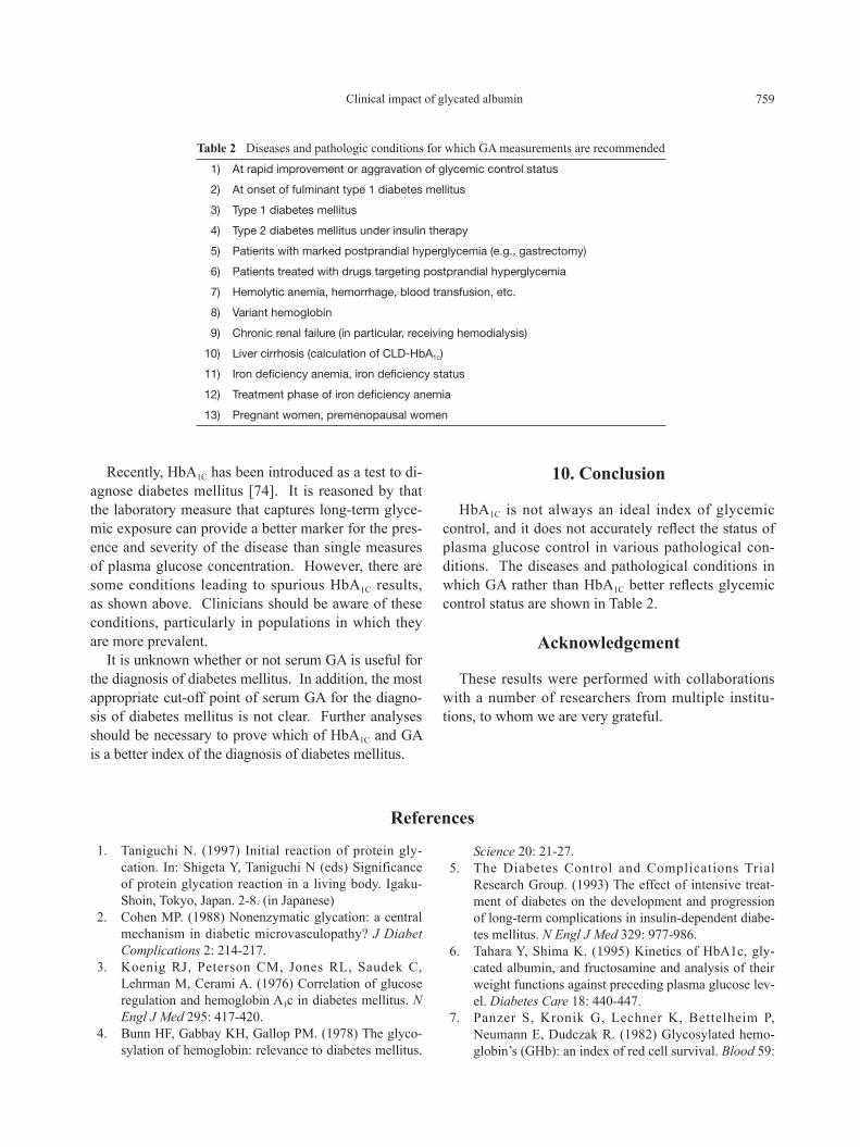
10. Conclusion
HbA1C is not always an ideal index of glycemic control, and it does not accurately reflect the status of plasma glucose control in various pathological con-ditions. The diseases and pathological conditions in which GA rather than HbA1C better reflects glycemic control status are shown in Table 2.
Acknowledgement
These results were performed with collaborations with a number of researchers from multiple institu-tions, to whom we are very grateful.
Recently, HbA1C has been introduced as a test to di-agnose diabetes mellitus [74]. It is reasoned by that the laboratory measure that captures long-term glyce-mic exposure can provide a better marker for the pres-ence and severity of the disease than single measures of plasma glucose concentration. However, there are some conditions leading to spurious HbA1C results, as shown above. Clinicians should be aware of these conditions, particularly in populations in which they are more prevalent.
It is unknown whether or not serum GA is useful for the diagnosis of diabetes mellitus. In addition, the most appropriate cut-off point of serum GA for the diagno-sis of diabetes mellitus is not clear. Further analyses should be necessary to prove which of HbA1C and GA is a better index of the diagnosis of diabetes mellitus.
table 2 Diseases and pathologic conditions for which GA measurements are recommended1) At rapid improvement or aggravation of glycemic control status
2) At onset of fulminant type 1 diabetes mellitus
3) Type 1 diabetes mellitus
4) Type 2 diabetes mellitus under insulin therapy
5) Patients with marked postprandial hyperglycemia (e.g., gastrectomy)
6) Patients treated with drugs targeting postprandial hyperglycemia
7) Hemolytic anemia, hemorrhage, blood transfusion, etc.
8) Variant hemoglobin
9) Chronic renal failure (in particular, receiving hemodialysis)
10) Liver cirrhosis (calculation of CLD-HbA1c)
11) Iron deficiency anemia, iron deficiency status
12) Treatment phase of iron deficiency anemia
13) Pregnant women, premenopausal women
References
1. Taniguchi N. (1997) Initial reaction of protein gly-cation. In: Shigeta Y, Taniguchi N (eds) Significance of protein glycation reaction in a living body. Igaku-Shoin, Tokyo, Japan. 2-8. (in Japanese)
2. Cohen MP. (1988) Nonenzymatic glycation: a central mechanism in diabetic microvasculopathy? J Diabet Complications 2: 214-217.
3. Koenig RJ, Peterson CM, Jones RL, Saudek C, Lehrman M, Cerami A. (1976) Correlation of glucose regulation and hemoglobin A1c in diabetes mellitus. N Engl J Med 295: 417-420.
4. Bunn HF, Gabbay KH, Gallop PM. (1978) The glyco-sylation of hemoglobin: relevance to diabetes mellitus.
Science 20: 21-27. 5. The Diabetes Control and Complications Trial
Research Group. (1993) The effect of intensive treat-ment of diabetes on the development and progression of long-term complications in insulin-dependent diabe-tes mellitus. N Engl J Med 329: 977-986.
6. Tahara Y, Shima K. (1995) Kinetics of HbA1c, gly-cated albumin, and fructosamine and analysis of their weight functions against preceding plasma glucose lev-el. Diabetes Care 18: 440-447.
7. Panzer S, Kronik G, Lechner K, Bettelheim P, Neumann E, Dudczak R. (1982) Glycosylated hemo-globin’s (GHb): an index of red cell survival. Blood 59:
760 Koga et al.
2072-2077.21. Chiasson JL, Josse RG, Gomis R, Hanefeld M, Karasik
A, Laakso M; STOP-NIDDM Trial Research Group. (2003) Acarbose treatment and the risk of cardiovascu-lar disease and hypertension in patients with impaired glucose tolerance: the STOP-NIDDM trial. JAMA 290: 486-494.
22. Yoshiuchi K, Matsuhisa M, Katakami N, Nakatani Y, Sakamoto K, Matsuoka T, Umayahara Y, Kosugi K, Kaneto H, Yamasaki Y, Hori M. (2008) Glycated albu-min is a better indicator for glucose excursion than gly-cated hemoglobin in type 1 and type 2 diabetes. Endocr J 55: 503-507.
23. Koga M, Murai J, Saito H, Kasayama S. (2010) Glycated albumin and glycated hemoglobin are differ-ently influenced by endogeneous insulin secretion in patients with type 2 diabetes mellitus. Diabetes Care 33: 270-272.
24. Suwa T, Ohta A, Matsui T, Koganei R, Kato H, Kawata T, Sata Y, Ishii S, Kondo A, Murakami K, Katabami T, Tanaka Y. (2010) Relationship between clinical mark-ers of glycemia and glucose excursion evaluated by continuous glucose monitoring (CGM). Endocr J 57: 135-140.
25. Koga M, Saito H, Mukai M, Matsumoto S, Kasayama S. (2010) Glycated albumin levels are higher relative to glycated haemoglobin levels in gastrectomized sub-jects. Ann Clin Biochem 47: 39-43.
26. Virtue MA, Nuttall FQ, Furne JK, Levitt MA. (2004) Relationship between GHb concentration and erythro-cyte survival determined from breath carbon monoxide concentration. Diabetes Care 27: 931-935.
27. Cohen RM, Holmes YR, Chenier TC, Joiner CH. (2003) Discordance between HbA1c and fructosamine: evi-dence for a glycosylation gap and its relation to diabet-ic nephropathy. Diabetes Care 26: 163-167.
28. Iberg N, Fluckiger R. (1986) Nonenzymatic glycosyla-tion of albumin in vivo. Identification of multiple gly-cosylated sites. J Biol Chem 261: 13542-13545.
29. Tessari P, Kiwanuka E, Millioni R, Vettore M, Puricelli L, Zanetti M, Gucciardi A, Tosolini M, Cogo P, Carnielli V, Tiengo A, Barazzoni R. (2006) Albumin and fibrinogen synthesis and insulin effect in type 2 di-abetic patients with normoalbuminuria. Diabetes Care 29: 323-328.
30. Lu L, Pu LJ, Xu XW, Zhang Q, Zhang RY, Zhang JS, Hu J, Yang ZK, Lu AK, Ding FH,Shen J, Chen QJ, Lou S, Fang DH, Shen WF. (2007) Association of serum levels of glycated albumin, C-reactive protein and tu-mor necrosis factor-alpha with the severity of coronary artery disease and renal impairment in patients with type 2 diabetes mellitus. Clin Biochem 40: 810-816.
31. Pu LJ, Lu L, Shen WF, Zhang Q, Zhang RY, Zhang JS, Hu J, Yang ZK, Ding FH, Chen QJ, Shen J, Fang DH, Lou S. (2007) Increased serum glycated albumin lev-
1348-1350. 8. Jeffcoate SL. (2004) Diabetes control and complica-
tions: the role of glycated haemoglobin, 25 years on. Diabet Med 21: 657-665.
9. Bry L, Chen PC, Sacks DB. (2001) Effects of hemo-globin variants and chemically modified derivatives on assays for glycohemoglobin. Clin Chem 47: 153-163.
10. Armbruster DA. (1987) Fructosamine: structure, analy-sis, and clinical usefulness. Clin Chem 33: 2153-2163.
11. Guthrow CE, Morris MA, Day JF, Thorpe SR, Baynes JW. (1979) Enhanced nonenzymatic glucosylation of human serum albumin in diabetes mellitus. Proc Natl Acad Sci USA 76: 4258-4261.
12. Kouzuma T, Usami T, Yamakoshi M, Takahashi M, Imamura S. (2002) An enzymatic method for the mea-surement of glycated albumin in biological samples. Clin Chim Acta 324: 61-71.
13. Kouzuma T, Uemastu Y, Usami T, Imamura S. (2004) Study of glycated amino acid elimination reaction for an improved enzymatic glycated albumin measurement method. Clin Chim Acta 346: 135-143.
14. Takahashi S, Uchino H, Shimizu T, Kanazawa A, Tamura Y, Sakai K, Watada H, Hirose T, Kawamori R, Tanaka Y. (2007) Comparison of glycated albumin (GA) and glycated hemoglobin (HbA1C) in type 2 diabetic patients: usefulness of GA for evaluation of short-term changes in glycemic control. Endocr J 54: 139-144.
15. Kohzuma T, Koga M. (2010) Lucica GA-L glycated albumin assay kit: A new diagnostic test for diabetes mellitus. Mol Diagn Ther 14: 49-51.
16. Imagawa A, Hanafusa T, Miyagawa J, Matsuzawa Y, Osaka IDDM Study Group. (2000) A novel subtype of type 1 diabetes mellitus characterized by a rapid onset and an absence of diabetes-related antibodies. N Engl J Med 342: 301-307.
17. Koga M, Murai J, Saito H, Kasayama S, Imagawa A, Hanafusa T, Kobayashi T, the Members of the Japan Diabetes Society’s Committee on Research on Type 1 Diabetes. (2010) Serum glycated albumin to hemoglo-bin A1C ratio is a suitable index for diagnosis of fulmi-nant type 1 diabetes mellitus. Ann Clin Biochem 47: 313-317.
18. The DECODE study group. (1999) Glucose tolerance and mortality: comparison of WHO and American Diabetes Association diagnostic criteria. Lancet 354: 617-621.
19. Tominaga M, Eguchi H, Manaka H, Igarashi K, Kato T, Sekikawa A. (1999) Impaired glucose tolerance is a risk factor for cardiovascular disease, but not impaired fasting glucose. The Funagata Diabetes Study. Diabetes Care 22: 920-924.
20. Chiasson JL, Josse RG, Gomis R, Hanefeld M, Karasik A, Laakso M; STOP-NIDDM Trail Research Group. (2002) Acarbose for prevention of type 2 diabetes mel-litus: the STOP-NIDDM randomised trial. Lancet 359:
761Clinical impact of glycated albumin
44. Hashimoto K, Osugi T, Noguchi S, Morimoto Y, Wasada K, Imai S, Waguri M, Toyoda R, Fujita T, Kasayama S, Koga M. (2010) A1C but not serum gly-cated albumin is elevated because of iron deficiency in late pregnancy in diabetic women. Diabetes Care 33: 509-511.
45. Tetsuo M, Hamada T, Yoshimatsu K, Ishimatsu J, Matsunaga T. (1990) Serum levels of 1,5-anhydro-D-glucitol during the normal and diabetic pregnancy and puerperium. Acta Obstet Gynecol Scand 69: 479-485.
46. Kilpatrick ES, Keevilt BG, Richmond KL, Newland P, Addison GM. (1999) Plasma 1,5-anhydroglucitol con-centrations are influenced by variations in the renal threshold for glucose. Diabet Med 16: 496-499.
47. Kingston ME, Ali MA, Atiyeh M, Donnelly RJ. (1984) Diabetes mellitus in chronic active hepatitis and cirrho-sis. Gastroenterology 87: 688-694.
48. Giampaolo B, Giulio M, Marco Z, Elizabetta B, Andrea F, Emilio P. (1994) Prognostic significance of diabetes in patient with cirrhosis. Hepatology 20: 119-125.
49. Nomura Y, Nanjo K, Miyano M, Kikuoka H, Kuriyama S, Maeda M, Miyamura K. (1989) Hemoglobin A1 in cirrhosis of the liver. Diabetes Res 11: 177-180.
50. Trenti T, Cristani A, Cioni G, Pentore R, Mussini C, Ventura E. (1990) Fructosamine and glycated hemoglo-bin as indices of glycemic control in patiente with liver cirrhosis. Res Clin Lab 20: 261-267.
51. Bando Y, Tachibana Y, Fukuoka K, Toya T, Tanaka N, Yanagi S. (1997) Clinical study of long-term glycemic control makers in patients with chronic liver disease. J Jpn Diab Soc 40: 17-24.
52. Yamagishi S, Ohta M. (1998) Serum 1,5-anhydro-D-glucitol levels in liver cirrhosis. Acta Diabetol 35: 65-66.
53. Rohlfing CL, England JD, Wiedmeyer HM, Tennill A, Little RR, Goldstein DE. (2002) Defining the rela-tionship between plasma glucose and HbA1c. Diabetes Care 25: 275-278.
54. Koga M, Kasayama S, Kanehara H, Bando Y. (2008) CLD (chronic liver disease)-HbA1c as a novel indica-tor for estimation of mean plasma glucose in the pa-tients with chronic liver disease. Diabetes Res Clin Pract 81: 258-262.
55. Bando Y, Kanehara H, Toya D, Tanaka N, Kasayama S, Koga M. (2009) Association of serum glycated albu-min to glycated haeglobin A1c ratio with hepatic func-tion tests in patients with chronic liver disease. Ann Clin Biochem 46: 368-372.
56. Inaba M, Okuno S, Kumeda Y, Yamada S, Imanishi Y, Tabata T, Okamura M, Okada S, Yamakawa T, Ishimura E, Nishizawa Y; Osaka CKD Expert Research Group. (2007) Glycated albumin is a better glycemic indicator than glycated hemoglobin values in hemo-dialysis patients with diabetes: effect of anemia and erythropoietin injection. J Am Soc Nephrol 18: 896-
el is associated with the presence and severity of coro-nary artery disease in type 2 diabetic patients. Circ J 71: 1067-1073.
32. Coban E, Ozdogan M, Timuragaoglu A. (2004) Effect of iron deficiency anemia on the levels of hemoglobin A1c in nondiabetic patients. Acta Haematol 112: 126-128.
33. Kim C, Bullard KM, Herman WH, Beckles GL. (2010) Association between iron deficiency and A1C Levels among adults without diabetes in the National Health and Nutrition Examination Survey, 1999-2006. Diabetes Care 33:780-785.
34. Koga M, Morita S, Saito H, Mukai M, Kasayama S. (2007) Association of erythrocyte indices with glycated haemoglobin in pre-menopausal women. Diabetic Med 24: 843-847.
35. Koga M, Saito H, Mukai M, Matsumoto S, Kasayama S. (2009) Influence of iron metabolism indices on gly-cated haemoglobin but not glycated albumin levels in premenopausal women. Acta Diabetol DOI: 10.1007/s00592-009-0123-6.
36. Gram-Hansen P, Eriksen J, Mourits-Andersen T, Olesen L. (1990) Glycosylated haemoglobin (HbA1c) in iron- and vitamin B12 deficiency. J Inter Med 227: 133-136.
37. Koga M, Murai J, Saito H, Matsumoto S, Kasayama S. (2009) Usefullness of glycated albumin as a glycemic control marker after iron treatment for diabetic patients with iron deficiency anemia. J Jpn Diab Soc 52: 341-345. (in Japanese)
38. Evers IM, de Valk HW, Mol BW, ter Braak EW, Visser GH. (2002) Macrosomia despite good glycaemic con-trol in type I diabetic pregnancy: results of a nation-wide study in the Netherlands. Diabetologia 45: 1484-1489.
39. Lauenborg J, Mathiesen E, Ovesen P, Westergaard JG, Ekbom P, Mølsted-Pedersen L, Damm P. (2003) Audit on stillbirths in women with pregestational type 1 dia-betes. Diabetes Care 26: 1385-1389.
40. Phelps RL, Honig GR, Green D, Metzger BE, Frederiksen MC, Freinkel N. (1983) Biphasic changes in haemoglobin A1c concentrations during normal hu-man pregnancy. Am J Obstet Gynecol 147: 651-653.
41. Worth R, Potter JM, Drury J, Fraser RB, Cullen DR. (1985) Glycosylated haemoglobin in normal pregnan-cy: a longitudinal study with two independent methods. Diabetologia 28: 76-79.
42. Sanaka M. (2006) Management of pregnant patients with diabetes mellitus. Diabetes J 34: 127-135. (in Japanese)
43. Hashimoto K, Noguchi S, Morimoto Y, Hamada S, Wasada K, Imai S, Murata Y, Kasayama S, Koga M. (2008) A1C but not serum glycated albumin is elevat-ed in late pregnancy owing to iron deficiency. Diabetes Care 31: 1945-1948.
762 Koga et al.
66. Koga M, Otsuki M, Matsumoto S, Saito H, Mukai M, Kasayama S. (2007) Negative association of obesity and its related chronic inflammation with serum gly-cated albumin but not glycated hemoglobin levels. Clin Chim Acta 378: 48-52.
67. Bulló M, Garcia-Lorda P, Megias I, Salas-Salvadó J. (2003) Systemic inflammation, adipose tissue tumor necrosis factor, and leptin expression. Obes Res 11: 525-531.
68. Don BR, Kaysen G. (2004) Serum albumin: relation-ship to inflammation and nutrition. Semin Dial 17: 432-437.
69. Koga M, Saito H, Mukai M, Otsuki M, Kasayama S. (2009) Serum glycated albumin levels are influenced by smoking status, independent of plasma glucose lev-els. Acta Diabetol 46: 141-144.
70. Koga M, Murai J, Saito H, Mukai M, Kasayama S. (2010) Serum glycated albumin, but not glycated he-moglobin, is low in relation to glycemia in hyperurice-mic men. Acta Diabetol 47: 173-177.
71. Koga M, Murai J, Saito H, Mukai M, Kasayama S. (2010) Serum glycated albumin, but not glycated he-moglobin, is low in relation to glycemia in men with hypertriglyceridemia. J Diabetes Invest DOI: 10.1111/j.2040-1124.2010.00049.x, 2010.
72. Koga M, Murai J, Saito H, Mukai M, Kasayama S. (2010) Serum glycated albumin levels, but not glycat-ed hemoglobin, is low in relation to glycemia in non-diabetic men with nonalcoholic fatty liver disease with high alanine aminotransferase levels Clin Biochm 43: 1023-1025.
73. Takei I, Kuwa K, Umemoto M, Hoshino T, Tominaga M, Okahashi M, Tani W, Nakayama T, Sanke T, Igarashi M, Ishibashi M, Miyashita T, Koka K, Atsumi Y, Amemiya S, Sugo A, Nagamine Y. (2008) Japanese guideline for reporting HbA1c results reported in IFCC units and JDS units. Rinsho Kagaku 37: 393-409. (in Japanese)
74. The International Expert Committee. (2009) International Expert Committee report on the role of the A1C assay in the diagnosis of diabetes. Diabetes Care 32: 1327–1334.
903.57. Peacock TP, Shihabi ZK, Bleyer AJ, Dolbare EL, Byers
JR, Knovich MA, Calles-Escandon J, Russell GB, Freedman BI. (2008) Comparison of glycated albumin and hemoglobin A(1c) levels in diabetic subjects on hemodialysis. Kidney Int 7: 1062-1068.
58. Chujo K, Shima K, Tada H, Oohashi T, Minakuchi J, Kawashima S. (2006) Indicators for blood glucose con-trol in diabetics with end-stage chronic renal disease: GHb vs. glycated albumin (GA). J Med Invest 53: 223-238.
59. Freedman BI, Shenoy RN, Planer JA, Clay KD, Shihabi ZK, Burkart JM, Cardona CY, Andries L, Peacock TP, Sabio H, Byers JR, Russell GB, Bleyer AJ. (2010) Comparison of glycated albumin and hemo-globin A1c concentrations in diabetic subjects on peri-toneal and hemodialysis. Perit Dial Int 30: 72-79.
60. Fukuoka K, Nakao K, Morimoto H, Nakao A, Takatori Y, Arimoto K, Taki M, Wada J, Makino H. (2008) Glycated albumin levels predict long-term surviv-al in diabetic patients undergoing haemodialysis. Nephrology 13: 278-283.
61. Matsumoto S, Murai J, Saito H, Kasayama S, Miyazaki A, Koga M. (2009) Classification and diagnosis of variant hemoglobin by mechanisms causing abnor-mal HbA1C values. J Jpn Diab Soc 52(Suppl 1): S-294. (Abstract; in Japanese)
62. Schleicher ED, Olgemoller B, Wiedenmann E, Gerbitz KD. (1993) Specific glycation of albumin depends on its half-life. Clin Chem 39: 625-628.
63. Koga M, Murai J, Saito H, Matsumoto S, Kasayama S. (2009) Effect of thyroid hormone on serum glycated al-bumin levels: Study on non-diabetic subjects. Diabetes Res Clin Pract 84: 163-167.
64. Nishimura R, Kanda A, Sano H, Matsudaira T, Miyashita Y, Morimoto A, Shirasawa T, Kawaguchi T, Tajima N. (2006) Glycated albumin is low in obese, non-diabetic children. Diabetes Res Clin Pract 71: 334-338.
65. Koga M, Matsumoto M, Saito H, Kasayama S. (2006) Body mass index negatively influences glycated albu-min, but not glycated hemoglobin, in diabetic patients. Endocr J 53: 387-391.