1 Chapter 24: Disorders of the facial nerve Mark May - Famona Site
44
1 Chapter 24: Disorders of the facial nerve Mark May The material presented is based on the author's experience in managing over 2000 patients over a period of 20 years. The emphasis is on management in terms of diagnosis, prognosis, and treatment. The presentation begins with applied basic science and progresses to clinical evaluation, stressing pathophysiology, differential diagnosis, special tests, natural history, and treatment of specific disorders. For more details the reader is referred to May (1986). Embryology Normal and abnormal presentations of the facial nerve can best be understood through an awareness of its embryonic development (Gasser, 1967a, b). The main pattern of the nerve's complex course, branching pattern, and relationships is established during the first 3 months of prenatal life. During this period the muscles of expression also differentiate, become functional, and actively contract. Important steps in facial nerve development occur throughout gestation and the nerve is not fully developed until approximately 4 years after birth (Table 24.1). Table 24.1. Time during gestation that anatomical structures appear Week of gestation Structures noted Week 3 Collection of neural crest cells to become seventh cranial nerve identifiable Week 5 Chorda tympani, greater petrosal, VII motor nucleus Week 6 External genu, postauricular branch, branch to posterior belly digastric Week 7 Geniculate ganglion, nervus intermedius Week 8 Stapedius nerve, temporofacial and cervicofacial part of extracranial facial nerve becomes apparent End of week 8 Rest of terminal branches of VII form Week 7-8 Myoblasts that will form the facial muscles are noted Week 12 All facial muscles are identifiable. Congenital anomalies can be understood by relating them to embryological development. The facial nerve develops within the second pharyngeal arch during the time that closely adjacent derivatives of the first arch and first external groove and internal pouch are forming the external and middle ear regions. Anomalies of the facial nerve within the temporal bone should therefore be anticipated whenever there is an associated malformation of the external or middle ear. If the stapes or incus is deformed the surgeon should be on guard for a possibly misplaced and exposed facial nerve; a soft tissue mound over the footplate of the stapes or the promontory may actually be the facial nerve (Jahrsdoerfer, 1981).
Transcript of 1 Chapter 24: Disorders of the facial nerve Mark May - Famona Site
1
Chapter 24: Disorders of the facial nerve
Mark May
The material presented is based on the author's experience in managing over 2000patients over a period of 20 years. The emphasis is on management in terms of diagnosis,prognosis, and treatment. The presentation begins with applied basic science and progressesto clinical evaluation, stressing pathophysiology, differential diagnosis, special tests, naturalhistory, and treatment of specific disorders. For more details the reader is referred to May(1986).
Embryology
Normal and abnormal presentations of the facial nerve can best be understood throughan awareness of its embryonic development (Gasser, 1967a, b). The main pattern of thenerve's complex course, branching pattern, and relationships is established during the first 3months of prenatal life. During this period the muscles of expression also differentiate,become functional, and actively contract. Important steps in facial nerve development occurthroughout gestation and the nerve is not fully developed until approximately 4 years afterbirth (Table 24.1).
Table 24.1. Time during gestation that anatomical structures appear
Week of gestation Structures noted
Week 3 Collection of neural crest cells to become seventh cranial nerveidentifiable
Week 5 Chorda tympani, greater petrosal, VII motor nucleusWeek 6 External genu, postauricular branch, branch to posterior belly
digastricWeek 7 Geniculate ganglion, nervus intermediusWeek 8 Stapedius nerve, temporofacial and cervicofacial part of
extracranial facial nerve becomes apparentEnd of week 8 Rest of terminal branches of VII formWeek 7-8 Myoblasts that will form the facial muscles are notedWeek 12 All facial muscles are identifiable.
Congenital anomalies can be understood by relating them to embryologicaldevelopment. The facial nerve develops within the second pharyngeal arch during the timethat closely adjacent derivatives of the first arch and first external groove and internal pouchare forming the external and middle ear regions. Anomalies of the facial nerve within thetemporal bone should therefore be anticipated whenever there is an associated malformationof the external or middle ear. If the stapes or incus is deformed the surgeon should be onguard for a possibly misplaced and exposed facial nerve; a soft tissue mound over thefootplate of the stapes or the promontory may actually be the facial nerve (Jahrsdoerfer,1981).

2
A great variety of facial nerve arrangements have been encountered within thetemporal bone (Proctor and Nager, 1982). The nerve may course with the chorda tympaninerve, bifurcate, trifurcate, or take innumerable other aberrant pathways within the temporalbone. When a large chorda tympani nerve is encountered it may be carrying motor fibres tothe face. In such instances, the vertical segment of the facial nerve just distal to the pointwhere the chorda tympani nerve branches off may dwindle to a fibrous strand and lie in anarrowed fallopian canal. This condition has been encountered in children born with facialparalysis. The nerve may be dehiscent and it may herniate into the middle ear cavity (Johnsonand Kingsley, 1970). This unusual presentation of the facial nerve, when encountered duringotological surgery, must not be confused with a facial nerve schwannoma. Excision or biopsyof such a structure would cause iatrogenic facial paralysis which would have to be repairedby surgery.
Anatomy
A general knowledge of the anatomy of the seventh cranial nerve is essential fordiagnosis and treatment of facial nerve disorders. For example, specific differential diagnosticpossibilities can be derived by localizing the site of the lesion (Table 24.2) and, in the eventthat surgical therapy is inappropriate, defining the level of facial nerve involvement is critical.
Table 24.2. Signs indicating probable diagnosis of lesions of the facial nerve atvarious levels
Levels SignsProbable diagnosis
(I) SupranuclearCortex and internal capsule Tone and upper face intact, loss of volitional movement with
intact spontaneous expression, slurred speech (tongueweakness), hemiparesis (arm greater than leg) on side of facialinvolvement.Paresis of upper extremity begins with involvement of thumb,finger and hand movement.
Lesion of motor cortex or internal capsule on oppositeside of facial involvement.Paresis upper extremity usually middle cerebral artery.Paresis lower extremity usually anterior cerebral artery.
Opercular syndrome Voluntary facial and lingual movements impaired, emotionaland automatic movements preserved or exaggerated.speech is dysarthric, laqrngeal, sternocleidomastoid, andtrapezius muscles involved.Weakness of the tongue, pharynx, jaws, neck muscles, andupper extremity may occur.EEG may not be abnormal because of depth of lesion inoperculum (insula or insland of Reil complex).Upper face usually not spared as with other motor cortexlesions.
Vascular, neoplastic, encephalitic or traumatic lesions.
3
Extrapyramidal Increased salivary flow, spontaneous facial movement impaired,volitional facial movement intact.Masked face of parkinsonism or dystonia, progressivehemifacial spasm.Grimacing and choreiform movements.
Tumour or vascular lesion of basal ganglia.Parkinsonism.Meige's syndrome (cervical facial dystonia).
Midbrain Involvement of face and oculomotor roots; loss of pupillaryreflexes, external strabismus, and oculomotor paresis onopposite side of facial paresis.
Unilateral Weber's syndrome (vascular lesion).Bilateral facial paresis with other cranial nerve deficits,emotional liability, hyperactive gag reflex, marked hyperflexiaassociated with hypertension.
Pseudobulbar palsy associated with multiple infarcts.Pontine nucleus Involvement of cranial nerves VII and VI on side of lesion with
gaze palsy on side of facial paresis.Contralateral hemiparesis, ataxia, cerebellovestibular signs.
Involvement of pons at level of VII and VI nuclei bypontine glioma, multiple sclerosis, encephalitis,infection, or polio.
Contralateral hemiplegia with ipsilateral facial palsy.Possible lesion just above pontine facial nucleus, belowdecussation of corticobulbar tract.
Internal strabismus may be present on side of facial palsy.Millard-Gubler syndrome, Foville's syndrome.
Deficits of cranial nerves VII and VI noted from time of birthwith or without other congenital anomalies.Facial motor involvement usually incomplete sparing of cornerof mouth or lower lip common.Another type of presentation is involvement of the lower lipwith complete or partial sparing of upper face.Anomalies of the pinna, canal or mandible associated withfacial palsy indicate developmental defect of facial nerve.
Developmental facial palsy (noted at birth).Oculofacial syndrome or Moebius' syndrome.Thalidomide toxicity.Non-developmental facial palsy due to facial orabducens nerve anomalies are most often due toinfranuclear lesions.
(II) Infranuclear intracranialCerebellopontine angle(CPA) Impairment of hearing, especially discrimination out of
proportion to pure tone scores.Possible ataxia, abnormalities of tearing or taste, stapes reflexdecay, decreased corneal sensation.Facial motor deficit (late sign).
4
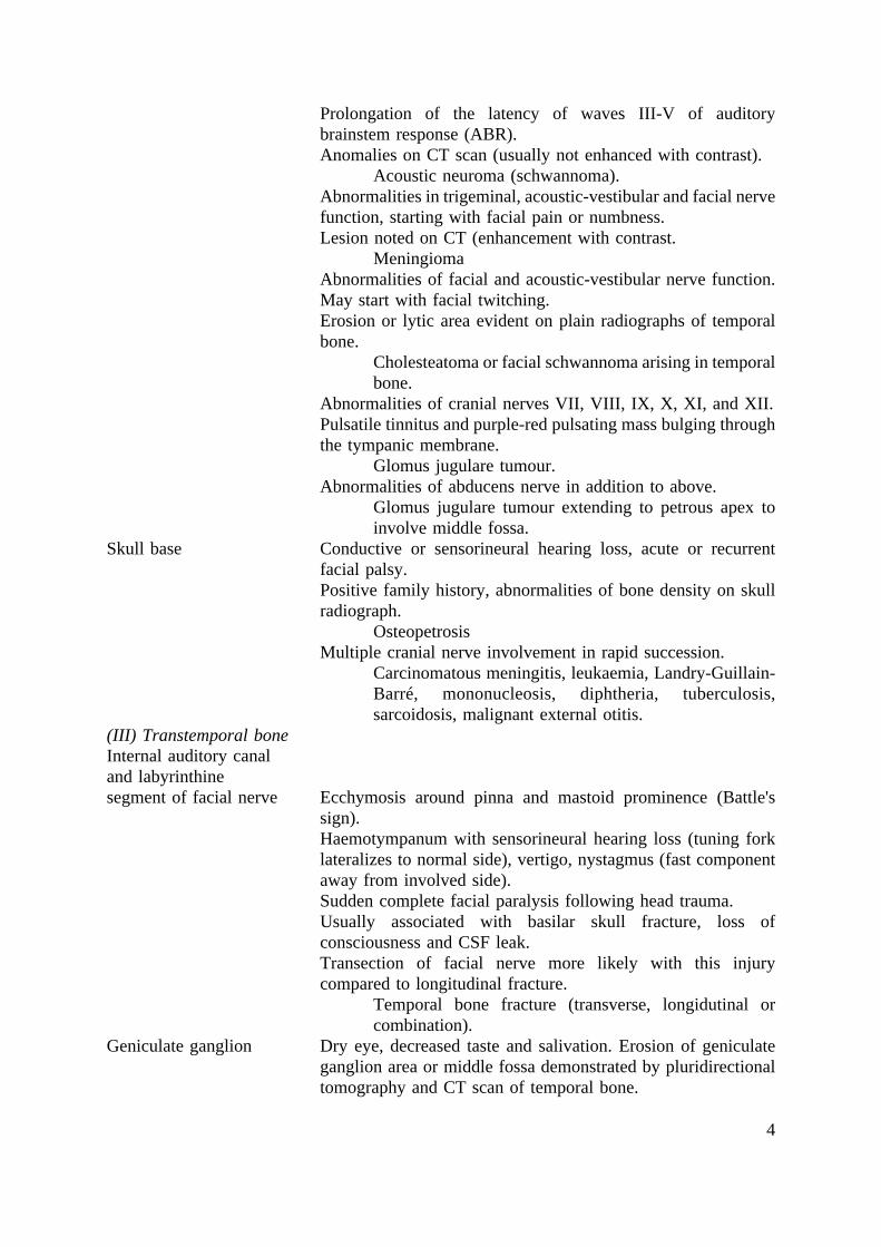
Prolongation of the latency of waves III-V of auditorybrainstem response (ABR).Anomalies on CT scan (usually not enhanced with contrast).
Acoustic neuroma (schwannoma).Abnormalities in trigeminal, acoustic-vestibular and facial nervefunction, starting with facial pain or numbness.Lesion noted on CT (enhancement with contrast.
MeningiomaAbnormalities of facial and acoustic-vestibular nerve function.May start with facial twitching.Erosion or lytic area evident on plain radiographs of temporalbone.
Cholesteatoma or facial schwannoma arising in temporalbone.
Abnormalities of cranial nerves VII, VIII, IX, X, XI, and XII.Pulsatile tinnitus and purple-red pulsating mass bulging throughthe tympanic membrane.
Glomus jugulare tumour.Abnormalities of abducens nerve in addition to above.
Glomus jugulare tumour extending to petrous apex toinvolve middle fossa.
Skull base Conductive or sensorineural hearing loss, acute or recurrentfacial palsy.Positive family history, abnormalities of bone density on skullradiograph.
OsteopetrosisMultiple cranial nerve involvement in rapid succession.
Carcinomatous meningitis, leukaemia, Landry-Guillain-Barré, mononucleosis, diphtheria, tuberculosis,sarcoidosis, malignant external otitis.
(III) Transtemporal boneInternal auditory canaland labyrinthinesegment of facial nerve Ecchymosis around pinna and mastoid prominence (Battle's
sign).Haemotympanum with sensorineural hearing loss (tuning forklateralizes to normal side), vertigo, nystagmus (fast componentaway from involved side).Sudden complete facial paralysis following head trauma.Usually associated with basilar skull fracture, loss ofconsciousness and CSF leak.Transection of facial nerve more likely with this injurycompared to longitudinal fracture.
Temporal bone fracture (transverse, longidutinal orcombination).
Geniculate ganglion Dry eye, decreased taste and salivation. Erosion of geniculateganglion area or middle fossa demonstrated by pluridirectionaltomography and CT scan of temporal bone.
5
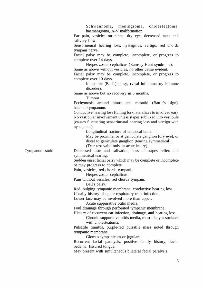
Schwannoma, meningioma, cholesteatoma,haemangioma, A-V malformation.
Ear pain, vesicles on pinna, dry eye, decreased taste andsalivary flow.Sensorineural hearing loss, nystagmus, vertigo, red chordatympani nerve.Facial palsy may be complete, incomplete, or progress tocomplete over 14 days.
Herpes zoster cephalicus (Ramsay Hunt syndrome).Same as above without vesicles, no other cause evident.Facial palsy may be complete, incomplete, or progress tocomplete over 10 days.
Idiopathic (Bell's) palsy, (viral inflammatory immunedisorder).
Same as above but no recovery in 6 months.Tumour
Ecchymosis around pinna and mastoid (Battle's sign),haematotympanum.Conductive hearing loss (tuning fork lateralizes to involved ear).No vestibular involvement unless stapes subluxed into vestibule(causes fluctuating sensorineural hearing loss and vertigo withnystagmus).
Longitudinal fracture of temporal bone.May be proximal or at geniculate ganglion (dry eye), ordistal to geniculate ganglion (tearing symmetrical).(Tear test valid only in acute injury).
Tympanomastoid Decreased taste and salivation, loss of stapes reflex andsymmetrical tearing.Sudden onset facial palsy which may be complete or incompleteor may progress to complete.Pain, vesicles, red chorda tympani.
Herpes zoster cephalicus.Pain without vesicles, red chorda tympani.
Bell's palsy.Red, bulging tympanic membrane, conductive hearing loss.Usually history of upper respiratory tract infection.Lower face may be involved more than upper.
Acute suppurative otitis media.Foul drainage through perforated tympanic membrane.History of recurrent ear infection, drainage, and hearing loss.
Chronic suppurative otitis media, most likely associatedwith cholesteatoma.
Pulsatile tinnitus, purple-red pulsatile mass noted throughtympanic membrane.
Glomus tympanicum or jugulare.Recurrent facial paralysis, positive family history, facialoedema, fissured tongue.May present with simultaneous bilateral facial paralysis.

6
Melkersson-Rosenthal syndrome.(IV) Extracranial Incomplete facial nerve paresis.
Hearing, balance, tearing, stapes reflex, taste, salivary flowspared.
Penetrating wound of face; sequelae of parotid surgery;malignancy of parotid, tonsil or oronasopharynx; rarely,with benign lesion of parotid gland compressing facialnerve.
Uveitis, salivary gland enlargement, fever.Sarcoidosis (Heerrfordt's syndrome), lymphoma.
(V) Sites variable Bilateral facial paralysis from birth.Moebius' syndrome.
Bilateral facial paralysis, acquired.Landry-Guillain-Barré syndrome, sarcoidosis,mononucleosis, leukaemia, idiopathic (Bell's) palsy.
Facial paralysis, especially simultaneous bilateral facialparalysis with symmetrical ascending paralysis, decreased deeptendon reflexes, minimal sensory changes.Abnormal spinal fluid (protein and few cells, albumino-cytological dissociation).
Landry-Guillain-Barré syndrome.Deficits of cranial nerves VI and VII or VII, VI, and III,possibly in association with other neurological signs.
Carcinoma of nasopharynx, metastatic carcinoma frombreast, ovary, prostate, meningitis, leukaemia, diabetesmellitus.
(VI) Pseudobulbar palsy Inappropriate or exaggerated laughing or crying.May be associated with marked increase in jaw jerk, or gagreflex.
Polyneuritis.Toxic, viral or vascular lesion involving bilateralcorticobulbar pathways.
Cortex and internal capsule
Anatomy of the pyramidal system from the cortex to the pontine nucleus is illustrated.Facial motor nerves are represented with the forehead uppermost and the eyelids, midface,and lips located sequentially below the representation of the forehead. Note that the tracts tothe lower face are crossed while innervation to the forehead is both crossed and uncrossed.Sparing of the forehead movement is considered to be characteristic of a cortical lesion.However, it is also possible to have forehead sparing with a lesion of the pontine facialnucleus, with selective lesions within the temporal bone, or even in association with an injuryto the nerve in its distribution in the face. Since preservation of forehead function is notsufficient to make a diagnosis of a central lesion, other neurological signs must be looked for(Table 24.3).
7
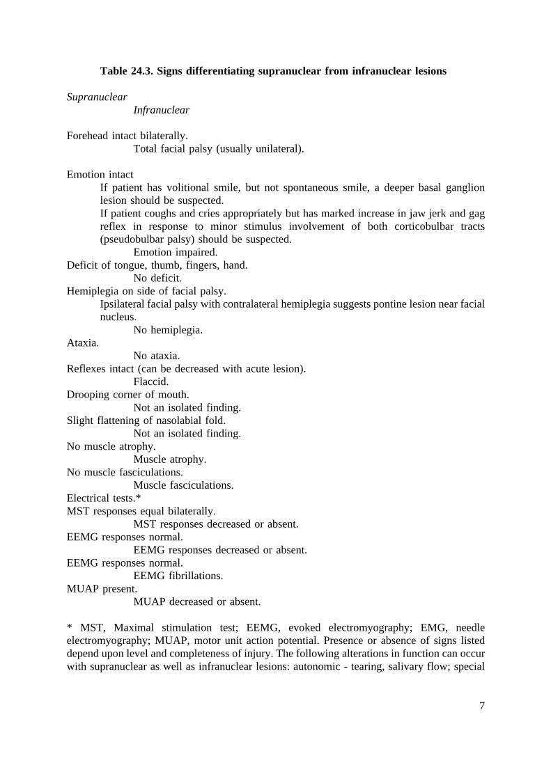
Table 24.3. Signs differentiating supranuclear from infranuclear lesions
SupranuclearInfranuclear
Forehead intact bilaterally.Total facial palsy (usually unilateral).
Emotion intactIf patient has volitional smile, but not spontaneous smile, a deeper basal ganglionlesion should be suspected.If patient coughs and cries appropriately but has marked increase in jaw jerk and gagreflex in response to minor stimulus involvement of both corticobulbar tracts(pseudobulbar palsy) should be suspected.
Emotion impaired.Deficit of tongue, thumb, fingers, hand.
No deficit.Hemiplegia on side of facial palsy.
Ipsilateral facial palsy with contralateral hemiplegia suggests pontine lesion near facialnucleus.
No hemiplegia.Ataxia.
No ataxia.Reflexes intact (can be decreased with acute lesion).
Flaccid.Drooping corner of mouth.
Not an isolated finding.Slight flattening of nasolabial fold.
Not an isolated finding.No muscle atrophy.
Muscle atrophy.No muscle fasciculations.
Muscle fasciculations.Electrical tests.*MST responses equal bilaterally.
MST responses decreased or absent.EEMG responses normal.
EEMG responses decreased or absent.EEMG responses normal.
EEMG fibrillations.MUAP present.
MUAP decreased or absent.
* MST, Maximal stimulation test; EEMG, evoked electromyography; EMG, needleelectromyography; MUAP, motor unit action potential. Presence or absence of signs listeddepend upon level and completeness of injury. The following alterations in function can occurwith supranuclear as well as infranuclear lesions: autonomic - tearing, salivary flow; special

8
sensory - taste; motor function - stapes reflex (stapes nucleus separate from pontine facialmotor nucleus).
Extrapyramidal system
The extrapyramidal system consists of the basal ganglia and the descending motorprojections other than the fibres of the pyramidal or corticospinal tracts. This system providesfor automatic associated movements and spontaneous, emotional, mimetic human faciallanguage which accompanies the more precise voluntary responses. The interplay between thepyramidal and extrapyramidal system accounts for tonus and stabilizes the motor responses.The affect of parkinsonism is known to be the result of extrapyramidal pathway destruction,and the facial dystonia of Meige's syndrome, a rare clinical entity, is thought to be due tobasal ganglion disease. The severe progressive hemifacial spasm that accompanies Meige'ssyndrome will be discussed further under central nervous system facial nerve disorders.
Emotion is another function of the extrapyramidal cortical system and is mediated bydischarges passing through the cingulate, orbital, and other frontal cortical areas and thebasolateral portion of the amygdala.
Upper midbrain
A lesion in the upper midbrain will involve the oculomotor pathways and result inipsilateral loss of direct and consensual pupillary light reflexes, ipsilateral external strabismus,and oculomotor paresis. In addition, paresis of contralateral muscles of the head and body willbe noted. This symptom complex is referred to as unilateral Weber's syndrome.
Lower midbrain
A lesion in this region that is above the facial nerve nucleus involves the tracts of theabducens and may cause contralateral paresis of the face and muscles of the extremities,ipsilateral abducens paresis, and internal strabismus. A lesion that extends far enough laterallyto include the emerging facial nerve fibres may present as peripheral ipsilateral facialparalysis associated with loss of taste and papillae on the anterior two-thirds of the tongue,and a dry eye on the same side. In addition, salivary flow from the submaxillary gland on theside of the lesion may be greatly diminished or absent.
It is important to emphasize that the peripheral topognostic tests for tearing, taste, andlacrimal flow can be altered by supranuclear lesions. However, a lesion in this region of thebrainstem would involve other neural functions as well, and would be highly unlikely toinvolve only facial function.
Pontine nucleus
The facial motor nucleus contains approximately 7000 neurons and is seated in thelower third of the pons, beneath the fourth ventricle. The neuronal processes that leave thenucleus pass around the abducens nucleus (cranial nerve VI) before emerging from thebrainstem. A peripheral seventh nerve paralysis, an internal strabismus on the same side, andinability to turn the non-paralysed eye toward the nose when asked to look toward the

9
paralysed side of the face, suggest a single lesion near the floor of the fourth ventricleinvolving the sixth and seventh cranial nerves. A lesion near the ventricle at the level of thesuperior salivary nucleus causes peripheral facial paralysis, a dry eye, paralysis of voluntarymuscles, loss of following gaze toward the side of the facial paralysis, and often vertical orrotatory nystagmus.
Cerebellopontine angle
The facial nerve emerges from the brainstem with a more slender nerve, the nerve ofWrisberg or nervus intermedius. Because of the association of the facial nerve with the nervusintermedius and the vestibuloacoustic nerve at the level of the cerebellopontine angle and inthe internal auditory canal, tearing, taste, submandibular salivary flow, and hearing andbalance may be disturbed with a facial nerve lesion at this level. Large lesions filling thecerebellopontine angle may compress other cranial nerves and cause deficits of the fifthcranial nerve and later the ninth, tenth, and eleventh cranial nerves. Lesions that may occurin the area include temporal bone fractures, acoustic neuromata (schwannomata),meningiomata, primary cholesteatomata, and perhaps hyper- and hypokinetic disorders fromvascular cross-compression of cranial nerves.
Transtemporal bone portion of the facial nerve
An understanding of the gross and microscopic anatomical relationships between thefacial, acoustic, and vestibular nerves, described by Silverstein and Norrell (1980), is essentialfor performing a retrolabyrinthine vestibular neurectomy. The intracranial segment of thefacial nerve from the brainstem to the fundus of the internal acoustic meatus is covered onlyby a thin layer of glia, which makes it quite vulnerable to any type of surgical manipulationbut also quite resistant to a slow process of stretching or compression. Thus, the facial nervein this region can become quite elongated and spread out over the surface of a sizeable butslow-growing vestibular nerve schwannoma without any gross evidence of facial weakness.
Fallopian canal
The course of the facial nerve through the fallopian canal is unique. No other nervein the body covers such a long distance through a bony canal. The nerve is also remarkablefor the Z shape of its infratemporal portion, in that it has a ganglion, and that the length ofits course is 28-30 mm. The nerve in the fallopian canal can be divided into three segments:labyrinthine, tympanic, and mastoid. The labyrinthine segment is the thinnest part of the facialnerve within the fallopian canal. The narrowest part is at its entrance, where it averages 0.68mm in diameter (Fisch and Esslen, 1972). Fisch (1977) feels that this bottleneck at theentrance of the fallopian canal predisposes the nerve to strangulation in cases of oedematousswelling. The observation is supported by post-mortem findings reported by Fowler (1963)and by Proctor, Corgill and Proud (1976). The blood supply to the nerve in this region isunique; this is the only segment of the facial nerve in which there are no anastomosingarterial arcades.
The labyrinthine segment of the facial nerve includes the geniculate ganglion. Thesomatosensory (pain), and special sensory (taste) fibres are afferent fibres that synapse in thegeniculate ganglion, while the autonomic secretomotor fibres to the lacrimal gland pass
10
through the geniculate ganglion and form the first branch of the facial nerve, the greaterpetrosal nerve. The secretory fibres to the parotid gland are carried with the ninth cranialnerve. They travel through the tympanic plexus and form the lesser petrosal nerve. There arecommunications with the nervus intermedius, which provides an alternate route for theparasympathetic fibres to reach the parotid, thus bypassing the tympanic plexus and the ninthcranial nerve branch of Jacobson. This might explain why sectioning Jacobson's nerve, inmany cases, have little effect on parotid salivary flow.
In the region of the geniculate ganglion there are ample alternative pathways andconnections for parasympathetic fibres to reach their terminations. Such alternative pathwaysexplain how lacrimal flow may be unaffected by slow-growing lesions at or proximal to thegeniculate ganglion, and the spontaneous recovery of tearing following resection of thegeniculate ganglion or nervus intermedius, such as might occur with posterior fossa surgery.The geniculate ganglion lacks a bony covering in approximately 15% of temporal bones, anarrangement which makes the facial nerve quite vulnerable to injury during surgery involvingthe middle cranial fossa, especially in children. Further, the bone of the tegmen tympani andmiddle fossa plate over this region may be quite thin.
In the author's experience with temporal bone fractures, this is the area of the facialnerve most often compressed by crushed, thin, bony fragments. The change in direction takenby the facial nerve at the genu is another reason why this site is the most common focus ofinjury when severe traction is applied to the nerve along the axis of its tympanic segment, asmay occur in longitudinal fracture of the petrous pyramid. The fact that the arachnoid piamater extends to the geniculate ganglion, as well as the complex embryological developmentof this portion of the nerve, may explain why this area of the facial nerve is so often the siteof primary cholesteatomata, vascular malformations, meningiomata, and schwannomata (Fisch,1977).
The geniculate ganglion marks the proximal end of the tympanic portion, and from thispoint the nerve courses 3-5 mm, before passing just behind the cochleariform process and thetensor tympani tendon. The cochleariform process is a useful landmark to find the facial nervewhen other landmarks are obscured by granulation tissue or cholesteatoma, or in cases oftrauma. The entire tympanic segment is approximately 8-11 mm long and the tympanic wallof this part of the fallopian canal is thin and easily fractured. In addition, dehiscences occurfrequently, allowing the uncovered nerve to prolapse into the oval window niche, partly orcompletely concealing the footplate of the stapes; this makes the nerve subject to traumaduring stapes surgery. The tympanic segment is divided from the mastoid portion by thepyramidal eminence.
At this point the fallopian aqueduct makes another turn downward, forming the secondgenu. The latter is another area where the facial nerve is vulnerable to injury during mastoidsurgery. The distal aspect of the tympanic segment is found by the surgeon through themastoid approach by entering the suprapyramidal recess (retrofacial recess). Here, the facialnerve is lateral and distal to the pyramidal process. In the presence of chronic infection, caremust be taken not to confuse a pathological dehiscence of the facial nerve in this region witha mound of granulation tissue. The best way to avoid this is to identify the nerve proximaland distal to the area that looks suspicious. The second genu, which marks the beginning ofthe mastoid segment, is lateral and posterior to the pyramidal process, which houses the
11
stapedius muscle that lies on the deep side of the facial nerve; this explains the fact that thefacial nerve lies lateral to the pyramidal process. The nerve continues vertically down theanterior wall of the mastoid process to the stylomastoid foramen. The distance from thebeginning of the second genu to the stylomastoid foramen varies between 10 and 14 mm. Thissegment of the facial nerve has three branches:
(1) the nerve to the stapedius muscle(2) the chorda tympani nerve(3) the nerve from the auricular branch of the vagus.
The nerve to the stapedius muscle arises from small neurons within the pons, locatedoutside the main facial nerve nucleus, which interface with the rostral end of the facialnucleus and the caudal end of the lateral superior salivatory nucleus (Lyon, 1978; Joseph etal, 1985). Although Lyon (1978) studied cats and Joseph et al (1985) studied rabbits todetermine the location of the motor neurons relative to the stapedius muscle, it is quite likelythat these neurons lie in a similar location in man. If so, this may help to explain whyalterations in the middle ear reflex occur when a brainstem lesion is present. Further, theseparate nucleus for the stapedius muscle innervation provides the anatomical basis forsparing of the stapedius muscle in patients with congenital facial palsy such as Moebius'syndrome.
Surgical landmarks to identify the facial nerve
The facial nerve will usually be found just deep to the short process of the incus, ina line between the short process of the incus and the anterior extent of the digastric ridge. Thefacial nerve is thus posterior to the chorda tympani nerve and just lateral to the ampullary endof the posterior semicircular canal. Skeletonizing the posterior canal is helpful in order toavoid fenestrating this part of the labyrinth. The tympanomastoid suture line is another usefullandmark since it lies just anterior to the facial nerve and close to the course of the chordatympani nerve. The chorda tympani nerve and facial nerves are deep to this suture. The facialnerve lies anterior to the sigmoid sinus and leaves the temporal bone through the stylomastoidforamen just anterior and lateral to the sigmoid sinus, where the digastric ridge turns and runsin the direction of the stylomastoid foramen.
Facial nerve sheath
The sheath that surrounds the facial nerve through its course in the fallopian canalconsists of periosteum, epineurium, and perineurium. Although surgical decompression andopening of the perineurium of the facial nerve are controversial in the management of Bell'spalsy and herpes zoster cephalicus, opening the sheath is imperative in cases of suspectedtumour or trauma. A tumour of the facial nerve may be discovered when the sheath is opened,or a traumatic haematoma may be found compressing the nerve deep to the sheath. Finally,when the nerve has been disrupted, it is necessary to open the sheath to find the proximal anddistal ends for repair.

12
Spatial orientation
Agreement is lacking, in spite of efforts to determine it, as to whether or not the facialnerve is spatially oriented in its extra-axial course from the brainstem to the periphery, as itis in the cortex and pontine nucleus. Evidence against topographical organization of the facialnerve fibres has come from several investigators who have found that the fibres destined foreach peripheral branch are diffusely located in the facial nerve trunk (Sunderland and Cossar,1953; Harris, 1968; Sade, 1975; Thomander, Aldskogius and Grant, 1982).
Thomander, Aldskogius and Grant (1982) exposed the individual peripheral facialnerve branches to horseradish peroxidase, permitting retrograde transport of the tracer todemonstrate the location of these fibres in the cat facial nerve trunk. The study indicated thatthe fibres to each peripheral branch were diffusely arranged in the facial nerve trunk at leastas far proximally as the tympanic segment.
Gacek and Radpour (1982) studied the cross-sectional anatomy of the facial nervethrough its course in the temporal bone by making discrete lesions in the facial nerve of thecat proximal to the geniculate ganglion and documenting anterograde wallerian degeneration.Gacek and Radpour (1982) discovered degenerated myelin sheaths in all three of theperipheral branches studied, regardless of whether the lesion involved the rostral, caudal ormiddle fascicles of the facial nerve. They concluded that small fascicles of the facial nerveat the level of the internal auditory meatus carried motor fibres to all peripheral branches, andthat motor axons of the facial nerve in the cat are not topographically arranged int he facialnerve trunk, as had previously been proposed. Jannetta (1975) described 31 patients withhemifacial spasm treated by removing a vessel compressing the facial nerve in thecerebellopontine angle. In those cases where the compressing vessel was found on thecephalic aspect of the nerve, the spasm was more severe in the upper part of the face. Incases where the vessel compressed the caudal aspect of the nerve, the spasm began in thelower face in an atypical fashion. This observation lends support to the existence of spatialorientation of the nerve in its most proximal intracranial portion.
Considering all the evidence, it is likely that there is some degree of spatialorganization of facial nerve fibres, especially at the level at which the axon processes leavethe brainstem nucleus and course toward the periphery. Accepting the fact that the peripheralfacial nerve is at best only partially topographically oriented, with some axons carried withthe upper division terminating in muscle groups of the lower face and vice versa, it isunderstandable that regeneration following facial nerve injuries usually results in some degreeof mass movement and synkinesis.
Blood supply
The nerve receives its nourishment from the anterior inferior cerebellar artery, whichenters the internal auditory meatus in close association with the seventh and eighth cranialnerves, the petrosal branch of the middle meningeal artery which runs along with the greaterpetrosal nerve, and the stylomastoid branch of the postauricular artery, which enters the facialcanal at the stylomastoid foramen. The territories supplied by the three arteries tend to overlapat any given level. As mentioned previously, the anastomosis between the arterial systems isimmediately proximal to the geniculate ganglion, making this segment of the facial nerve

13
vulnerable to ischaemia from oedema. This might have bearing on the pathogenesis of facialparalysis following embolization of the middle meningeal artery (Metson and Hanson, 1983).
Extracranial segment of the facial nerve
The facial nerve leaves the fallopian canal at the stylomastoid foramen. In newbornsand in children up to 2 years of age, the facial nerve as it exits the skull is just deep to thesubcutaneous tissue underlying the skin. After 2 years of age, as the mastoid tip and tympanicring form, the facial nerve takes a deeper position and, in an adult, it may be up to 5 cmbelow the level of the skin. Beyond the age of 2 years, the facial nerve is protected by thetympanic bone, the mastoid tip, the ascending ramus of the mandible, and the fascia betweenthe parotid and cartilaginous external canal.
The position of the facial nerve in the young child must be kept in mind by theotologist and head and neck surgeon. To avoid unintentional injury to the facial nerve, apostauricular incision should be modified to avoid coursing near the junction of the tympanicring and mastoid tip, and this area should be protected by placing a finger over the area atthe time the incision is made. The surgeon is cautioned not to depend upon a nerve stimulatorto find the facial nerve in the region of the stylomastoid foramen. A muscle response may benoted in spite of the fact that the stimulator is not directly on the facial nerve, or thestimulator may give no response when on the facial nerve, if a thin layer of connective tissueis insulating the nerve. The nerve must therefore be identified by its anatomical location andappearance.
The main trunk may be identified entering the substance of the parotid and thenbifurcating into an upper and a lower division. The facial nerve passes through the parotidgland and emerges over the fascia of the masseter muscle. There are communications betweenthe upper and lower divisions in the majority of patients, and these form a variety of patterns.The rich plexus of nerve filaments that forms in the peripheral zone, just before entering theundersurface of the facial muscles, provides for free intermingling between branches carriedby the upper and lower divisions, which may explain the diffuse distribution of axons withinthe main trunk of the facial nerve throughout its course from the brainstem.
Communications of the facial nerve
There are diffuse intra-axial connections within the central nervous system and, inaddition, the facial nerve communicates with the vestibulocochlear nerve within the internalauditory meatus, with the otic ganglion and sympathetic fibres in the area of the geniculateganglion and, just before it leaves the stylomastoid foramen, with the auricular branch of thevagus nerve. Outside the stylomastoid foramen the facial nerve communicates with theglossopharyngeal nerve, the vagus nerve, the great auricular nerve, and the auriculotemporalnerve. The peripheral branches communicate behind the ear with the lesser occipital, on theface with branches of the trigeminal, and in the neck with the cervical cutaneous nerve. Theserelationships have been documented by the meticulous dissections of Bischoff (1977). Thefact that myriads of strands of the facial nerve interconnect with the fifth, seventh, eighth,ninth, tenth, eleventh, and twelfth cranial nerves, and with the cervical cutaneous nerves, mayhelp to explain the symptoms of many syndromes; head and face pain, and ear, throat,eustachian tube, and neck pain. These syndromes are extremely hard to treat when the cause

14
is malignant disease or a functional imbalance such as that which causes cluster headachesor atypical facial neuralgia. These interconnections also explain mastoid, ear, face or neckpain associated with Bell's palsy and herpes zoster cephalicus, the presence of residual facialsensation after the trigeminal nerve has been cut, preservation of taste and tearing after facialnerve severance, and the occurrence of pain with skull base cancer after resection of the fifth,seventh, ninth, or tenth cranial nerve, the first or second cervical nerve, or the nervusintermedius.
Spontaneous recovery of facial nerve function
This free intermingling fibres of the facial nerve with fibres of other neural structures(particularly the fifth cranial nerve) has been proposed as the mechanism of spontaneousreturn of facial nerve function after peripheral injury to the nerve. Although spontaneousrecovery of facial function was noted in approximately 25% of patients studied by Martin andHelsper (1957), this potential should not be relied upon for spontaneous reanimation of theface following resection of the facial nerve. There is no question that appropriate nerve repairat the earliest possible time following injury yields the best results. Nevertheless, spontaneousrecovery does occur and may play a part in some of the cases in which the results of surgicalreanimation are superior.
One other mechanism for the spontaneous recovery of facial function should bediscussed. The plasticity hypothesis was first proposed by Cajal (1894), and was discussedin detail by Kandel (1977). This hypothesis offers the most plausible explanation, not onlyfor spontaneous recovery of facial function following facial nerve sectioning, but also forrepair after interruption of infranuclear pathways. The plasticity hypothesis, according toCajal, is based on pre-existing connections between groups of cells that are reinforced bymultiplication of terminal branches of protoplasmic appendices and nerve collateral, thusbringing about functional transformations in particular systems of neurons as the result ofappropriate stimuli or their combinations.
Neuropathophysiology
Nerve injury
The facial nerve carries approximately 10 000 fibres, of which 7000 are myelinatedmotor axons that reach the facial muscles (Van Buskirk, 1945). It must be understood that nonof the various injuries and disorders involving the facial nerve causes an all-or-nothing lesion,but rather each of the fibres is capable of being spared or injured to a different degree at anyone time.
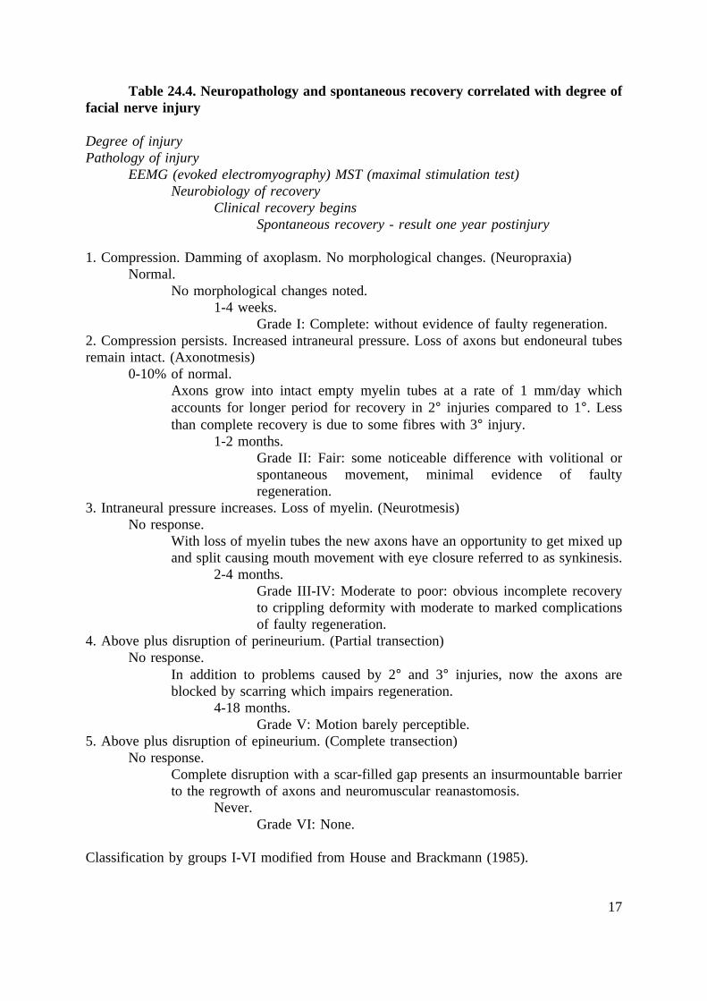
Classification of injury and recovery
Sunderland (1978) described five possible degrees of injury that a peripheral nervefibre might undergo. This classification system is depicted diagrammatically and is morecomprehensive than the classification of Seddon (1943), which described only neuropraxia,axonotmesis, and neurotmesis. The Table 24.4 shows the pathological changes that occur inthe nerve and the anticipated responses of the nerve to electrical testing, as well as the typeof recovery that might be expected with the various types of injuries. The span of possibilities

15
in terms of electrical responses, as well as recovery, reflects the possible mixtures of degreeof injury which might occur. The five degrees of injury suggested by Sunderland describevery nicely the pathophysiological events associated with all types of disorders that afflict thefacial nerve. The first three degrees of injury can occur with the viral inflammatory immunedisorders, such as Bell's palsy and herpes zoster cephalicus. The fourth and fifth degrees ofinjury occur when there is disruption of the nerve, as in transection, which might occur duringsurgery, as a result of a severe temporal bone fracture, or from a rapidly growing benign ormalignant tumour.
Fortunately, the pathological processes causing facial paralysis in patients with Bell'spalsy and herpes zoster cephalicus usually do not progress past the first or second degree ofinjury, which accounts for the fact that most individuals recover satisfactorily. A similarprocess causes facial paralysis due to acute suppurative otitis media, chronic otitis mediaassociated with a cholesteatoma, slow-growing benign neoplasms, and temporal bonefractures. In each of these disorders, the nerve is usually not transected, but rathercompressed. In acute otitis media and trauma, compression may be sudden or slowlyprogressive, evolving over 5-10 days, just as is noted with Bell's palsy and herpes zostercephalicus. However, unlike the process that occurs with Bell's palsy or herpes zostercephalicus, and these other disorders pressure is exerted on the nerve from without rather thanfrom within the intraneural space; nevertheless, the results of compression of the nerve arethe same. Eventually, axoplasm is dammed up, compression of venous drainage leads tofurther compression of the nerve and loss of axons, and eventually loss of endoneural tubeswhich leads to third-degree injury. In fourth- or fifth-degree injury, since most or all of theendoneural tubes have been disrupted, as well as the perineurium in the fourth-degree injuriesand the perineurium and epineurium in the fifth-degree injuries, recovery even under idealconditions is never as good as with the first three degrees.
Correlation of degree of injury, morphological changes in the nerve, and expectedtype of recovery.
- First degree: compression.
- Second degree: interruption of axoplasm and myelin.
- Third degree: disruption of endoneurium.
- Fourth degree: disruption of endoneurium and perineurium.
- Fifth degree: transection of nerve.
- Regeneration: as the degree of injury becomes more severe the quantity and qualityof recovery become worse.
(From May (1986)).
16
Altered function of the facial nerve following injury
Three major changes that occur in the axon following regeneration may contribute toa combination of hypo- and hyperkinesis:
(1) the distance between the nodes of Ranvier is altered(2) the newly formed axons are covered with myelin that is much thinner than the
normal axon(3) there is a splitting and crossing of axons that reinnervate denervated muscle
groups without necessarily corresponding to the cell body-motor unitarrangement that was present prior to degeneration.
As a result of these factors a tic or involuntary twitching occurs. In addition,inappropriate movement may be noted, such as movement of the mouth with blinking, orclosing of the eye with smiling. Another cause of abnormal facial movements followingregeneration may be changes that occur at the myoneural junction. In addition to these factors,it is quite likely that there are changes within and around the facial nerve nucleus in thebrainstem, as well as alterations in central connections to the cell body. The combination ofthese factors may lead to spasms that occur on the involved side of the face, causing the eyeto close and the corner of the mouth to pull. These spasms may be quite painful.
17
Table 24.4. Neuropathology and spontaneous recovery correlated with degree offacial nerve injury
Degree of injuryPathology of injury
EEMG (evoked electromyography) MST (maximal stimulation test)Neurobiology of recovery
Clinical recovery beginsSpontaneous recovery - result one year postinjury
1. Compression. Damming of axoplasm. No morphological changes. (Neuropraxia)Normal.
No morphological changes noted.1-4 weeks.
Grade I: Complete: without evidence of faulty regeneration.2. Compression persists. Increased intraneural pressure. Loss of axons but endoneural tubesremain intact. (Axonotmesis)
0-10% of normal.Axons grow into intact empty myelin tubes at a rate of 1 mm/day whichaccounts for longer period for recovery in 2° injuries compared to 1°. Lessthan complete recovery is due to some fibres with 3° injury.
1-2 months.Grade II: Fair: some noticeable difference with volitional orspontaneous movement, minimal evidence of faultyregeneration.
3. Intraneural pressure increases. Loss of myelin. (Neurotmesis)No response.
With loss of myelin tubes the new axons have an opportunity to get mixed upand split causing mouth movement with eye closure referred to as synkinesis.
2-4 months.Grade III-IV: Moderate to poor: obvious incomplete recoveryto crippling deformity with moderate to marked complicationsof faulty regeneration.
4. Above plus disruption of perineurium. (Partial transection)No response.
In addition to problems caused by 2° and 3° injuries, now the axons areblocked by scarring which impairs regeneration.
4-18 months.Grade V: Motion barely perceptible.
5. Above plus disruption of epineurium. (Complete transection)No response.
Complete disruption with a scar-filled gap presents an insurmountable barrierto the regrowth of axons and neuromuscular reanastomosis.
Never.Grade VI: None.
Classification by groups I-VI modified from House and Brackmann (1985).
18
Facial hyperkinesis may be due to another mechanism referred to as ephaptictransmission. This term describes facial hyperkinesis or a hemifacial spasm that seems tooccur spontaneously, without any discoverable cause. It is theorized that depolarization at thesite of injury acts as a stimulus to the intact portion of the fibre, and that the action potentialin one fibre is capable of exciting adjacent fibres in the area of injury. Granit, Leksell andSkoglund (1944) demonstrated ephaptic transmission at the site of compression in a nerve thatwas still capable of transmitting impulses across the site, and Kugelberg and Cobb (1951)demonstrated an acute, reversible phenomenon in the peripheral nerve of man. Afterproducing ischaemia by means of pneumatic cuffs, Kugelberg demonstrated the developmentof foci of spontaneous, repetitive, and synchronized discharges, both during the ischaemia andafter release of the cuff.
Synkinesis
This is an abnormal synchronization of movement, occurring with voluntary and reflexactivity of muscles that normally do not contract together. This phenomenon may be grosslydeforming and debilitating. In its worst form, mass movement of all parts of the involved sideof the face occurs; the patient is unable to move each part of the face separately. In itssubtlest form it may consist of no more than a tiny twitch of the chin accompanying blinkingon the side of the involvement. This may be the only sign of previous facial paralysis, andto detect it requires very close observation.
Crocodile tears
Increased unilateral lacrimation on the involved side associated with eating may occurwith a severe denervating lesion when it involves the facial nerve at or above the site of thegeniculate ganglion or along the greater petrosal nerve. This phenomenon is probably theresult of faulty regeneration of parasympathetic fibres, which innervate the lacrimal glandinstead of the salivary glands.
Stapedius tendon contraction
This is a hyperkinetic syndrome which occurs with faulty regeneration and causesfullness or roaring in the ear. The complaint is noted with facial movements and oftencoincides with facial spasm. The diagnosis can be confirmed on tympanometric recordingsemploying the electroacoustic bridge; sectioning the stapedius tendon through a tympanotomyapproach has been effective in relieving the spasm.
Hemifacial spasm
Unilateral facial nerve hyperactive dysfunction is characterized by the onset of mildintermittent spasms in the orbicularis oculi muscle that gradually increase in severity andfrequency and spread downward to include all of the muscles of facial expression, includingthe platysma. The most common cause is cross-compression from vessels in the posteriorfossa. On rare occasions this syndrome may be mimicked by benign lesions in the parotidgland (Horne, Crumley and Schindley, 1981), temporal bone (Brokeen, Pulec and Haleberg,1969; Jackson et al, 1980), or cerebellopontine angle tumours such as meningiomata,cholesteatomata, and schwannomata (May and Hardin, 1977). The most effective treatment
19
of hemifacial spasm is vascular decompression of the nerve at its root entry zone by theretromastoid approach (Jannetta et al, 1977).
Facial myokymia
A continuous fine fibrillary or undulating movement of the facial muscles gives theface an appearance suggesting a 'bag of worms'. This condition has been associated withmultiple sclerosis and intrinsic tumours of the brainstem.
Blepharospasm
Involuntary spasmodic eye closure may start on one side but classically it is a bilateraldisorder. It is characterized by symmetry and the electromyogram shows that individualcontractions are asynchronized. This condition has not been noted in children. Treatmentinvolves selective neurolysis or myolysis or, more recently, injections of botulinum A toxin(Biglan, May and Walden, 1986).
Psychogenic or habit tic
This condition is usually noted in children. The movements are repetitive and mayinvolve muscles outside the distribution of the seventh nerve. There is a compulsion toperform facial movements and they are under voluntary control. These movements are notobserved during sleep, as are the movements of hemifacial spasm, facial myokymia, andblepharospasm. Psychological evaluation and treatment are indicated for this disorder.
Focal cortical seizures
These movements involve the face and are usually tonic, often spreading beyond thedistribution of the seventh nerve. After a seizure, there may be transient postictal facialparalysis of the supranuclear type that spares the forehead muscles. The results ofelectroencephalographic recording during a seizure are diagnostic of this condition.
Evaluation of facial nerve function
Differential diagnosis
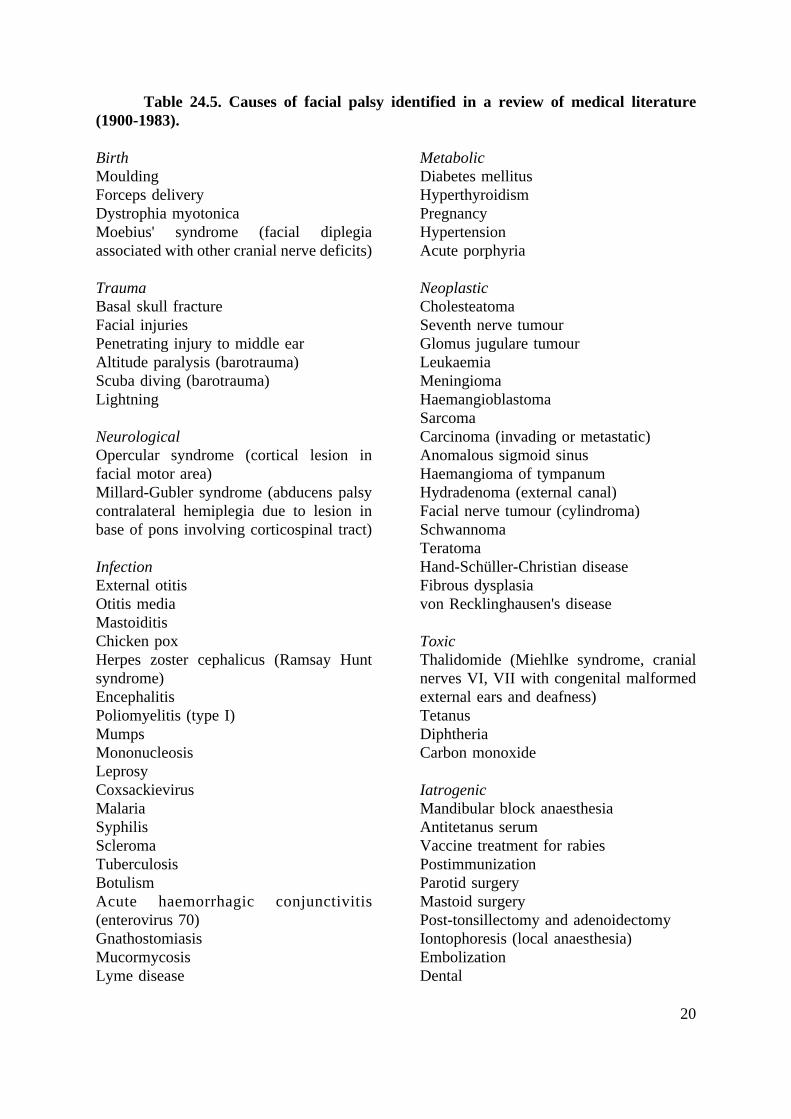
Peripheral facial paralysis is a diagnostic challenge. Every effort must be made todetermine the aetiology, since often a treatable cause can be found. The differential diagnosticpossibilities are numerous (Table 24.5). However, diagnostic clues are obtained from acarefully taken history, from the findings upon physical examination, and from the results ofspecial tests (Tables 24.6 and 24.7). The relative incidence of the variety of causes in theauthor's experience can be noted in Table 24.8. In spite of the fact that, in the majority ofpatients, a cause cannot be found and their condition is labelled idiopathic (Bell's palsy), theclinician must not be discouraged from taking the time required to make an accuratediagnosis, since without this approach a treatable, progressive, or life-threatening disorder maybe overlooked. It must be emphasized that Bell's palsy is a diagnosis by exclusion (Table24.9).
20
Table 24.5. Causes of facial palsy identified in a review of medical literature(1900-1983).
BirthMouldingForceps deliveryDystrophia myotonicaMoebius' syndrome (facial diplegiaassociated with other cranial nerve deficits)
TraumaBasal skull fractureFacial injuriesPenetrating injury to middle earAltitude paralysis (barotrauma)Scuba diving (barotrauma)Lightning
NeurologicalOpercular syndrome (cortical lesion infacial motor area)Millard-Gubler syndrome (abducens palsycontralateral hemiplegia due to lesion inbase of pons involving corticospinal tract)
InfectionExternal otitisOtitis mediaMastoiditisChicken poxHerpes zoster cephalicus (Ramsay Huntsyndrome)EncephalitisPoliomyelitis (type I)MumpsMononucleosisLeprosyCoxsackievirusMalariaSyphilisScleromaTuberculosisBotulismAcute haemorrhagic conjunctivitis(enterovirus 70)GnathostomiasisMucormycosisLyme disease
MetabolicDiabetes mellitusHyperthyroidismPregnancyHypertensionAcute porphyria
NeoplasticCholesteatomaSeventh nerve tumourGlomus jugulare tumourLeukaemiaMeningiomaHaemangioblastomaSarcomaCarcinoma (invading or metastatic)Anomalous sigmoid sinusHaemangioma of tympanumHydradenoma (external canal)Facial nerve tumour (cylindroma)SchwannomaTeratomaHand-Schüller-Christian diseaseFibrous dysplasiavon Recklinghausen's disease
ToxicThalidomide (Miehlke syndrome, cranialnerves VI, VII with congenital malformedexternal ears and deafness)TetanusDiphtheriaCarbon monoxide
IatrogenicMandibular block anaesthesiaAntitetanus serumVaccine treatment for rabiesPostimmunizationParotid surgeryMastoid surgeryPost-tonsillectomy and adenoidectomyIontophoresis (local anaesthesia)EmbolizationDental

21
IdiopathicBell's, familialMelkersson-Rosenthal syndrome (recurrentalternating facial palsy, furrowed tongue,faciolabial oedema)Hereditary hypertrophic neuropathy(Charcot-Marie-Tooth disease, Déjérine-Sottas disease)Autoimmune syndrome
Temporal arteritisThrombotic thrombocytopenic purpuraPolyarteritis nodosaLandry-Gui l la in-Barré syndrome(ascending paralysis)Multiple sclerosisMyasthenia gravisSarcoidosis (Heerfordt syndrome -uveoparotid fever)Osteopetrosis.
History
The type of onset of facial palsy is not diagnostic, whether incomplete, complete,sudden, or delayed. All of these patterns of onset have been noted with idiopathic (Bell's)palsy, as well as with other conditions in which the facial nerve may be compressed orinvaded within its anatomical course from the brainstem to the parotid. These other conditionsinclude herpes zoster cephalicus, temporal bone fractures, parotid or otological surgery,infections, and neoplasms. However, the type of onset may have prognostic significance.Complete recovery will most likely occur in cases of incomplete palsy that do not progressto complete palsy. The exception is the patient who does not begin to recover in 3-6 weeksor if the paresis progresses for more than 3 weeks; in such cases a tumour must be consideredas the underlying cause. Although slow progression beyond 3 weeks is diagnostic of a tumour,progression that occurs within the first 10 days of onset has been noted with idiopathic(Bell's) palsy, herpes zoster cephalicus, external blunt trauma, and surgical trauma to the facialnerve within the parotid, temporal bone, or posterior fossa.
Half of the patients with Bell's palsy present with a sudden complete onset of facialparalysis. In spite of this, it is not diagnostic of Bell's palsy since the onset was noted to besudden and complete in 40% of patients with confirmed tumours involving the facial nerve.In half of these patients the tumour was malignant. A sudden complete onset associated withtrauma may indicate that the facial nerve has been transected, while a history of a delayedonset or a slowly progressive onset would rule out nerve transection.
Facial paralysis has been noted to recur with idiopathic (Bell's) palsy, Melkersson-Rosenthal syndrome, and tumours. The incidence of recurrence in the author's experience withBell's palsy was 12%, with 36% on the same side and 64% on the opposite side. Theincidence of patients with idiopathic (Bell's) palsy who had ipsilateral recurrence was 4%. Ofthe total number of patients in this study who had ipsilateral recurrent facial palsy, 17% hadtumours. Thus, the onset of facial palsy is not, of itself, diagnostic; tumours, like Bell's palsy,can present with incomplete, complete, sudden, delayed, or recurrent ipsilateral peripheralfacial palsy.
In contrast to recurrent facial paralysis on the same side, recurrence involving theopposite side is almost always diagnostic of idiopathic (Bell's) palsy, since alternatingrecurrent facial paralysis has been noted only rarely with other disorders.

22
Melkersson-Rosenthal syndrome is the most common example of a rare disorder thatis characterized by recurrent alternating facial palsy. This syndrome is characterized by:
(1) recurrent alternating facial palsy(2) recurrent oedema of the lips, face, and eyelids(3) cheilitis(4) fissured tongue.
Most authors agree that the presence of any two of these four manifestations permitsthe diagnosis. The syndrome may be accompanied by migraine phenomena (Stevens, 1965).
Malignancies
A history of cancer, particularly involving the breast, lung, thyroid, kidney, ovary, orprostate, associated with a facial paralysis suggests that a metastatic lesion is causing thepalsy. Appropriate radiographic and laboratory studies are indicated to search for the primarysite as well as to localize the site of facial nerve involvement. In some cases, surgicalexploration of the temporal bone and extracranial course of the facial nerve is recommendedto locate the lesion.
Bilateral simultaneous palsy
Bilateral facial nerve paresis may be a medical emergency and presents a specialdiagnostic and therapeutic challenge. The therapeutic challenge is early diagnosis andappropriate treatment of a potentially progressive and life-threatening disorder. The mostcommon cause of acute simultaneous bilateral palsy in the author's series was Guillain-Barrésyndrome. Other less common causes included idiopathic (Bell's) palsy, leukaemia, bulbarpalsy, sarcoidosis, skull fracture, Moebius' syndrome, and myotonic dystrophy. Guillain-Barrésyndrome, acute leukaemia, and bulbar palsy due to rabies immunization presented as life-threatening medical problems.
Differential diagnosis of bilateral facial palsy by physical findings
Guillain-Barré syndrome
Guillan-Barré syndrome is an acute inflammatory polyradiculoneuropathy evolving asa paralytic disease of unknown cause. The characteristic pathological feature of Guillain-Barrésyndrome is a lymphocytic cellular infiltration of peripheral nerves and destruction of myelin.The major complaint is weakness with the severity of the motor weakness covering a widecontinuous spectrum from mild ataxia to total paralysis of every motor and cranial nerve. Inmost instances it is noticed first in the legs, but can begin in the arms. Tendon reflexes areabolished in the affected areas and facial diplegia is seen in at least half of the cases.Weakness can evolve to total motor paralysis and, when respiratory muscles become involved,respiratory embarrassment may lead to death. Abnormal cerebrospinal fluid findings arecharacteristic of this disorder, although in the first few days cerebrospinal fluid may benormal. After several days, the cerebrospinal fluid protein begins to rise and may becomevery high and peak at approximately 4-6 weeks after the onset of clinical symptoms. Cellsin the cerebrospinal fluid are not prominent. The absence of cells in conjunction with an

23
elevated protein level is the 'albumino-cytological dissociation' which at one time was thoughtto be characteristic of the disease. Guillain-Barré syndrome is a recognizable disease entity;its diagnosis is based on clinical, laboratory, and electrodiagnostic findings. In the author'sexperience, the prognosis for spontaneous recovery in Guillain-Barré syndrome is the sameas for idiopathic (Bell's) palsy.
Infectious mononucleosis
Infectious mononucleosis is characterized by fluctuating fever, sore throat, andlymphadenopathy. Uncommonly, unilateral, recurrent, and simultaneous bilateral facialparalysis has been caused by this disorder. The syndrome of infectious mononucleosis, causedby Epstein-Barr virus, has a classical presentation and can often be diagnosed on clinicalgrounds. The prodrome lasts from 3 to 5 days, and consists of headache, malaise, myalgia,and fatigue. Sore throat occurs in the first week and is the most common feature of infectiousmononucleosis. A greyish-white exudative tonsillitis is practically pathognomonic, persists for7-10 days, and is present in approximately 50% of cases. Petechiae located near the borderof the hard and soft palates are observed in about one-third of patients towards the end of thefirst week of illness. Lymph node enlargement is a hallmark of infectious mononucleosis. Theonset is gradual, and anterior and posterior cervical lymph node chains are the mostcommonly involved. Infectious mononucleosis resembles a number of febrile disorderscharacterized by fever, sore throat, adenopathy, and lymphocytosis. It may be difficult todistinguish from the early stages of other forms of febrile exudative pharyngotonsillitis, suchas streptococcal infections, and exudative tonsillitis of viral aetiology. The differentiationdepends upon the results of throat cultures as well as haematological and serological featurescharacteristic of infectious mononucleosis.
Sarcoidosis
A patient presenting with bilateral facial paralysis and uveitis should be suspected ofhaving sarcoidosis. Sarcoidosis is a granulomatous disease of undetermined origin thatinvolves multiple systems. Although there is no single laboratory test that is absolutelydiagnostic, sarcoidosis is characterized by an elevation in serum and urinary calcium levels,an increase in serum globulin, and an elevated serum angiotensin-converting enzyme level.A chest X-ray may demonstrate hilar adenopathy or diffuse pulmonary infiltrates, andexamination of the eye grounds may indicate uveitis, supporting the diagnosis of sarcoidosis.The diagnosis is made on the basis of clinical findings together with biopsy of tissue involvedby the sarcoid. Such tissue will contain a non-caseating granuloma with giant cells. Facialpalsy is the most commonly seen clinical neurological deficit to accompany sarcoidosis.Uveitis occurs four times more commonly in patients with neurological symptoms than inthose without. The peripheral neuropathy associated with sarcoidosis has been shown to bedue to perineural inflammatory changes, with the nerve fibres themselves undamaged. Thismight account for the favourable prognosis with steroid therapy.
Lyme disease
Lyme disease has also been reported to cause bilateral facial paralysis (Clark et al,1985). This disease is characterized by erythema chronicum migrans, tick-bornemeningopolyneuritis, myocardial conduction abnormalities, and Lyme arthritis. The disorder

24
was first recognized in 1975 by close geographical clustering of children with arthritis in thesmall community of Lyme, Connecticut. This spirochaete disorder is transmitted by anarthropod vector. The disease should be suspected if the patient has been along the north-eastern coast in the USA, in the mid-west (Wisconsin and Minnesota), or in California orOregon during the summer or early autumn months. These are the geographical locationswhere the tick vector is found. People are most likely to be out of doors and thus exposed toa tick bite in the warmer months of the year. This disorder has been recognized in Europe andAustralia as well. In Europe, the disease complex is referred to as Bannwarth's syndrome.
The disease is characterized by a skin lesion that begins as a red macule or papule andexpands to form a large red ring with partial central clearing. This lesion typically lasts about3 weeks. Associated symptoms include malaise, fatigue, chills and fever, headache, stiff neck,backache, myalgias, nausea, vomiting, and sore throat. Some patients may develop a spectrumof neurological symptoms. The diagnosis can be confirmed by sending a blood sample forserological examination to detect characteristic cryoglobulins and circulating immunecomplexes. In the report by Clark et al (1985), the incidence of facial palsy was over 10%of all patients with Lyme disease and one-quarter of these patients had bilateral paralysis. Theprognosis for recovery was excellent. Only one of the 124 palsies in this series had significantsequelae. Tetracycline is considered the drug of choice, with penicillin and erythromycin asacceptable alternatives. The antibiotic therapy is directed at concurrent symptoms and toprevent serious late complications of Lyme disease. The antibiotics did not alter the courseof the paralysis.
Idiopathic (Bell's) palsy
One must consider a diagnosis of idiopathic (Bell's) palsy for those patients in whomno cause of facial palsy can be found. If vesicles are present, herpetic neuropathy may be thecause. Other physical findings which may help to define the cause of facial palsy as Bell'spalsy include the presence of a red chorda tympani nerve or vascular flaring in the posteriorsuperior aspect of the tympanomeatal area, pain and numbness, hyperacusis, dizziness, lossof tearing, and taste.
Significance of special tests
Trying to localize the site of a lesion using the results of tests for tearing (Zilstorff-Pedersen, 1965), taste (Kvarup, 1958), and salivary flow (Blatt, 1965), popularized byTschiassny (1953), has been found to be of limited value when the lesion is acute and of littleor no value in long-standing facial paralysis. This is true for the prognostic value of thesetests as well, in contradistinction to a previous report by the author (May, Blumenthal andTaylor, 1981).
The lack of correlation between test results and the location of the lesion is related toa number of variables:
(1) the anatomy of the facial nerve and its branches is quite variable, allowing fora variety of alternate pathways for the axons to reach their termination

25
(2) the lesion responsible for the paralysis may not be sharply localized to aparticular level, since a lesion may affect different components of the nerve atvarious levels and with different degrees of severity
(3) recovery of the various components may occur at different times
(4) the techniques used to measure the various facial nerve functions may not becompletely reliable.
Electrical tests
Whereas tearing, salivary flow, and taste have not been useful as diagnostic andprognostic tests, the prognosis in acute facial palsy can be accurately determined by serialelectrical testing. The time course of the degree of loss of response can be plotted. Thesteeper the line within the first 10 days the poorer the prognosis. Therefore, prognosis isbased upon not only the absolute level in 10 days, but also the acceleration of the loss withinthat period of time (Fisch, 1984). The response to electrical tests has been found to be mosthelpful in the first 5 days after onset (Esslen, 1977). A study by May, Klein, and Taylor(1985) showed that, if a response to maximal stimulation or evoked electromyography(EEMG) of 25% of normal or greater is maintained up to the tenth day after onset, the patienthas a 98% chance of having a satisfactory recovery. If the response remains at 11-24% withinthe first 10 days, there is an 84% chance of having a satisfactory recovery when the responseto maximal stimulation or evoked electromyography drops to 0-10% within the first 10 days.
Reporting results - facial function recovery
A standardized, internationally acceptable system for reporting recovery of facialfunction after injury to the facial nerve has been established (House and Brackmann, 1985)(Table 24.10). From a clinical point of view, patients who fell into grades I and II wereconsidered to have a satisfactory recovery compared to those who fell into grades III and IV.The latter group was considered to have an unsatisfactory recovery. Patients with recoverygrades I or II can be separated easily from those in grades III and IV by the absence of theability to lift the eyebrow or the presence of obvious synkinesis on the involved side.
This five degrees of injury suggested by Sunderland (Table 24.4) describe very nicelythe pathological events associated with all types of disorders that afflict the facial nerve.Further, the five degrees of injury fit in very nicely with the clinical classification of recoveryreported by House and Brackmann (1985).
General management of facial palsy
Office (outpatient) medical management of acute facial palsies
Patients and their families were satisfied if answers could be provided to threequestions:
(1) What is the cause (diagnosis)?(2) When can recovery be expected (prognosis)?
26
(3) What can be done to promote recovery (treatment)?
In most patients who present with an acute facial palsy these three questions can beanswered after a thorough evaluation is performed during the initial office visit. When nospecific cause such as trauma, infection, or tumour can be identified and the patient'ssymptoms fit the picture of idiopathic (Bell's) palsy as described previously (see Table 24.6),the patient is told that the facial nerve weakness was most probably caused by a viralinflammatory immune disorder often referred to as Bell's palsy. The prospects for recoveryfrom this disorder are excellent, and the patient should be reassured that he or she has not hada stroke, and will not be permanently deformed. Next, the time and degree of likely recoveryare predicted by evaluating:
(1) the completeness of the palsy(2) the response to the maximal stimulation test or evoked electromyography(3) the time recovery first begins.
The degree of recovery can be categorized (Table 24.10) as grade I (complete, withno detectable difference between the normal and the involved side), grade II (a very subtledeficit remains), or grade III or IV (incomplete recovery marred by more or less severe signsof faulty regeneration, such as synkinesis and spasm as well as facial weakness). Almostevery patient with idiopathic (Bell's) palsy or acute facial palsy due to trauma or infectionwho maintains some facial movement beyond 14 days after onset will have a satisfactoryrecovery from this disorder (grade I or II recovery).
Nevertheless, patients must be followed carefully, both in order to document recoveryand to watch for signs of progression that indicate a worse prognosis. The prognosis in acutefacial palsy can be accurately determined by serial electrical testing, as noted previously.
Management plan
As long as patients maintain an incomplete palsy, and have been evaluated within thefirst 14 days of onset, they can be given an appointment to return in 3 weeks for furtherevaluation. However, they should be told to return sooner if the palsy progresses asdetermined by daily evaluation of facial movement. This can be accomplished by the patientstanding in front of a mirror or having a family member observe the effects of raising theeyebrows, squeezing the eyes closed, wrinkling the nose, attempting to whistle, blowing outthe cheeks, and grinning so as to show the teeth. As long as facial function does not worsen,the patient should have satisfactory return of function with no further treatment. However, ifthe patient with persistent incomplete palsy does not begin to recover in 6 weeks or theparesis worsens rather than shows improvement, a tumour should be suspected.
When a patient presents with a complete facial motor deficit one must rely upon theresponse to maximal stimulation or evoked electromyography and the time post-onset thatbeginning of facial recovery is first noted to determine prognosis and develop a managementplan. Early recovery of facial function, within the first 3 weeks, is a reliable indication thatrecovery will be satisfactory, but this prediction should be supported by electrical testsperformed every other day up to the tenth day. If facial paralysis persists and response toevoked electromyography remains above 11% of normal, the patient is re-evaluated every

27
other day up to the fourteenth day post-onset. If on the fourteenth day the response tomaximal stimulation persists or evoked electromyography remains above 11% of normal, thepatient is informed that the prognosis for early and ultimately satisfactory recovery isexcellent. On the other hand, if the response to evoked electromyography drops below 11%of normal or is lost completely within the first 14 days, then the prognosis for satisfactoryrecovery drops to 21%.
Table 24.6. Differential diagnosis of aetiology of facial palsy by history andphysical findings
Bell's palsy(1) Acute onset of unilateral facial palsy(2) Numbness or pain of ear, face, neck, ortongue (50%)(3) Viral prodroma (60%)(4) Recurrent facial palsy (12%) (ipsilateral36%, alternating 64%)(5) Positive family history (14%)(6) Loss of ipsilateral tearing and/orsubmandibular salivary flow (10%)(7) Decrease in or loss of ipsilateral stapesreflex (90%)(8) Red chorda tympani nerve (noted in40% of patients evaluated in first 10 daysin whom the chorda tympani could beseen; also noted with herpes zostercephalicus and Guillain-Barré syndrome)(9) Self-limiting and spontaneouslyremitting.
Herpes zoster cephalicus(1) Same as for Bell's, except pain morecommon and severe(2) Vesicles on pinna, face, neck, or oralcavity (100%)(3) Sensorineural hearing loss and/orvertigo (40%)
Tumour(1) Sudden complete onset similar toBell's; EEMG results abnormal (10%within 5 days)(2) Recurrent same side (17%)(3) Slowly progressive weakness beyond 3weeks (59%)(4) No recovery after 6 months(5) Twitching with paresis
(6) Mass in parotid, submandibular gland,or neck(7) Mass between ascending ramus andmastoid tip(8) Progression of other motor cranialnerve deficits(9) Some of branches of facial nervespared(10) History of cancer
Bilateral simultaneous facial palsy(1) Guillain-Barré(2) Moebius' syndrome(3) Sarcoidosis(4) Myotonic dystrophy(5) Skull trauma(6) Infectious mononucleosis(7) Cytomegalovirus(8) Acute porphyrias(9) Botulism(10) Lyme disease(11) Bell's - herpes simplex
Birth(1) Congenital diplegia (Moebius'syndrome, thalidomide toxicity)(2) Lower lip palsy (developmental)(3) Trauma(4) Tumour
TraumaSkull fracture (acute or delayed)
Infection(1) Bulbar palsy (viral meningitis,encephalitis, or immune reaction)(2) Postinfluenza, rabies, or poliomyelitisimmunization

28
(3) Infectious mononucleosis(4) Botulism(5) Tetanus(6) Syphilis(7) Malaria(8) Lyme disease(9) Herpes zoster cephalicus(10) Otitis media (acute or chronic, with orwithout cholesteatoma)(11) Leprosy
MetabolicAcute porphyria
NeoplasticAcute leukaemia
IatrogenicBilateral arterial embolizationIdiopathic(1) Guillain-Barré syndrome(2) Sarcoidosis (Heerefordt syndrome -uveoparotid fever)(3) Polyarteritis nodosa(4) Bell's palsy
Melkersson-Rosenthal syndrome(1) Recurrent alternating facial palsy(2) Fissured tongvue(3) Labial-periorbital facial oedema(4) Non-specific labial granuloma(5) Positive family history.
Once the prognosis has been established, patients are asked to return in 3 months, 6months, and finally one year for final evaluation of facial function employing the system ofHouse and Brackmann (1985). However, while waiting for recovery to begin, medicaltreatment is recommended, and precautions must be taken to prevent possible sequelae offacial nerve paralysis.
Medical treatment
There are three main types of treatment for acute facial palsy: physical,pharmacological, and psychophysical.
Physical therapy includes heat, massage, and exercises performed twice a day. Patientsare advised to wet a Turkish towel with hot water, wring it out, and place the hot towel onthe face until the towel cools. Then the patient should massage facial cream into the skinaround the eyes and mouth and over the midface for a few minutes, ideally using an electricvibrator. Finally, the patient should stand in front of a mirror and watch the face while raisingthe eyebrows, squeezing the eyes closed, wrinkling the nose, whistling, blowing out thecheeks, and grinning. Even though no facial movement may be noted, intact nerve fibres willbe activated, and the exercises will help to maintain muscle tone.
Although several medications, including steroids, have been used to treat facialparalysis, none has been shown to be efficacious.
Psychophysical modalities such as motor sensory re-education have been useful(Schram and Burres, 1984; Balliet, 1984). In the acute phase, integrated electromyographictracings of motor strength can often be displayed on an oscilloscope, offering a patientsignificant encouragement at a time when no visible movement can be seen. The course ofthe recovery can be followed since there is a relationship between the response of voluntaryeffort recorded on the oscilloscope and actual recovery. During the post-acute phase, whenrecovery has begun, the patient can benefit from a combination of strategies usingbiofeedback, working in front of a mirror, and touching the face while attempting movements.
29
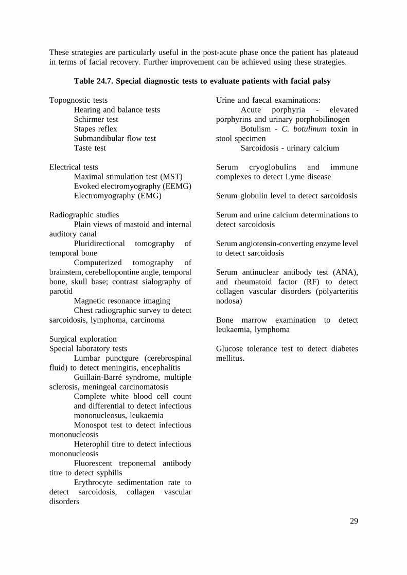
These strategies are particularly useful in the post-acute phase once the patient has plateaudin terms of facial recovery. Further improvement can be achieved using these strategies.
Table 24.7. Special diagnostic tests to evaluate patients with facial palsy
Topognostic testsHearing and balance testsSchirmer testStapes reflexSubmandibular flow testTaste test
Electrical testsMaximal stimulation test (MST)Evoked electromyography (EEMG)Electromyography (EMG)
Radiographic studiesPlain views of mastoid and internal
auditory canalPluridirectional tomography of
temporal boneComputerized tomography of
brainstem, cerebellopontine angle, temporalbone, skull base; contrast sialography ofparotid
Magnetic resonance imagingChest radiographic survey to detect
sarcoidosis, lymphoma, carcinoma
Surgical explorationSpecial laboratory tests
Lumbar punctgure (cerebrospinalfluid) to detect meningitis, encephalitis
Guillain-Barré syndrome, multiplesclerosis, meningeal carcinomatosis
Complete white blood cell countand differential to detect infectiousmononucleosus, leukaemiaMonospot test to detect infectious
mononucleosisHeterophil titre to detect infectious
mononucleosisFluorescent treponemal antibody
titre to detect syphilisErythrocyte sedimentation rate to
detect sarcoidosis, collagen vasculardisorders
Urine and faecal examinations:Acute porphyria - elevated
porphyrins and urinary porphobilinogenBotulism - C. botulinum toxin in
stool specimenSarcoidosis - urinary calcium
Serum cryoglobulins and immunecomplexes to detect Lyme disease
Serum globulin level to detect sarcoidosis
Serum and urine calcium determinations todetect sarcoidosis
Serum angiotensin-converting enzyme levelto detect sarcoidosis
Serum antinuclear antibody test (ANA),and rheumatoid factor (RF) to detectcollagen vascular disorders (polyarteritisnodosa)
Bone marrow examination to detectleukaemia, lymphoma
Glucose tolerance test to detect diabetesmellitus.

30
Depression
Patients who suddenly suffer complete facial paralysis of acute onset initially fear thatthey have a permanent deformity or have suffered a stroke. Once patients have been reassuredthat they did not have a stroke, the obvious facial deformity often leads to depression. If theprognosis is favourable for early recovery, the patient should be encouraged by this news, butif recovery is not expected for 2-4 months, the patient should be informed of this openly andsupported sympathetically. Patient counselling and group therapy have been effective inhelping patients to deal with this deformity, especially when patients are selected to be of thesame sex and similar age, and the patient counsellor has had a satisfactory recovery or learnedto deal with the problem in a positive way.
Table 24.8. Causes of facial nerve disorders in 1989 patients seen over 20 years(1963-1985) by one clinician
Cause PatientsNo %
Bell's palsy 1082 55Herpes zoster cephalicus 146 7Trauma 375 19Tumour 127 6Infection 78 4Birth 62 3Hemifacial spasm 45 2Central nervous system (axial) disease 21 1Other 41 2Tumour suspect 12 1
Total 1989 100
Physical pain
Approximately half of patients with acute idiopathic (Bell's) palsy and almost all withherpes zoster cephalicus have pain. In most cases pain can be controlled with a non-narcoticanalgesic, although in rare instances a narcotic my be required.
Eye care
Efforts should be directed towards keeping the globe moist to prevent keratitis andcorneal breakdown. The patient should voluntarily close the eyelids on the involved sidewhenever the eye feels irritated or burns, about two to four times a minute, and drops shouldbe used during the day and ointment at night. In addition, a moisture chamber should be wornover the involved eye whenever the patient is out of doors or the eye becomes irritated.Surgery to reanimate the paralysed eyelids should be considered if medical treatment isineffective, in particular when patients lack Bell's phenomenon, have corneal Anaesthesia, andlack tears or have a Dry eye - the BAD syndrome. A tarsorrhaphy should be a last resort andrevised later using one of the more effective reanimation techniques. A tarsorrhaphy produces
31
a cosmetic blight, limits vision, and often does not protect the exposed cornea. In the eventthat the tarsorrhaphy can be reversed, sequelae often result including notching of the lidmargin and trichiasis. Implantation of a gold weight or eyelid spring for the upper lid andlower lid tightening procedures have been so effective that a tarsorrhaphy is rarely indicated.
Table 24.9. Bell's palsy - diagnosis of exclusion
The palsy is not Bell's if one of the following is presentSigns of tumourBilateral simultaneous palsyVesiclesInvolvement of multiple motor cranial nervesHistory and findings of traumaEar infectionSigns of central nervous system lesionFacial palsy noted at birthTriad of infectious mononucleosis (fever, sore throat, cervical lymphadenopathy).
Surgical management of facial nerve paralysis
Indications for surgery
The benefit of facial nerve surgical decompression through the transmastoid or middlefossa route has not been established for idiopathic (Bell's) palsy, or herpes zoster cephalicus.Further, surgical decompression for acute suppurative otitis media, necrotizing external otitis,or facial paralysis following iatrogenic or external temporal bone trauma is indicated only inselected cases (Maiman et al, 1985). However, facial paralysis due to an ongoing process suchas chronic suppurative otitis media with or without cholesteatoma can only be relieved byeradicating the primary process. It should be performed prior to electrical denervation to givethe most satisfactory facial function recovery, and must not be delayed if the palsy hasprogressed from incomplete to complete over a period of hours or days and if the responseto evolved electromyography is less than 25% of normal or dropping precipitously after thethird day following onset.
In addition, there are two situations where surgery is absolutely indicated in managingfacial nerve disorders: facial nerve transection and tumour infiltration. Further, there are timeswhen nerve transection or tumour infiltration can only be established by surgical exploration,in particular when the temporal bone has fractured or a tumour is suspected.
Technique of transmastoid surgical exploration of facial nerve -labyrinthine segment to stylomastoid foramen
The postaural approach offers direct access to the tympanomastoid, geniculate, anddistal labyrinthine segments of the facial nerve. The technique for this approach preserveshearing. When exposure of the geniculate ganglion and labyrinthine segment is required andpreservation of hearing need not be considered, the translabyrinthine route is preferred.

32
Table 24.10. Classification system for reporting results of recovery from facialparalysis
Degree of injury Grade Definition
Normal (1°) I Normal symmetrical function in all areasMild dysfunction II Slight weakness noticeable only on close (barelynoticeable) (1°-2°) inspection.
Complete eye closure with minimal effort.Slight asymmetry of smile with maximal effort.Synkinesis barely noticeable, contracture, or spasmabsent.
Moderate dysfunction III Obvious weakness, but not disfiguring.(obvious difference) (2°-3°) May not be able to lift eyebrow.
Complete eye closure and strong but asymmetricalmouth movement with maximal effort.Obvious, but not disfiguring synkinesis, massmovement or spasm.
Moderately severe dysfunction (3°) IV Obvious disfiguring weakness.Inability to lift brow.Incomplete eye closure and asymmetry of mouthwith maximal effort.Severe synkinesis, mass movement, spasm.
Severe dysfunction (3°-4°) V Motion barely perceptible.Incomplete eye closure, slight movement cornermouth.Synkinesis, contracture, and spasm usually absent.
Total paralysis VI No movement, loss of tone, no synkinesis,contracture, or spasm.
The transmastoid approach to the facial nerve consists of a preliminary mastoidectomywith removal of air cells from the antrum downward to the mastoid tip, and defining the ridgeof the digastric groove. Further, cells are removed from the antrum forward to the root of thezygoma until the upper edge of the incus and the prominence of the bony horizontal canal areidentified. Care is taken not to disturb the ossicles. The bony meatal wall, while thinned, isleft intact. The landmark for the vertical mastoid portion of the facial nerve is the posteriortip of the incus above and the anterior end of the digastric groove below. Under the operatingmicroscope, the periosteum of the digastric groove is exposed and followed forward andupward until the stylomastoid foramen is exposed. Then the bone between the foramen andthe horizontal semicircular canal is thinned with a diamond burr, used parallel to the courseof the nerve, under continuous irrigation with Ringer's solution or Tis-U-Sol to remove bonedust and blood and prevent overheating, As the nerve is approached, it begins to appearthrough the paper-thin bone as a pink streak. Brisk bleeding may be encountered from theartery that enters the fallopian canal at the pyramidal bend. With the diamond burr usedgently, the final thinning of bone over the nerve is accomplished. Thus, the fallopian canalis exposed from its tympanic portion to the stylomastoid foramen, with care being taken notto disturb the incus or to open the horizontal semicircular canal. With right and left dental
33
curettes the thinned bone covering the facial epineurium is lifted off, exposing the contentsof the fallopian canal. With magnification provided by the operating microscope, this can beaccomplished safely without injuring the nerve.
When the horizontal segment is involved, decompression is carried out through atriangle bounded by the facial nerve medially, chorda tympani and tympanic annulus laterally,and short process of the incus superiorly which provides the surgical guidelines for thisapproach. In constructing this triangular window into the facial recess, it is advisable to leavea small pillar of bone over the fossa incudis to prevent accidental brushing of the incus bythe burr, which could result in serious, irreversible acoustic trauma. When the hearing isnormal and the entire horizontal segment must be decompressed, it may be necessary todisarticulate the incus. In most cases this manoeuvre can be done quite safely through thefacial recess by gently separating the capsules of the incudostapedial and malleoincudal jointleaving the short process of the incus attached to the fossa incudis. The incus can thus berotated toward the middle ear to facilitate dissection over the proximal tympanic, geniculate,and distal labyrinthine segments. The incus can then be rotated towards the mastoid so thatthe mid-tympanic portion of the facial nerve can be dissected without concern for transmittingvibrations from the incus to the stapes into the inner ear. Following decompression, the incusis replaced in its natural anatomical relationship where it will remain providing the fossaincudis has been preserved.
Decompression of the facial nerve is completed by slitting the sheath vertically on itsposterior aspect with a disposable Beaver knife. If bleeding is troublesome it should becontrolled with Surgical. Use of electric cautery is discouraged, as even a wet-field bipolarcautery may create an unwanted injury by the transmission of heat at the site of application.If it is absolutely necessary to use the bipolar wet-field cautery, it must be done while thearea is being irrigated.
Exploration and decompression or repair for traumatic facial palsy
When facial paralysis follows trauma, either surgical or otherwise, the site of injurymust be exposed. Since this is most often in the tympanic or pyramidal portion of the nerve,it is possible to expose and decompress this segment by following the nerve into the facialrecess through the postauricular approach. In some cases it may be necessary to take downthe posterior canal wall for adequate exposure. The tympanic portion of the fallopian canalis examined under the operating microscope to determine the site and extent of injury. If theossicles are intact, the incus may need to be dislocated from the stapes and later replaced togain access to the tympanic fallopian canal, as described previously. The nerve is exposed atthe site of injury and for at least 5 mm in both directions until normal-appearing nerve isencountered. If the nerve is intact but swollen or compressed by a depressed bone fragment,the latter is removed and the sheath slit. If the nerve has been partially torn, the intact fibresare carefully preserved and the torn fibres approximated or replaced by a small free graft ifthey cannot be approximated. If the nerve is completely severed, it must be repaired byapproximation or by a free nerve graft as described later.
34
Repair of severed facial nerve by approximation
Theoretically, it might seem that regeneration of the facial nerve would be moresatisfactory across a single junction than across two junctions at either end of a free graft. Forthis reason it is tempting to reapproximate the facial nerve when there are just a fewmillimetres between the two ends. However, re-routing the facial nerve to gain length toaccomplish an end-to-end approximation in the horizontal and vertical segments by mobilizingthe proximal and distal ends is not the procedure of choice. Often, the more the nerve is freedup, the more it seems to shorten and the more the blood supply to the nerve is jeopardized.
Additional length can be obtained for end-to-end approximation if the injury to thefacial nerve is proximal to the geniculate ganglion and hearing and balance function havebeen destroyed. In this case the facial nerve van be freed from its first genu, separating thenerve from the geniculate ganglion and mobilizing the vertical and horizontal segmentsposteriorly towards the internal auditory canal. Length can also be achieved for the purposeof an end-to-end anastomosis with injuries of the facial nerve in the region of the parotidgland. However, as a general rule, it is much better to put a nerve graft between severedportions rather than to try to mobilize the ends of the nerve to accomplish an end-to-endanastomosis. Based upon the author's experience, the results were as good or better when afree graft was introduced as when a nerve was re-routed. If the ends cannot be broughttogether without tension, then a free graft must be inserted. Lack of tension at the site ofapproximation is the best guarantee that the repair will be a success.
Repair of a severed facial nerve by graft
When there is a gap between the cleanly cut ends of a severed facial nerve, so that thedistal segment of the nerve cannot be brought up to establish contact with the proximal endwithout tension, a nerve graft should be inserted. The great auricular and sural cutaneousnerves are most suitable for facial nerve grafting. The great auricular is ideal for grafts up to10 cm in length and the sural cutaneous for longer grafts.
A segment of nerve, measured so as to be slightly longer than the gap to be bridged,is removed, and its ends are cut sharply at right angles with a safety razor blade against awooden tongue depressor. The nerve graft is handled carefully to avoid pinching or othertrauma. Under a microscope, the nerve graft is carefully approximated to the distal andproximal stumps using 10-0 monofilament suture, employing the technique illustrated. Byaccomplishing a fascicular anastomosis, the graft need not be protected by covering it witha vein graft or silastic tubing. As long as the two ends of the graft lie within the temporalbone and to not extend outside the stylomastoid foramen or into the internal auditory canalsuturing has not been necessary.
Results of facial nerve graft repair
The best results were achieved when the central nerve stump was connected to theperipheral system of the facial nerve within one year of injury. When the central stump wasnot available or the time between injury and repair was between one and 2 years, theprocedure of choice was a hypoglossal-facial anastomosis. When repair was performedbetween 2-4 years after injury, the distal stump of the facial nerve was biopsied and, if it was

35
fibrotic, a muscle transposition procedure was performed. If injury had occurred more than4 years previously, or if the facial nerve and muscle system were not suitable for theprocedures just described, temporalis muscle transposition was the preferred reanimationtechnique for the mouth, and separate eye reanimation techniques were combined with mouthreanimation.
Knowing the cause of the facial paralysis may be critical in determining how best torestore function. Most facial palsies evaluated for possible rehabilitation will be the result oftrauma, either surgical or accidental. If the injury was acute and the nerve was severed, thebest results are achieved when repair is performed within 30 days of injury.
The time following onset of the paralysis must be taken into account. No irreversibleprocedure such as a nerve graft, facio-facial cross-face graft, or hypoglossal-facial anastomosisshould be undertaken while there remains the possibility of spontaneous recovery. If the facialnerve was spared following acoustic tumour surgery, it is advisable not to perform aprocedure that interrupts the integrity of the facial nerve less than 12 months from the timeof injury, in order to allow adequate time for evidence of spontaneous recovery to occur.Twelve months is a good waiting period before performing such nerve repair procedures,since it has been the author's experience that if no recovery has been noted in this period oftime spontaneous recovery of useful function is unlikely.
However, time is of the essence in achieving the best possible results when the nervehas been injured, since an eightfold decrease in axon diameter occurs over 3 months(Sunderland and Bradley, 1950b). The decrease is due to shrinkage and later gradualthickening of the collagen of the endoneurial sheath (Sunderland and Bradley, 1950b). Thissuggests that nerve repair be undertaken without delay, so that axons which regenerate earlycan grow into the collapsing tubes, thus re-inflating them and suppressing collagenizationbefore it progresses too far. In general, the sooner re-innervation begins, the better. The idealtime for nerve grafting is within 30 days and not later than one year following injury; beyondone year the results of nerve grafting by any technique have been disappointing. The bestresults with a hypoglossal-facial anastomosis were achieved when surgery was performedwithin the first 2 years after injury, although satisfactory results were noted following surgeryperformed up to 4 years after injury (Conley, 1975). Recovery has been noted with laterrepairs in cases where part of the peripheral system was spared or spontaneously regenerated.
There are a number of factors that influence results following nerve repair. Technicalflaws that might downgrade recovery include tension at the suture line, residual tumour, lackof suitable nerve ends, and the presence of infection. Other factors, such as the timing of thesurgery following injury, the cause of the injury, the site of the injury, the number ofanastomotic sites and the length of the graft, have been considered as variables influencingresults. Nevertheless, the author's experience has shown that the most important factor istiming after injury.
The first sign of returning function is improving tonus of the paralysed side of theface, actually before there is any voluntary movement. Even in a long graft from the internalauditory meatus to the extracranial segment of the facial nerve, returning motion has beendetected as early as 4 months, but it may be delayed as long as 24 months, the averageinterval being 10 months. In cases of nerve repair, maximum recovery requires 2 years and

36
improvement may continue over a period of 5 years. Under ideal conditions satisfactoryrecovery following nerve repair can be expected in over 90% of cases.
Other techniques for facial reanimation
Results following facial nerve grafting or hypoglossal nerve anastomosis can beaugmented employing reanimation techniques directed to the eye and mouth. Brow lift, goldweight or eyelid spring implantation and lower lid tightening combined with temporalismuscle transposition provide immediate eye reanimation with mouth symmetry and voluntarymovement within 3-6 weeks after the procedure. Free muscle neurovascular repair techniquesshould be reserved for cases where the techniques already mentioned are not possible. Thefree muscle techniques are still evolving and greater experience is required before preciseindications and anticipated results can be proposed.
Management of idiopathic (Bell's) palsy,herpes zoster cephalicus and other facial
nerve disorders of viral origin
Bell's palsy
Bell's palsy is a term used to designate acute peripheral facial palsy of unknown cause,although accumulating evidence supports a viral inflammatory immune mechanism. Thedisorder is self-limiting, non-progressive, non-life-threatening, spontaneously remitting, andat this time can be neither prevented nor cured. The incidence varies between 15 and 40 per100.000 population (Hauser et al, 1971; Adour et al, 1978; Peitersen, 1982).
Clinical features
Bell's palsy is characterized by a viral prodrome (60%), which is accompanied by painaround the ear (50%), facial numbness (40%), changes in taste (50%), and numbness of thetongue (20%) (May and Hardin, 1977). A positive family history was obtained in 14% ofpatients, and the syndrome recurred in 12%. Of those with a history of recurrence, the sameside was involved in 36%, while in the remaining 64% the palsy recurred on the other side.The common involvement of stapedius reflex, and salivary flow indicates that the segmentmost often involved is the tympanomastoid portion of the facial nerve (May and Hardin,1977).
Predicting outcome
By studying the results of evoked electromyography and evaluating the completenessof the palsy, the patient's prognosis for recovery of facial function can be predicted with ahigh degree of accuracy. More than 90% of patients will have a satisfactory recovery,provided the palsy is incomplete and response to evoked electromyography remains greaterthan 10% beyond the first 14 days after onset. Patients with a complete palsy and responseto evoked electromyography of 10% or less within the firs 5-10 days have an 80% chance ofan unsatisfactory recovery. It is this latter group that requires the greatest attention in termsof treatment directed toward improving the natural history of facial palsy and preventingcomplications of nerve degeneration.

37
Natural history
Peitersen (1982) studied the natural history of over 1000 patients with Bell's palsy seenover a 15-year period, and found that in 84% recovery was satisfactory; 71% recoveredwithout sequelae, and 13% had defects that were barely noticeable. The other 16% of patientshad obvious incomplete recovery of facial function, but sequelae were crippling in only 4%,and there was not a single patient who did not have some recovery. Peitersen noted that 85%of the patients in his study began to recover facial function within 3 weeks of the palsy,which in 31% was incomplete at onset. Peitersen concluded that there is a relationshipbetween the degree of injury and ultimate recovery and the time that recovery is first noted;the earlier recovery is noted, the better the prognosis for a satisfactory and speedy recovery.
Treatment for Bell's palsy is supportive, and involves eye care, pain control,reassurance, heat, massage, and facial exercises. Steroids and surgery have not been shownto alter the natural history.
Herpes zoster cephalicus (Ramsey Hunt syndrome)
Hunt first described a syndrome, now called herpes zoster cephalicus, or herpes zosteroticus, which is characterized by a viral prodrome, with severe pain in and around the ear,and vesicles involving the pinna (Hunt, 1907, 1908). In its mildest form there may not be anyneurological signs, but in a more severe form it may be accompanied by a sensorineuralhearing loss and disturbed vestibular function, and even viral encephalitis. The vesicles inRamsey Hunt syndrome may occur over the ear, face, and neck down to the shoulder, andmay also involve the tongue, larynx, or buccal mucosa. The distribution of the vesiclesdepends on which sensory afferent fibres are involved by the viral eruption, but all of thenerves that communicate with the facial nerve may be involved, including cranial nerves V,VII, VIII, IX, and X and the cervical plexus arising from cervical nerves 2, 3, and 4. The signcommon to all forms of herpes zoster cephalicus, and necessary to establish the diagnosis, isthe vesicles.
Groves (1976) has presented a comprehensive review of facial nerve disorders,including a review of the literature on the history, aetiology, and treatment of Ramsey Huntsyndrome. Herpes zoster cephalicus is similar to Bell's palsy, except that in the former thevesicles are present, and there is a higher incidence of auditory vestibular involvement,postherpetic pain, and a poorer prognoses. Response to maximal stimulation or evokedelectromyography may remain in the normal range beyond 10 days, then be lost by thefourteenth to the twenty-first day. This is in contrast to Bell's palsy, in which electricalresponse may become abnormal by the tenth day. A sensorineural hearing loss was noted in10% of patients and a decreased response to electronystagmography in 40% of patients withherpes zoster cephalicus. Bilateral vestibular suppression has been noted with this disorder,perhaps an indication of brainstem involvement.
Natural history
The natural history of herpes zoster cephalicus differs from that of Bell's palsy inseveral ways, perhaps reflecting the difference in behaviour of herpes simplex type I andvaricella-zoster viruses. Bell's palsy recurs in 12% of cases, but herpes zoster cephalicus

38
rarely recurs. In addition, the acute phase of the infection, as measured by electrical responseand progression of palsy, peaks in 5-10 days with Bell's palsy but in 10-14 days with herpeszoster cephalicus. Lastly, 84% of individuals suffering from Bell's palsy have a satisfactoryrecovery of facial function, but only 60% of those with herpes zoster cephalicus recover asatisfactory degree of facial function.
Treatment
Treatment of herpes zoster is similar to that for Bell's palsy, with the exception thatgreater attention must be devoted to control of pain and the vesicular eruption. Often narcoticsare required and a steroid antibiotic cream is effective for treating the vesicular eruption. Useof antiviral agents may hold promise, but must be subjected to a prospective, controlledrandomized study (see Chapters 13 and 17). The availability of a vaccine to prevent chickenpox has been announced but has not yet been released. Prevention of chicken pox wouldeliminate herpes zoster since it is a reactivation of chicken pox virus.
Other viral disorders
Other viruses in the herpes virus group can cause facial nerve disorders. The Epstein-Barr virus is the known cause of infectious mononucleosis, and has also been isolated in casesof Guillain-Barré syndrome. The cytomegalovirus has also been isolated in patients withGuillain-Barré syndrome, suggesting that multiple viral agents are capable of producing thisdisorder. The Melkersson-Rosenthal syndrome is still another clinical entity that maymasquerade as Bell's palsy. However, although it is possible that a viral agent may play a rolein this last disorder, there is strong evidence that it is actually a hereditary autoimmunedisorder. It is characterized by recurrent alternating facial palsy, tongue plication, facialswelling, granulomatous labial submucosal lesions, a positive family history, and migraineheadaches. The presence of two of these factors associated with recurrent alternating facialpalsy satisfies the necessary criteria for diagnosis.
Facial nerve disorders in the newborn and children
Facial disorders in children can be due to a variety of causes and should not beassumed to be of the Bell's type. Further, the type of treatment and ultimate outcome dependon early, accurate diagnosis of the cause of the palsy. The principles of managing facialparalysis in children are the same as those for adults with a few exceptions, and these willbe noted. The information presented is based on the diagnosis and management of facialparalysis in 332 patients, from newborn to 18 years, seen between 1963 and 1985. The causesof facial palsy in these children were similar to those in adults, with the exception of paralysisnoted at birth and the number of cases due to acute otitis media (Table 24.11).
Facial paralysis noted at birth
The differential diagnosis and treatment of facial paralysis in the newborn has beenreviewed by May et al (1981) and Harris et al (1983). The two main differential diagnosticpossibilities are developmental and traumatic; the factors that aid in differentiating betweenthem are listed in Table 24.12.

39
The most common finding associated with congenital facial paralysis was the presenceof one or more other anomalies. Weakness of the lower lip has particular significance in thatit may be associated with multiple congenital anomalies (Pape and Pickering, 1972).Developmental bilateral facial palsy is frequently incomplete, with the lower portion of theface usually less affected than the upper part. This distinguishes it from facial palsy due totrauma, which is rarely bilateral and in which the upper and lower parts of the face areequally involved. Bilateral immobility of the face may not be apparent at birth and may bemanifested by incomplete eyelid closure when asleep, an open mouth, and/or inability to suck.
Syndromes associated with congenital facial paralysis
Moebius' syndrome
This is a rare congenital disorder which usually includes bilateral facial palsy,unilateral or bilateral abducens palsy, anomalies of the extremities, absence of variousmuscles, particularly the pectoral group, and involvement of other cranial nerves, particularlythe last four and especially the hypoglossal.
Dystrophia myotonica
This disorder is a steadily progressive familial distal myopathy with associatedweakness of the muscles of the face, jaw, neck, and levators of the eyelids. Children withmuscular dystrophy usually present at birth with congenital facial diplegia, although withoutabducens paralysis, and only later evidence the progressive nature of the myopathy (Hansonand Rowland, 1971). Congenital facial diplegia associated with dystrophia myotonica thatappears at birth is the earliest manifestation of the disease in its severest form. UnlikeMoebius' syndrome, there is muscle wasting, particularly of the sternocleidomastoid, temporal,and facial muscles, creating an expressionless face which is so characteristic that it is referredto as the myopathic facies. Extramuscular dystrophies such as cataract, premature frontalbaldness, and testicular atrophy are also present, and the neck is usually described as swan-like. This latter defect is due to wasting of the muscles of mastication and thesternocleidomastoid muscle.
Thalidomide embryopathy
Phocomelia (seal-like limbs) has apparently been known since Babylonian times, andwas described by Ballantyne (1904), but the sudden increased incidence of this rare deformitybetween 1958 and 1962 focused attention on the disorder. Investigations discovered that thesedative thalidomide taken by the mother between the twenty-eighth and forty-second day ofpregnancy led to thalidomide embryopathy with associated arrested development of the earand abducens paralysis (Miehlke, 1965).
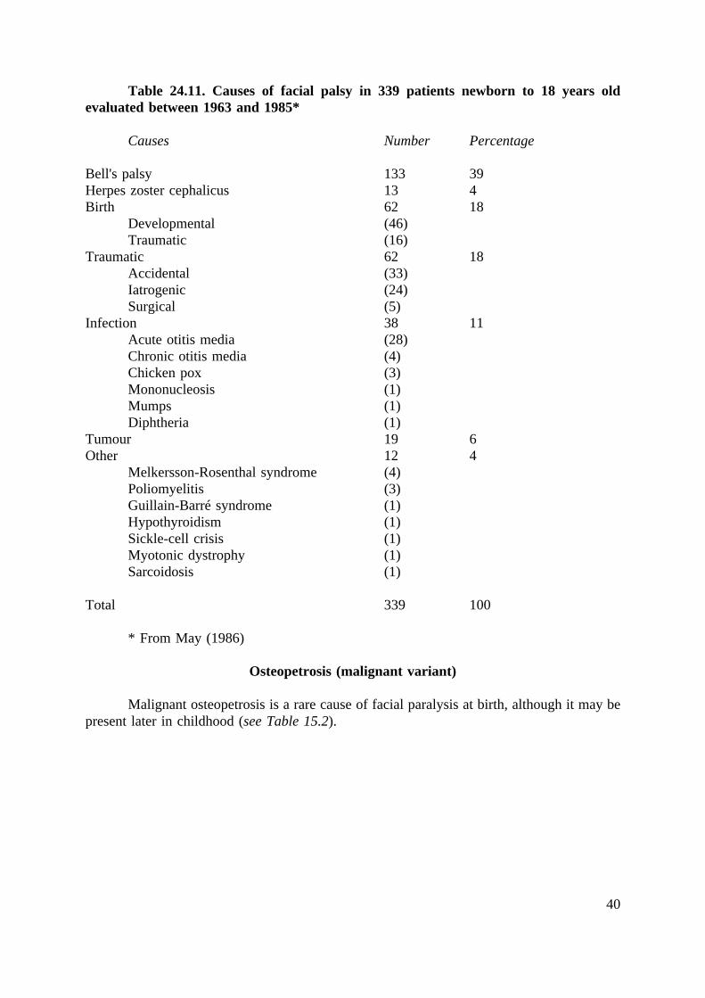
40
Table 24.11. Causes of facial palsy in 339 patients newborn to 18 years oldevaluated between 1963 and 1985*
Causes Number Percentage
Bell's palsy 133 39Herpes zoster cephalicus 13 4Birth 62 18
Developmental (46)Traumatic (16)
Traumatic 62 18Accidental (33)Iatrogenic (24)Surgical (5)
Infection 38 11Acute otitis media (28)Chronic otitis media (4)Chicken pox (3)Mononucleosis (1)Mumps (1)Diphtheria (1)
Tumour 19 6Other 12 4
Melkersson-Rosenthal syndrome (4)Poliomyelitis (3)Guillain-Barré syndrome (1)Hypothyroidism (1)Sickle-cell crisis (1)Myotonic dystrophy (1)Sarcoidosis (1)
Total 339 100
* From May (1986)
Osteopetrosis (malignant variant)
Malignant osteopetrosis is a rare cause of facial paralysis at birth, although it may bepresent later in childhood (see Table 15.2).

41
Table 24.12. Facial palsy at birth: differential diagnosis*
Developmental Traumatic
HistoryNo recovery of facial function after birth Total paralysis at birth with some
recovery noted subsequentlyfamily history of facial and other anomalies
PhysicalOther anomalies, bilateral palsy, lower lip Haemotympanum, ecchymosis, tics,or upper face palsy synkinesisOther cranial nerve deficits
Radiograph of temporal boneAnomalous external, middle, or inner ear; Fracturemandible; or vertical segments of facial nerve
Maximal stimulation / evoked electromyographyResponse decreased or absent and without Normal at birth, then decreasing to possiblechange on repeat testing loss of response
ElectromyographyReduced or absent response, no evidence of Normal at birth, then loss of spontaneousdegeneration motor units and 10-21 days later
appearance of fibrillations and giant motorunit potentials
Auditory brain stem responseAbnormality in waves III-V Normal, providing hearing is normal.
* From May (1986)
Management of developmental facial paralysis
At the present time, with the exception of free muscle neurovascular transplantationwhich is still investigational, there is no effective way to restore facial function in thenewborn or young child with facial paralysis due to a congenital anomaly. It is in the child'sbest interest to delay reanimation surgical procedures until the patient reaches his or heradolescent years. Therefore, management of the newborn or young child with a congenitalfacial paralysis should be directed towards preventing complications and performing animationtechniques which have very low morbidity. The main area of concern is the eye. Childrenwith facial paralysis from birth usually do not have problems with keratitis and cornealscarring. However, this may occur, particularly if the child has poor Bell's phenomenon,decreased tearing, or entropion with irritation of the globe from eyelashes rubbing against thecornea. The child should be evaluated periodically by an ophthalmologist and, if there is anyevidence of irritation or corneal keratitis, medical and perhaps surgical measures should beconsidered to correct the deformities.
Trauma
Facial nerve trauma can be either accidental (external trauma), surgical (unavoidableinjury during surgery), or iatrogenic (unintentional surgical injury); the incidence of each is
42
approximately equal in the author's experience. Facial paralysis has been noted followinginfiltration of local anaesthetics to the ear, face, and oral cavity. The mechanism may beeither direct infiltration of the trunk or branch of the facial nerve with the anaesthetic orprecipitation of an inflammatory immune disorder, as noted with Bell's palsy. The time ofonset after injection is most helpful from a differential diagnostic point of view. Anunresolved facial paralysis following injection is most probably related to the needlepenetrating the nerve; while when a patient recovers from the anaesthesia and then days laterdevelops a palsy, the most likely type is Bell's (idiopathic). Treatment for a patient withparalysis following local infiltration involves all of the recommendations made for Bell'spalsy. Prognostic signs include the completeness of the palsy, electrical test results, and thetime that recovery is first noted. If the injury is incomplete (first through third degree), thenthe treatment is the same as that described for Bell's palsy.
Surgical management
There is only one absolute indication for surgical exploration and repair in acutetraumatic facial paralysis: when the nerve has been transected. In cases of temporal bonefracture in the author's series, the nerve was most likely to be transected if there was markeddisplacement of the temporal bone fragments seen on the computerized tomographic (CT)scan and electrical response to stimulation was lost by the fifth day.
If it is known that the nerve has been disrupted, it should be repaired at the earliestpossible moment; if the injury occurred during surgery, the ideal time for repair is at the timeof operation. However, repair may need to be delayed if the patient's general condition isunstable or the wound is contaminated, although it should be performed within 30 days afterinjury for best results. The ideal choice is nerve repair, when possible, since this is the onlytechnique that will provide any semblance of mimetic movement. The hypoglossal-facialnerve anastomosis is the procedure of choice when direct nerve repair is not possible,providing it is done within 2-4 years following the injury. Both nerve repair and hypoglossal-facial anastomosis techniques provide useful functional recovery when applied under theappropriate circumstances. However, both may result in mass movement and the inability toseparate eye from mouth movement.
Regional reanimation techniques may be used as primary or secondary procedures torestore facial expression when the other two techniques are unsuitable, when they have failed,or when further animation is required. Although spontaneous or mimetic facial languagecannot be restored with regional reanimation techniques, they have the advantage over nerverepair and hypoglossal-facial anastomosis of:
(1) giving immediate results(2) separating eye from mouth movement(3) being independent of the cause and location of injury and between injury and
reconstruction.
By applying the principles just discussed, it is possible to restore facial function tosome degree for all patients with facial paralysis following trauma.
43
Tumours involving the facial nerve
Tumour types
Tumours of the head and neck may lie in close proximity, envelop, or invade the facialnerve as it courses from the brainstem through the temporal bone and parotid gland to reachthe facial muscles. The benign lesion most frequently seen to involve the facial nerve is aschwannoma. Half of these tumours are acoustic neuromata (schwannomata) and are locatedin the cerebellopontine angle or internal auditory canal and, in the author's series, half werefound to involve the facial nerve or chorda tympani nerve within the temporal bone. Vascularlesions such as meningiomata, angiomata, interosseous haemangiomata, and arteriovenousmalformations were the second most common types of benign tumours causing facial palsyand, with few exceptions, involved the facial nerve extraneurally at or proximal to thegeniculate ganglion. Unlike schwannomata, these lesions often could be separated from thefacial nerve with preservation of residual function.
Malignant tumours may involve the facial nerve. The most common types ofmalignancy affecting the nerve in the author's series of patients have been adenoid cystic andmucoepidermoid carcinomata, and the most common site of origin of these lesions has beenthe parotid gland.
Reanimation immediately after tumour resection
Since brow function is rarely restored by grafting of the facial nerve or cranial nervesubstitution, a brow lift is often performed immediately after resection of a tumour mass.Closure of the upper eyelid may be restored by implantation of a gold weight or a spring, andthe lower eyelid may be tightened by a Bick (lid-tightening) procedure to protect the cornea.
The mouth region can be reanimated by transposing the temporalis muscle to thecorner of the mouth. By performing this procedure in conjunction with eye reanimationprocedures, the mouth and eye can move separately, which is not always possible with nervegrafting or cranial nerve substitution techniques. An additional advantage of this techniqueis that the results are immediate.
When facial paralysis has been long-standing preoperatively, or excision of the tumourmass leaves a facial nerve not amenable to grafting or even hypoglossal-facial nerveanastomosis, the techniques described for reanimating the eye and mouth may still beperformed.
Facial paralysis in acute and chronic otitis media
In spite of the frequency of acute otitis media, particularly in children, associatedfacial paralysis is quite uncommon. A spontaneous satisfactory recovery is the usual coursefollowing treatment with an appropriate antibiotic and a myringotomy. Surgical therapy isindicated if the infection does not respond to these measures. Surgical decompression, evenwith loss of electrical response, has not been shown to improve recovery of facial function.

44
Chronic suppurative infection of the middle ear has a different natural history and doescall for immediate surgical intervention when associated with a peripheral facial paralysis.Often the pathological process involves compression of an exposed nerve by cholesteatomaor chronically infected granulation tissue. Abscess and osteitis are not unusual findings at thetime of surgery. Surgery, for the best possible recovery of facial function, should be carriedout within 24 hours, provided that the patient's general condition permits.
Hyperkinetic disorders
The variety of hyperkinetic disorders has been mentioned under faulty regeneration.The three most common are essential hemifacial spasm, blepharospasm, and hemifacial spasmof the mass type or synkinetic variety following faulty regeneration. Primary hemifacial spasmis most effectively treated by retromastoid vascular decompression of the nerve. In cases thatfail or in patients who are not candidates for an intracranial procedure, selective peripheralneurolysis may be tried, with resulting weakness. Another alternative is injections ofbotulinum A toxin. The effects of one injection last from 6 weeks to 6 months, and these mayhave to be repeated. The injections relieve the spasm, but are also associated with weakness,although this is not so marked as with selective neurolysis. Blepharospasm does not respondto retromastoid vascular decompression but has been effectively relieved by peripheralselective neurolysis, or selective resection of muscles in and around the eyelids (Anderson,1982). Botulinum A toxin injections offer significant relief and are a much more conservativeapproach (Biglan, May and Walden, 1986). Severe hemifacial spasm secondary to faultyregeneration does not respond to retromastoid vascular decompression but can be relieved tosome degree by selective myectomy, neurolysis, or injections.