FOLLOW-UP OF THE ORTHOPAEDIC TREATMENT OF DEVELOPMENTAL DYSPLASIA OF THE HIP BY SONOGRAPHY
Upload
taryn-wearingCategory
view
217download
1
1
Anatomy of the Hip
Principles of Orthopaedic and Trauma Care
December 2008Faculty of Health & Life SciencesUWE

2
Aim: The aim of this learning package is to enable you to develop knowledge and understanding of the musculoskeletal anatomy of the hip joint to underpin professional practice and to facilitate education of individuals in your care and other healthcare workers
Learning Outcomes
• To identify the musculoskeletal and neurovascular tissues comprising the hip joint
• To describe how these structures affect the joint function and movement in health and disease / deformity
• To explain the significance of the vascular supply of the femoral head in relation to Proximal Femoral Fractures

3
Resources for learning
• Please log on to the anatomy.tv website to enable identification of the relevant anatomy and to make use of their excellent supplementary information in your answers.
• Select Interactive anatomy: Primary Hip Arthroplasty, then Hip Joint model, layer 1 to answer the following slides (unless otherwise stated).

4
Hip Joint
• The largest synovial joint
• A ball & socket joint
• Enables a wide range
of movement

5
The Hip joint
Function• Movement, the joint is surrounded by a large complex musculature that
enables movement of the limb in relation to the pelvis, • Balance and stability, the pelvis is supported on the femoral heads, stability comes from the shape of the articular surfaces, capsular and ligamentous strength and the muscles, which must be capable of maintaining set positions i.e sitting and immediate controlled power, ie running upstairs• Transmission of body weight, from the vertebral column, through the pelvis to the femora when standing and onto the ischial tuberosities when sitting

6
The Hip Joint
Structure: an articulation between the head of the
femur and the acetabulum
Acetabulum:• a socket formed by three bones comprising the
innominate bone. Identify these structures from the picture,
-
-
-
Calais Germain B (1993)
7
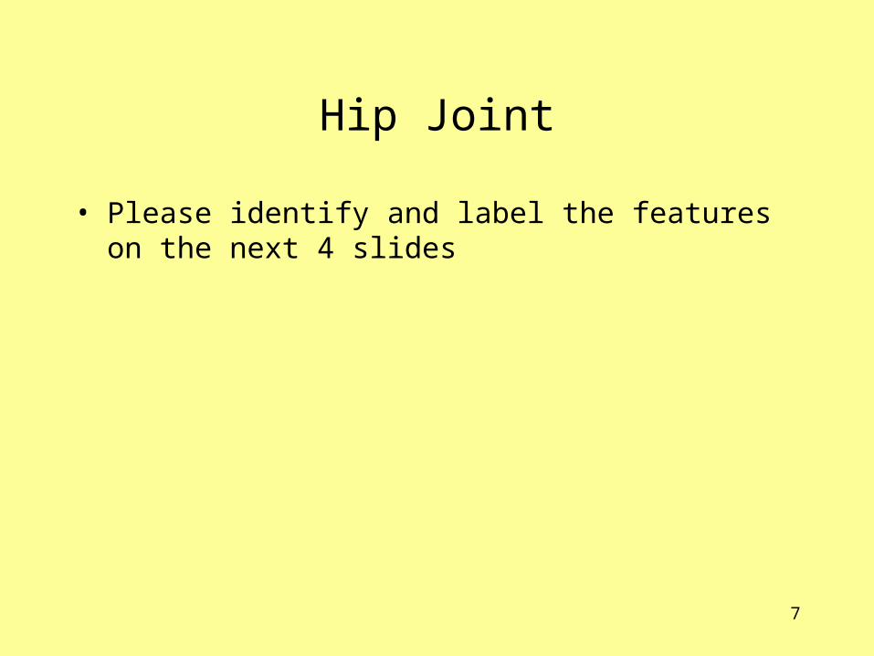
Hip Joint
• Please identify and label the features on the next 4 slides

8
Anterior Hip (layer 1)
D
C
A
B

9
Posterior Hip (layer 1)
A
B
C
D
10
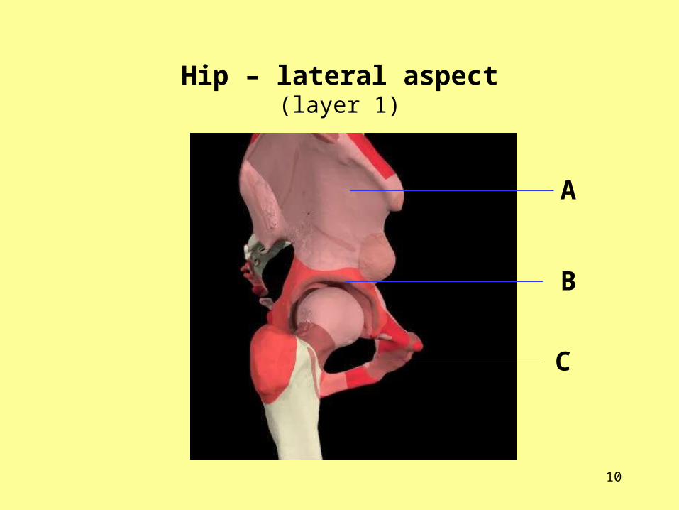
Hip – lateral aspect(layer 1)
A
B
C
11
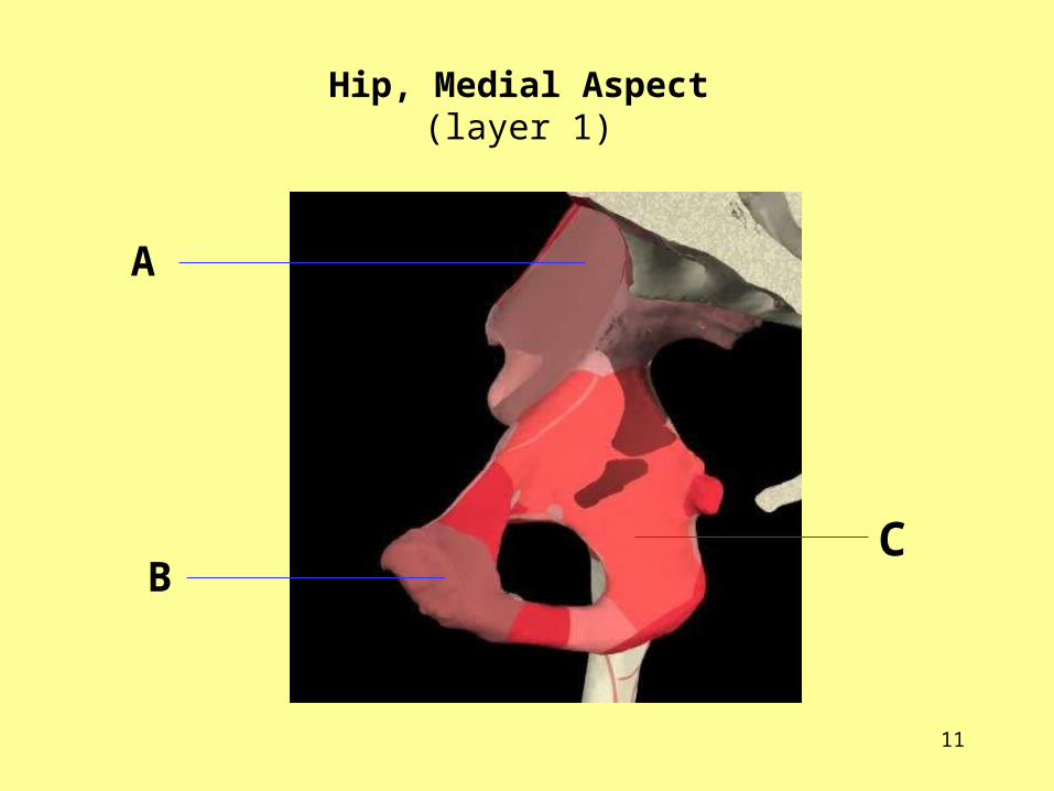
Hip, Medial Aspect(layer 1)
CB
A
12
Acetabulum • At birth, most structures are cartilaginous. At aged
10yrs, the 3 bones are separated by a tri-radiate strip of cartilage. Full ossification occurs around puberty
• Cartilage is horse shoe shaped in the acetabulum, the lower gap is bridged by the transverse ligament & is continuous with the fibrocartilage labrum which forms a rim & tight fit around the neck of femur.
• Consider the shape ? Acetabulum is the Latin term for ‘vinegar cup’
• Label structures A, B, C, D. What is their function ?

13
Acetabulum model
A
B
C
D

14
The Proximal Femur - anterior aspect (layer 1)
• Label structures A, B, C, D
• 2/3 of the surface is covered with Articular cartilage, thickest
towards centre & is thinnest at periphery, which promotes hip joint stability
• Head is obliquely set on femoral shaft
A B C
D

15
Proximal femur, medial aspect (layer 1)
Label these structures.
What connects at A ?
A

16
Questions
• What are the implications for individuals of an increase / decrease of femoral neck angles ? Read up or discuss with a clinician or physiotherapist.
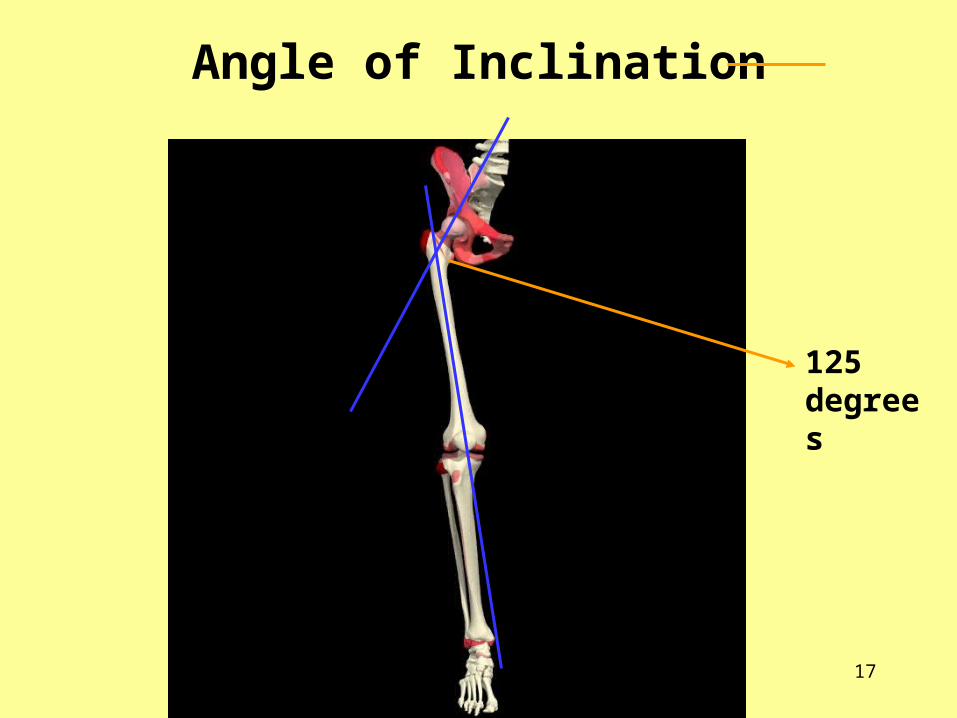
The Angle of Inclination
– angle between head of femur & shaft is approx’ 125 degrees, so
on standing with knees & ankles together, the femur is
angled / inclined towards the knee.
The Angle of Anteversion - is between the femoral neck & shaft, as seen from the view to
the head/neck along the shaft from the femoral condyles. The
neck emerges antero-medially – not at right angles.
17
Angle of Inclination
125 degrees

18
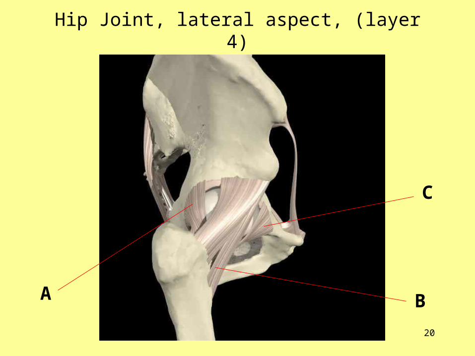
Extracapsular Ligaments
The hip joint has a fibrous capsule which is very strong,strengthened by fibres that pass around the joint in acircular way
In places the capsule is thickened to form 3 ligaments, these are referred to, by their attachments, as• Iliofemoral• Ischiofemoral• pubofemoralNB. These ligaments become taut in hip extension and relax in hip
flexion. What might be the significance of this following hip replacement?
• Label the 3 ligaments on the next two slides

19
Hip Joint, anterior aspect (layer 4)
A
B
C
20
Hip Joint, lateral aspect, (layer 4)
A B
C

21
Intracapsular Ligaments(acetabulum view)
• What is the Ligamentum Teres and where is it?
• How does it contribute to the blood supply of the femoral head?
• Is this significant ?

22
Bursae of the hip joint• What is a bursa (ae)?
• Paste this site into your browser for further information: http://orthoinfo.aaos.org/topic.cfm?topic=a00409
• Where are they in the hip joint?
• Medieval weavers repetitive movement lead some to develop Trochanteric bursitis (Weavers bottom) which they tried to avoid with heavily padded underwear.
It was noticeable enough for Shakespeare to pun on the name ‘Bottom’ for his weaver in Mid Summer nights Dream !

23
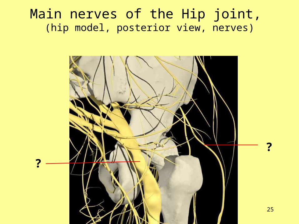
Main nerves of the Hip joint
Identify & label,
• the main nerves supplying the hip joint area in the following slides
• Consider the term sciatica - what does it mean?
• Femoral Nerve Block may be used for pain management following Proximal Femoral Fracture (PFF), discuss with the multi-professional team, how does it work?

24
Main nerves of the Hip joint, (hip model, anterior view, nerves)
?
?
?
?
25
Main nerves of the Hip joint, (hip model, posterior view, nerves)
?
?
26
Hip Joint Arterial Blood supply
Identify & label, • the main artery supplying the hip area
• the arteries that ascend the the neck of femur
• the arteries supplying the trochanters
Key Question:• What is the clinical significance of the blood supply to
the femoral head ?

27
Main arteries of the Hip joint, (hip model, anterior view, arteries)
??

28
?
?
?

29
Muscles of the hip joint
The hip joint can move in,• flexion, extension,• adduction, abduction• external rotation • internal rotation
The next slide gives a
summary of these
movements and the muscles
involved.
Use anatomy.tv to identify the muscles on slides
30 & 31. The side text gives the muscle actions

30
NB. The arrows represent the forces produced by the various muscles
Summary of movements at the hip jointCalais Germain (1993)

31
Muscles enabling hip function (Anatomy.tv: select Interactive Hip, Right Leg model, layer 10)
Anterior View Posterior View

32
Muscles enabling hip function (Anatomy.tv, select Interactive Hip, Right Leg model, layer 10)
Lateral View Medial View
33
Hip joint, Range of Movement
Enlist the help of a friend and obtain / borrow a
goniometer or a protractor.
Identify the normal range of movement demonstrated in the Hip in:• Flexion• Extension • Abduction• Adduction• Medial rotation• Lateral rotation

34
Summary:Through studying the anatomy of the hip, the student can relate hip structures to hip function and apply this knowledge and understanding in health and to pathology and deformities.
References:• www.anatomy.tv• Calais Germain B (1993) Anatomy of Movement
Seattle, Eastland Press• Palastanga N, Field D, Soames R (2006) Anatomy
and Human Movement 5th Edn. Butterworth Heinemann
.