1 Adhesion molecules and other secreted host-interaction determinants in Apicomplexa
80
1 Adhesion molecules and other secreted host-interaction determinants in Apicomplexa: insights from comparative genomics Vivek Anantharaman, Lakshminarayan. M. Iyer, S. Balaji, & L. Aravind* National Center for Biotechnology Information, National Library of Medicine, National Institutes of Health, Bethesda, MD 20894, USA *Address for correspondence LA ([email protected]) National Center for Biotechnology Information, National Library of Medicine, National Institutes of Health, Bethesda, MD 20894, United States of America Phone: 301-595-2445 Running title: Apicomplexan surface proteins
Transcript of 1 Adhesion molecules and other secreted host-interaction determinants in Apicomplexa

1
Adhesion molecules and other secreted host-interaction determinants in Apicomplexa: insights from comparative
genomics
Vivek Anantharaman, Lakshminarayan. M. Iyer, S. Balaji, & L. Aravind*
National Center for Biotechnology Information, National Library of Medicine, National Institutes of Health, Bethesda, MD 20894, USA
*Address for correspondence
LA ([email protected]) National Center for Biotechnology Information,
National Library of Medicine, National Institutes of Health,
Bethesda, MD 20894, United States of America Phone: 301-595-2445
Running title: Apicomplexan surface proteins
2
ABSTRACT .................................................................................................... 3 I. INTRODUCTION.......................................................................................... 4
A. Apicomplexa: overview of morphology, life-cycles, host and tissue range........ 5 B. Evolutionary history of Apicomplexa and implications for origins of parasitism . 7
II. COMPARATIVE GENOMICS AND APICOMPLEXAN BIOLOGY ............................... 9 A. Current state of apicomplexan genomics .................................................... 9 B. Genomic perspective on transmembrane (TM) and secreted proteins in apicomplexa and other eukaryotes ...............................................................12 C. Lineage-specific expansions and diversification of apicomplexan secreted and membrane proteins....................................................................................15
III. CONSERVED DOMAINS IN APICOMPLEXAN ADHESION AND HOST INTERACTION PROTEINS....................................................................................................18
A. Conserved domains of animal, bacterial and ancient eukaryotic provenance ...18 B. Globular domains, transmembrane and low-complexity segments of exclusively apicomplexan provenance...........................................................................24
1. Apicomplexa-specific globular domains ..................................................24 2. Apicomplexa-specific multi-TM domains .................................................27 3. Low-complexity protein families among apicomplexan surface proteins.......28
C. Secreted and cell-surface enzymatic domains in apicomplexans....................29 1. Peptidases .........................................................................................29 2. Secreted protein kinases and phosphatases ............................................30 3. Other secreted enzymes ......................................................................31 4. Membrane-embedded enzymes with possible role in survival, signaling or pathogenesis .........................................................................................32
IV. MATURATION, DEPLOYMENT AND EXPORT OF APICOMPLEXAN SURFACE PROTEINS....................................................................................................33
A. Surface protein maturation: glycosylation pathways....................................34 B. Surface protein maturation: protein processing ..........................................36 C. Surface protein deployment and connection with cytoskeleton......................38 D. Rhoptries, micronemes and protein extrusion during invasion ......................39 E. Protein export by apicomplexans during intracellular life cycle ......................42
1. The PV and the PEXEL/VTS system in Plasmodium ...................................42 2. Role of exported proteins in apicomplexan pathogenesis...........................44
V. REGULATION OF SURFACE PROTEIN GENE EXPRESSION.................................48 A. Chromatin-level controls, telomere effect and transcription regulation ...........48 B. Post-transcriptional or RNA-based regulation .............................................50
VI. GENERAL EVOLUTIONARY CONSIDERATIONS AND CONCLUSIONS ..................51 A. Evolution of parasitism and comparisons with other parasites.......................51 B. General Conclusions ...............................................................................54
REFERENCES................................................................................................62
3
ABSTRACT Apicomplexa, being an obligate parasitic clade of alveolate eukaryotes, have
developed numerous distinctive adaptations for invading and surviving within animal
cells. Our knowledge of these organisms has increased enormously as result of
genome sequencing efforts and associated large-scale physiological and biochemical
studies on taxa from different branches of the alveolate tree. Here we provide a
synthetic overview of the diversity and evolutionary history of cell-membrane-
associated, secreted and exported proteins related to apicomplexan parasitism. A
notable feature in this regard was the early acquisition of adhesion protein domains
and glycosylation systems through lateral transfer from animals. These were utilized
in multiple contexts, including invasion of host cells and parasite-specific
developmental processes. Apicomplexans possess a specialized version of the
ancestral alveolate extrusion machinery, the rhoptries and micronemes, which are
deployed in invasion and delivery of proteins into host cells. Each apicomplexan
lineage has evolved a unique spectrum of extruded proteins that modify host
molecules in diverse ways. Hematozoans, in particular, appear to have evolved novel
systems for export of proteins into the host organelles and cell membrane during
intracellular development. These exported proteins are an important aspect of the
pathogenesis of Plasmodium and Theileria, being involved in response to fever and in
leukocyte proliferation respectively. At the genome-scale, the complement of
apicomplexan surface proteins has primarily diversified via massive lineage-specific
expansions of certain protein families, which are often coded by subtelomeric gene
arrays. Many of these families have been found to central to immune evasion. At the
level of protein families, domain shuffling and accretion have resulted in adhesins
with new domain architectures. In terms of individual genes constant selective
pressures from the host immune response has resulted in extensive protein
polymorphisms and gene losses. Apicomplexans have also evolved complex
regulatory mechanisms controlling expression and maturation of surface proteins at
the chromatin, transcriptional, post-transcriptional and post-translational levels.
Evolutionary reconstruction suggests that the ancestral apicomplexan had
thrombospondin and EGF domain adhesins, which were linked to the parasite
cytoskeleton, and played a central role in invasion through formation of the moving
junction. It also suggests that the ancestral parasite had O-linked glycosylation of
surface proteins which was partially or entirely lost in hematozoan lineages.

4
I. INTRODUCTION
Apicomplexa are an extraordinary lineage of eukaryotic microorganisms that have
been recognized since the earliest microscopic observations – Leuwenhoek described
spores of Eimeria from infected rodents. To date approximately 2,400 species have
been described using microscopic observations (Levine, 1988; Vivier et al., 1990).
More recently, sampling of eukaryotic diversity via environmental sequencing of
rDNA and other phylogenetic markers suggests that traditional observations might
have considerably under-appreciated their actual diversity (Moon-van der Staay et
al., 2001). All well-studied apicomplexans are parasites of animals, and infect most
major invertebrate and vertebrate lineages. Apicomplexans are causative agents of
major human and veterinary diseases: 1) Malaria is caused by several Plasmodium
species in various mammals, including humans, reptiles and birds (Kreier, 1977). 2)
Two forms of theileriosis, namely East Coast fever and Tropical theileriosis in cattle
and water buffaloes are caused respectively by Theileria parva and T.annulata
(Dobbelaere et al., 2002).3) Babesiosis or Red-water fever is caused by Babesia
species in cattle, horses, dogs and rarely humans (Levine, 1988).4) Toxoplasma
gondii causes toxoplasmosis in several mammals including rodents, felids and
humans. In humans, especially, fetal infections can result in serious eye and brain
damage (Dubey et al., 1988). 5) Neospora caninum infects dogs, cattle and other
mammals and has been implicated as a major cause of abortion in cattle. 6)
Sarcocystis species infects muscles of various wild herbivores, such as hare and deer
thereby making them easier prey for their predators (Levine, 1988). 7) Eimeria
species infect birds, and are a major cause of morbidity in poultry. 8) Isospora is the
causative agent of an acute diarrhea in humans and other mammals and is
particularly common in immunosuppressed or young individuals. 9) Cryptosporidium
species have been implicated in gastro-intestinal disease that is a major cause of
morbidity in HIV-infected patients (Fayer, 1997). Several poorly studied
apicomplexans are also known to infect all major invertebrate lineages; most
common amongst these are gregarines which infect various organs of arthropods,
annelids and mollusks (Levine, 1988; Vivier et al., 1990). As yet uncharacterized
apicomplexans are also believed to infect unicellular foraminiferans (Kopecna et al.,
2006).
Despite a wealth of traditional morphological investigations using light and electron
microscopy, molecular studies on apicomplexa were slowed by difficulties posed by

5
complex life cycles, problems in maintaining growth or perpetuating development in
cell cultures and in several cases absence of effective genetic tools. However, the
recent avalanche of information coming from genome sequences, and associated
high-throughput proteomics and gene expression are rapidly improving our
understanding of every aspect of apicomplexan biology and providing new tools to
complement traditional approaches (Aravind et al., 2003; Roos, 2005). An area of
tremendous interest in light of the quest for anti-parasite treatments is the parasite
cell-surface and extracellular events related to host-parasite interactions (Miller et
al., 1998). Also relevant are recent developments in understanding regulatory
processes that allow stage-specific expression of particular surface proteins, as well
as the differential gene-expression mechanisms that allow immune evasion by
parasites (Ralph et al., 2005). In this article we synthesize these new data in an
evolutionary context to present an overview of proteins involved in interactions of
apicomplexan parasites with their hosts. In particular, we concentrate on
transmembrane and secreted or extruded proteins that are at the focus of the
parasite-host interface. We begin by providing a background on apicomplexan
biology, phylogenetics and the current state of genomics and use this as a scaffold to
explore developments relating to the core topics.
A. Apicomplexa: overview of morphology, life-cycles, host and tissue range
The motile form (the zoite) of the apicomplexan cell that initiates infection has a
highly polar cell structure. The apical end of the zoite contains a polar ring from
which emerge microtubules going back all the way into the cell. Also associated with
the apex are paired pedunculate organelles termed rhoptries, which are unique
extrusive organelles. Another distinctive organelle is the apicoplast, which is a
degenerate photosynthetic organelle descending from a eukaryotic secondary
endosymbiont, most probably of rhodophyte ancestry (Foth et al., 2003). The apical
end of the cell is also enriched in dense tubular bodies termed micronemes, which
contain molecules required for interacting with the host cell (Fig. 1). During invasion
they appear to fuse to the neck of the rhoptry to discharge their contents. Together,
these features form the distinctive apical complex that lends apicomplexa its name.
Apicomplexa are also characterized by a life cycle along with certain hallmark phases
that appear to be present in most studied members of the clade (Fig. 1). The zoite
initiates infection by entering the host cell or associating intimately with it. It then
grows by deriving nutrients from the host (the trophozoite stage) and often

6
undergoes a phase of repeated mitotic divisions termed schizogony or merogony.
Daughter zoites of this phase are generally termed merozoites and go on to infect a
new set of cells. There is also a sexual phase which typically results in sexually
dimorphic gametes. Fusion of gametes results in a zygote that is enclosed within an
oocyst. Within the oocyst there is a reductive division, which might be followed by
additional rounds of mitotic division, finally resulting in a new zoite termed the
sporozoite that is ready to infect new hosts. Divisions within the oocyst may result in
formation of resistant sporocysts that are shed to the exterior of the host and
transmitted to new hosts via the environment. Here, sporozoites emerge from
sporocysts to infect cells of the new host. While the above pattern is shared by all
apicomplexans, in actual life cycles of various members of this clade these phases
play out in a bewildering diversity of contexts with respect to the number of hosts,
type of hosts and tissue types they infect (Levine, 1988; Vivier et al., 1990) (Fig. 1).
The simplest life cycles involve a single host (monoxenous) and are typified by that
of Cryptosporidium (Fig. 1) and members of the gregarine grade of apicomplexa.
However, in many of the latter organisms, trophozoites are not located entirely
within host cells, but partially invade them, and are attached via structures known as
the epimerite or mucron. In addition to invading the gut epithelium several of these
parasites enter the coelom and invade a range of other tissues (e.g. Monocystis agilis
invades seminal vesicles of earthworms, whereas Ophriocystis invades insect
Malphigian tubules) (Levine, 1988; Vivier et al., 1990). Selective pressures from the
necessity of reaching new hosts appear to have favored inclusion of additional hosts,
resulting in heteroxenous life cycles (Lafferty, 1999) (Fig. 1). These additional hosts
or intermediate hosts typically serve as carriers that act as vehicles in delivering
the parasite to the definitive host in which the parasite’s sexual cycle is completed.
There have been multiple convergent innovations of heteroxenous cycles even within
apicomplexa that parallel similar life cycles in a variety of eukaryotic parasites, such
as kinetoplastids, various platyhelminth lineages, nematodes and acanthocephalans
(Lafferty, 1999). One such cycle, seen in Toxoplasma gondii and related coccidian
apicomplexan involves reaching a predatory mammalian definitive host via prey
which serves as the intermediate host (Fig. 1). T.gondii and several related
apicomplexans are also known or suspected to modify the behavior or health of
intermediate hosts such that they become more susceptible to predation by definitive
hosts and thereby perpetuate their life cycle. Similar predation-dependent

7
heteroxenous cycles are encountered in invertebrate-parasitic apicomplexans like
Aggregata eberthi that uses crabs as intermediate hosts and their molluscan
predators, cuttlefish, as definitive hosts. Hematozoans (hemosporidians and
piroplasms) appear to have evolved from precursors that infected arthropods by
adding vertebrate intermediate hosts (Fig. 1). Emergence of vertebrate hemophagy
in invertebrates appears to have been a driving force that led to using vertebrate
tissues, especially blood cells as the site for asexual development, thereby enabling
reentry into the definitive host (Levine, 1988; Vivier et al., 1990).
B. Evolutionary history of Apicomplexa and implications for origins of
parasitism
Like many other specialized parasites, apicomplexa exhibit drastic morphological
modifications with respect to other eukaryotes, but retain a degree of structural
conservatism within the clade (Leander et al., 2003). This resulted in considerable
confusion in traditional taxonomical schemes of relationships within apicomplexa.
However, several recent molecular phylogenies point to an emerging consensus on
intra-apicomplexan relationships, as well some clarity regarding their affinities to
other major eukaryotic lineages (Fig. 1). Within apicomplexa the basal-most lineage
recognized in most studies is the archigregarine clade, which is typified by
Selinidium, a parasite of marine worms. The next clade comprises of a large
assemblage that links Cryptosporidium with two major gregarine clades typified
respectively by Monocystis+Ophriocystis and Gregarina. The two correspond
approximately to neogregarines and eugregarines of classical taxonomy. While the
bootstrap support for this assemblage is weak, it is consistently recovered in most
studies supporting its reality (Kopecna et al., 2006; Leander et al., 2006). This is
followed by a crown group formed by classical coccidians including, among others,
Toxoplasma, Sarcocystis and Eimeria and hematozoans, which are specialized
vertebrate blood parasites. The hematozoan clade in turn contains piroplasms such
as Theileria, and hemosporidians containing various species of the genus
Plasmodium and related genera like Hemoproteus and Leucocytozoon (Yotoko et al.,
2006). Genome comparisons of Theileria and Plasmodium show regions of
microsynteny and also reveal several shared proteins sets to the exclusion of other
apicomplexans, thereby strongly supporting the monophyly of hematozoans (Pain et
al., 2005). Monophyly of the coccidian+hematozoan crown group is similarly strongly

8
supported by several uniquely shared protein sets that have become available from
genome sequencing efforts.
The closest sister clade of apicomplexa is a group of environmentally prevalent,
predatory, biflagellate protists, the colpodellids (typified by Colpodella) (Kuvardina et
al., 2002; Leander et al., 2003). Together these are in turn related to an assemblage
formed by dinoflagellates and their sister-group, the parasitic perkinsids. All these
groups further unify with ciliates to form the alveolate clade (Fig. 1). The alveolate
lineage is supported by several morphological synapomorphies, most of which
appear in some form in apicomplexans. These include presence of a system of inner
membranous sacs or alveoli, micropores of diverse morphologies that communicate
with the cell exterior and specific “extrusosome” organelles (Leander et al., 2003).
Additionally, it is believed that the common ancestor of alveolates, or an even earlier
ancestor shared by alveolates and the entire stramenopile (chromist) lineage
possessed an originally photosynthetic secondary endosymbiont of possible
rhodophyte provenance (Bhattacharya et al., 2004; Foth et al., 2003). This
endosymbiont, precursor of the apicomplexan apicoplast, appears to have been
repeatedly lost or at least become incapable of photosynthesis on multiple occasions
in alveolates. In apicomplexans, colpodellids, perkinsids and other poorly
characterized forms like Acrocoelus (a hemichordate gut parasite) and Cryptophagus
(a cryptomonad algal parasite) the primary extrusosome is the rhoptry. In ciliates
several morphologically distinct forms of extrusosomes, like ejectisomes and
trichocysts, are seen. These observations suggest that the common ancestor of
alveolates already possessed a specialized extrusion system that might have played
a role in interaction with other organisms and the environment, and a micropore that
possibly played a role in feeding (Leander et al., 2003). These ancestral pre-
adaptations aided repeated emergence of parasitism in several alveolate lineages.
Several dinoflagellates and their closest sister group perkinsids are parasites of
diverse eukaryotes. Both perkinsids, dinoflagellates like Paulsenella (an algal
parasite) and predatory colpodellids deploy their rhoptry secretions to respectively
invade host cells or penetrate them and directly draw up cytoplasmic contents. Close
morphological similarities of colpodellids and perkinsids implies that the common
ancestor of this entire sub-group of the alveolate clade was probably a similarly
modeled, actively motile predator that used rhoptry secretions to attack target cells,

9
penetrate them and draw their contents (Kuvardina et al., 2002; Leander et al.,
2003). Consistent with this proposal trophozoites of basal apicomplexans, like
Selenidium, directly imbibe nutrients by penetrating the host cell rather than being
an entirely intracellular parasite like crown group apicomplexans (Vivier et al., 1990).
Aspects of the ancestral predatory/parasitic life style continue to show up in invasion
strategies of extant forms like Plasmodium. Recent imaging studies on sporozoites
indicate that they actively glide against the blood flow to reach the hepatic sinusoidal
cell-layer. They then tightly adhere to it and pass through Kupffer cells to reach
hepatocytes. Sporozoites reach their target hepatocyte only after leaving a trail of
dead hepatocytes, which they penetrate on route to their destination (Frevert et al.,
2005). Thus, one could conceive evolution of apicomplexan parasites as an adaptive
series from colpodellid-like predators, to archigregarine-like parasites to completely
intracellular forms. On one hand this was accompanied by gain of adaptations for
intracellular parasitism, evasion of host defenses, and modification of host cell and
host behavior. On the other there was degeneration in form of loss of flagella and
decreased metabolic capabilities. Monophyly of alveolates is firmly established on
molecular and morphological grounds. The higher-order relationship of alveolates
and stramenopiles (the chromoalveolate clade), which include diverse photosynthetic
taxa like diatoms and brown algae, bicosoecid flagellates, oomycetes, while requiring
more investigation, has been supported by certain morphological and molecular
features (Bhattacharya et al., 2004). Comparative genome analysis and trees built
from concatenated multiple alignments of all ribosomal proteins suggest that the
chromoalveolate assemblage form a sister group to the crown group that includes
animal, fungi, amebozoans like Dictyostelium and plants. Outside of these are
kinetoplastids and diplomonads like Giardia forming successively earlier branching
eukaryotic lineages (Bhattacharya et al., 2004; Templeton et al., 2004).
II. COMPARATIVE GENOMICS AND APICOMPLEXAN BIOLOGY
A. Current state of apicomplexan genomics
Apicomplexan genomics was initiated with sequencing of the complete genome of
Plasmodium falciparum. Genomes of the human benign tertian malaria parasite
P.vivax and the murine parasite P.yoelii were also sequenced in parallel, although
their assembly and annotation is in a considerably poorer condition than that of
P.falciparum (Carlton et al., 2002; Gardner et al., 2005; Gardner et al., 2002).
Lower coverage sequencing and limited annotation have also become recently

10
available for genomes of P.berghei and P.chabaudi, which are rodent parasites widely
used as models for human malaria (Hall et al., 2005). The genome of early-
branching Cryptosporidium parvum was sequenced next, and more recently
complemented with a second species, C.hominis (Abrahamsen et al., 2004; Xu et al.,
2004). This was followed by sequencing of two piroplasm species, T.parva and
T.annulata (Gardner et al., 2005; Pain et al., 2005). All these genome sequences
have become available through the Genbank database of NCBI and the apicomplexan
database ApiDB. The genome of T.gondii has also been recently sequenced and has
been made available via ToxoDB/ApiDB, but to date there has not been a publication
on the genome sequence (Roos, 2005). These genomic efforts have also facilitated a
variety of high-throughput analyses, datasets from which are publicly available to
varying degrees. Of these, most detailed studies have been on P.falciparum, where
multiple groups have published high-throughput transcriptome and proteome
analysis of the intra-erthryocytic developmental cycle (IDC) (Bozdech et al., 2003;
Hall et al., 2005; Le Roch et al., 2003). Transcriptome analysis of febrile response of
P.falciparum is under way and that of intra-hepatocytic development of P.yoelii has
been published (Oakley et al., 2007; Wang et al., 2004). Likewise, the transcriptome
analysis of the blood-stages of T.parva has also been recently published (Bishop et
al., 2005). Transcriptome analysis has been complemented by in depth proteomic
analysis, at least in P.falciparum, and is promising to throw light on parasite proteins
targeted to the merozoite cell-surface as well as those routed to the infected
erythrocyte surface (Florens et al., 2004; Florens et al., 2002; Hall et al., 2005).
Additionally, a substantial subsection of the protein-protein interaction map of
P.falciparum has been determined through two-hybrid interaction mapping (LaCount
et al., 2005). However, this data is riddled with several false positives due to
presence of large low-complexity stretches in Plasmodium proteins.
Apicomplexan genomes are marked by striking base-compositional differences:
T.gondii has an even GC content (~50%), whereas those of Theileria (~34%),
Crytosporidium (30%) and Plasmodium (19.5%) have low GC content (Gardner et
al., 2005). The strikingly AT-rich genome of Plasmodium is also reflected in its
proteins in the form of enrichment for asparagine as well numerous low-complexity
inserts of asparagine-rich segments even within globular domains of proteins
(Aravind et al., 2003). General results of apicomplexan comparative genomics point
in the direction of considerable molecular specialization of each parasite, beyond a

11
core set of uniquely apicomplexan features. Predicted apicomplexan proteomes
range from approximately 4000 to over 7000 annotated proteins, which is
comparable to what is seen in free-living and pathogenic fungi with yeast-like
morphologies, Entamoeba, Giardia and some kinetoplastids. Apicomplexan genomes
are clearly larger than microsporidian genomes, but distinctly smaller than those of
free-living alveolates, namely the cilate Tetrahymena, and multicellular crown-group
eukaryotes (Eisen et al., 2006; Gardner et al., 2005; Templeton et al., 2004).
Apicoplast and mitochondrial genomes of all apicomplexans show considerable
reduction, with the extreme case being Cryptosporidium. In this organism the
apicoplast and its genome have been completely lost, whereas the mitochondrion is
degenerate and has lost its genome (Abrahamsen et al., 2004). The mitochondrial
genome of Theileria is reduced to a 7.1Kb DNA element with limited coding capacity
(Kairo et al.). Even though metabolic capabilities of all studied apicomplexans are
highly reduced relative to free-living eukaryotes with comparable genome sizes, they
display noticeable lineage-specific diversity. In T.gondii and Plasmodium and there is
evidence for several distinct functionally linked metabolic processes, such as fatty
acid synthesis, amino acid inter-conversions and carbohydrate metabolism,
happening in the cytoplasm, apicoplast and mitochondrion, with the apicoplast
playing a particularly major anabolic role (Ralph, 2005). On the other extreme,
practically all of these anabolic pathways appear to have been lost in
Cryptosporidium and it possesses very limited metabolic capabilities, implying heavy
dependence on the host (Abrahamsen et al., 2004). Theileria represents an
intermediate situation, having lost several enzymes seen in Plasmodium and
T.gondii, related to shikimate and type-II fatty acid metabolism (Gardner et al.).
Hereinafter, while referring to apicomplexans we mean the above-mentioned taxa
with completely sequence genomes. Functional partitioning of apicomplexans
proteomes show similarities as well as differences from free-living eukaryotes with
similar genome sizes. Numbers of protein kinases and phosphatases, GTPases and
basal chromatin and transcription complex components are similar suggesting that
they are likely to possess comparable signal transduction and global transcription
regulatory capabilities. Except for Cryptosporidium, which has lost a large number of
introns and spliceosomal components, apicomplexans possess a robust complement
of introns and a well-developed splicing apparatus. Apicomplexans markedly differ
from free-living eukaryotes in allocating a notable section of their proteomes for

12
parasitic adaptations (Templeton et al., 2004). Most significant of these proteins are
secreted and cell-surface transmembrane or membrane anchored proteins. These
proteins shall form the focus of this review and will be discussed in detail in the
remainder of the article.
B. Genomic perspective on transmembrane (TM) and secreted proteins in
apicomplexa and other eukaryotes
Eukaryotic TM and secreted proteins (collectively termed “surface proteins”) are
usually characterized by presence of signals that determine their localization or
topology (Nilsson et al., 2005). The situation is more complex in apicomplexans
because they spend a significant part of their life-cycle within host cells. Except
piroplasms, others reside within a parasitophorous vacuole (PV) membrane within
host cells. Hence, after secretion from the parasite membrane (PM) a protein might
technically localize to the PV lumen or exit it to reach the host cytoplasm or cell
membrane (Lingelbach et al., 1998). Additionally, in most apicomplexans there is a
subset of proteins with hydrophobic signal peptides followed by a substantially-sized
‘leader sequence’ that serves as a localization signal for apicoplast targeting rather
than secretion (Foth et al., 2003). Localization to the membrane is determined by
hydrophobic transmembrane (TM) helices that anchor proteins to cell-membrane,
and they are broadly classified as type I (N-terminus extracellular or luminal), type II
(C-terminus extracellular or luminal), multi-pass, lipid-anchored or GPI-anchored TM
proteins. In apicomplexans, a TM protein could potentially localize to the nuclear, ER,
mitochondrial, apicoplast or plasma membranes (Nilsson et al., 2005). Additionally,
in course of intracellular development they could localize to the PV membrane (PVM),
if present, or the host cell membrane (Lingelbach et al., 1998). However, a number
of lines of evidence, such as immunological/proteomics studies, conserved
localization signals and sequence conservation and phyletic patterns, suggest that
majority of membrane proteins coded by apicomplexan genomes appear to localize
to the external membranes rather than organellar or internal membranes (Florens et
al., 2002; Hall et al., 2005; Sam-Yellowe et al., 2004; Tonkin et al., 2006).
External membrane and secreted proteins, which are major determinants of survival
and pathogenesis, mediate a range of biological functions. General functional groups
of these proteins that will be considered in this article include: 1) TM proteins that

13
localize either to the parasite cell membrane or to the host-cell membrane (if the
parasite is residing in the host cell) and play a role in adhesion, immune-evasion,
host-cell remodeling and mobilizing nutrients. 2) Secreted proteins that might
perform a variety of functions like formation of protective cysts, degradation or
modification of host molecules or modification of host behavior. 3) Proteins that are
exported from the intracellular parasite into the host cell cytoplasm and influence
host physiology by interacting with components of different host-cell compartments.
There will be relatively limited discussion of membrane proteins localizing to internal
and organellar membranes. We also do not cover all aspects transporter evolution,
reviewing just those aspects relevant to specific facets of parasite-host interactions.
Systematic analysis of complements of predicted membrane and secreted proteins in
diverse eukaryotes ranging from early branching diplomonads to large genomes of
ciliates, animals and plants shows that their counts scale more or less linearly
(R2=.91) with proteome size (Fig. 2). Thus, most eukaryotes devote approximately
30% of their protein-coding capacity to secreted or TM proteins. TM and membrane
anchored proteins by themselves show a strong linear trend (R2=.92) and represent
approximately 15-20% of eukaryotic proteomes. Secreted proteins by themselves
also show a linear trend with respect to proteome size, but the correlation is lower
(R2=.8). Linear scaling of protein counts is also discernible for some of the largest
conserved superfamilies of membrane proteins observed in all eukaryotic genomes,
such as the major facilitator superfamily (MFS) and ABC transporters (R2=0.8-0.81);
these two transporters constituting around 6% of membrane proteins on average in
a eukaryotic proteome. Most noticeable instances of over-representation in terms of
overall counts of TM proteins are seen in Plasmodium and the nematode
Caenorhabditis elegans, suggesting that particular selective pressures of life style
might indeed produce deviations from the general trend. In all eukaryotes the most
frequent group of membrane proteins are single pass and GPI-anchored membrane
proteins and constitute about 45% of all the membrane proteins. Counts of multi-
pass proteins with 2 or more TM segments steadily decrease in number following a
roughly exponential decay, with the exception of animals that shown anomalous
counts for 7TM proteins (resulting from expansions of 7TM chemoreceptors) (Nilsson
et al., 2005) (Fig. 2).

14
A simple explanation for the striking linear scaling of surface proteins is that with
increasing genome size their numbers increased in direct proportion through
duplications. However, a more careful examination of these proteins suggests that
this is not the case. Most secreted proteins of eukaryotes are not conserved across
kingdoms or even within monophyletic lineages like apicomplexa. While some
membrane proteins like ABC transporters and MFS transporters, or proteins of inner
membrane trafficking systems contain lineages that are conserved throughout
eukaryotes (Igarashi et al., 2004; Saier et al., 2001), remaining classes of
membrane proteins show no evidence for widespread conservation. Even within
widespread conserved superfamilies like ABC and MFS there is no evidence that
majority of members show orthologous relationships across eukaryotes. This implies
that while there has been a relatively strong constraint on the total fraction of
membrane and secreted proteins coded by eukaryotes throughout their evolution,
actual evolutionary affinities and type of families of membrane and secreted proteins
contributing to the total fraction can widely vary across organisms. This also
suggests that the selective pressure imposing linear scaling of numbers of membrane
proteins probably arises from a fundamental cellular constraint. A possible constraint
is a strong limitation on the fraction of proteins being synthesized in a cell that can
be secreted or routed to membrane proteins at a given point. This idea is also
supported by the observation that organisms like Plasmodium or C.elegans that show
major deviations do not express their surplus of membrane proteins at the same
time. Rather, the surplus membrane proteins, namely rifins/stevors in Plasmodium
(see section III.B.2) and 7TM odorant receptors in C.elegans are expressed few at
time respectively in different cell cycles or different neurons (Florens et al., 2004;
Florens et al., 2002; Spehr et al., 2005).
Thus, parasitic adaptations of apicomplexa in general are not strongly reflected in
differences in the fraction of membrane and secreted proteins, but qualitatively in
terms of the distinctness of the types of such proteins in the proteome. Thus, the
key to investigating parasitism-related adaptations amongst surface proteins is
identification of innovations shared by apicomplexa as a whole and innovations
specific to internal branches of the clade. These are best understood in terms of
lineage-specific expansions and innovations in membrane and secreted protein
families, which supply molecular determinants of parasite-host interactions.
15
C. Lineage-specific expansions and diversification of apicomplexan secreted
and membrane proteins
Lineage-specific expansions (LSEs) are defined as the proliferation in number of a
particular protein family (protein domain) in a given lineage relative to a sister
lineage (Lespinet et al., 2002). For example, there is a single or few proteins with
Duffy binding-like (DBL) domains in various Plasmodium species like P.vivax, but in
P.falciparum codes for numerous PfEMP-1 proteins with multiple DBL domains (Singh
et al., 2006; Tolia et al., 2005) (Table I). Thus there is a LSE of the DBL domain
proteins in the latter lineage. Previous studies have shown that LSEs are a prevalent
feature of all eukaryotic genomes including Plasmodium species. The number of
lineage-specifically expanded clusters of a given member count have been shown to
follow a power-law scaling in various eukaryotes (Fig. 3) indicating that there are a
few massively expanded families of proteins in each lineage. Sequence analysis
shows that a major fraction of the expanded proteins might have no close homologs
in other lineages. Analysis, across diverse eukaryotes, of proteins that showed
massive LSEs suggested that they usually belong to a few general functional
categories: 1) transcription factors; 2) secreted/TM proteins that are required in
abundant quantity (e.g. for generation of extracellular protein matrices, or certain
secreted enzymes), or in diverse forms. This category includes both immune evasion
proteins of parasites and pathogen recognition proteins of hosts that exhibit diversity
or antigenic variation; 3) Proteins involved detoxification or recognition of
xenobiotics (Lespinet et al., 2002).
Most large LSEs in apicomplexans, which have been functionally characterized,
encode secreted or membrane proteins with several distinct roles related to host-
interaction, pathogenesis and immune evasion (Aravind et al., 2003; Templeton et
al., 2004). All apicomplexan genomes encode at least one major LSE of proteins that
might play a major role in cytoadhesion and antigenic variation (Table I). Examples
of these include unrelated PfEMP1 (var genes) of the Dbl superfamily, and
rifins/stevors of the rifin superfamily in P.falciparum, vir family in P.vivax and related
expansions such as the yir and bir families in P.yoelii and P.berghei (del Portillo et
al., 2001; Gardner et al., 1998). Comparable expansions are seen in other
apicomplexan lineages, namely the FAINT domain family in Theileria, the SRS/SAG1
domain family in T.gondii and mucin proteins in Cryptosporidium (Abrahamsen et al.,
2004; He et al., 2002; Pain et al., 2005) (Table I). It is likely that all these LSEs

16
have general functional properties comparable to var, rifin and vir families of
Plasmodium. There is evidence that in Plasmodium they exploit their high copy
number for differential expression in different cell-cycles or for diversification through
recombination. Thus, one driving force for LSE in these cases is probably the need to
have sufficient antigen variation because these exposed proteins, which might also
be required for cytoadherence, are likely to come under attack from the host
immune system (Freitas-Junior et al., 2000).
Examples of smaller LSEs amongst membrane proteins include different types of
solute transporters. For example, both Cryptosporidium and Theileria show an
expansion of a specific class of ABC transporters, whereas both Theileria and T.gondii
show an expansion of MFS transporters (Fig. 3). Like the above variant surface
proteins, many of the expanded transporters are also encoded in sub-telomeric
regions, suggesting the possibility that they are expressed differentially in different
cell-cycles to provide antigenic variation for immune evasion (Figueiredo et al.,
2002; Freitas-Junior et al., 2000) (Table I). However, as noted above the overall
fraction of transporters encoded by apicomplexa is on average comparable to the
other eukaryotes, and subtelomeric transporter gene expansions are also seen in
free-living eukaryotes like S.cerevisiae (Mortimer et al., 1992) (Fig. 2). Hence, these
transporters might instead represent specially adapted versions that expanded due
to efficient nutrient uptake properties in unique intra-host locations. Another
functional theme associated with LSEs of apicomplexan surface proteins is
exemplified by the oocyst wall protein (OWP) family in Cryptosporidium (Templeton
et al., 2004). In this case the multiple OWP genes probably play a role similar to
gene amplifications in providing sufficient templates for synthesizing large amounts
of protein, especially for production of thick-walled cysts that are required for
transmission to new hosts.
Another major group of proteins that show lineage-specific expansions are those that
are exported into the host cytoplasm. Several examples are seen in P.falciparum
(Fig. 3), such as the RESA-like specialized DNAJ-domain proteins, SERA proteases,
plasmepsin proteases and fatty acyl CoA synthetases (PfACS). Plasmepsins are
involved in degradation of hemoglobin and the PfACSs localize to the peripheral
erythrocyte skeleton and mobilize fatty acyl-coA for the parasite, which is unable to
carry out de novo fatty acid synthesis (Liu et al., 2006; Matesanz et al., 2003; Miller

17
et al., 2002). Lineage-specific TASH family of proteins from T.annulata includes
several proteins with DNA-binding AT-hook motifs, which localize to the host-cell
nucleus and appear to alter its chromatin structure and potentially transcription of
host genes (Pain et al., 2005; Shiels et al., 2004). A similar theme is suggested by
two independent groups of kinases that are respectively expanded in some
Plasmodium species like P.falciparum (the R45-kinase family) and T.gondii (ROP2-
like rhoptry kinases) at least some of which might directly phosphorylate host
proteins (El Hajj et al., 2006; Schneider et al., 2005) (Fig. 3, Table II). In these
cases two major selective pressures could have led to LSE: 1) LSEs might allow
targeting a diverse range of host proteins by means of a common biochemical
mechanism - similar catalytic (e.g. kinases) or protein-ligand interactions (e.g. AT-
hook-DNA interactions). Members of the R45 kinase expansion in P.falciparum show
differential expression patterns, whereas T.gondii ROP2 like kinases show major
differences in the active site, implying that they might target different host proteins
at different spatial or temporal points. 2) LSEs might also allow more efficient
channeling of host resources for the parasite. Thus, expansions of plasmepsins and
PfACS might respectively increase efficiency of hemoglobin digestion and fatty acid
mobilization.
Several striking examples of differential LSEs of surface proteins are seen even
between species of the genus Plasmodium with otherwise congruent life-cycles, hosts
and tissue specificity (Carlton et al., 2002; del Portillo et al., 2001; Gardner et al.,
1998). Further, members of several of these LSEs show evidence for rapid sequence
divergence or high non-synonymous versus synonymous codon substitution rates
(Plotkin et al., 2004). Hence, different members of expanded clusters appear to be
under strong selection to diversify either to acquire distinct roles or evade host
attack. One notable example of this diversification within LSEs is illustrated by the
sub-set of DBL domain proteins of P.falciparum involved in erythrocyte invasion (i.e.
EBA175 and its paralogs). Members of this LSE have acquired capabilities to bind
multiple erythrocyte receptors, thereby providing P.falciparum with multiple means
of invading erythrocytes, in contrast to P.vivax which is entirely dependent on
binding the Duffy antigen (Mayer et al., 2004). Thus, LSEs are not only of notable
adaptive significance at the level of diverse apicomplexan genera, but also in closely
related species within a genus. In conjunction with sequence analysis, these
observations also provide contextual precedence for deciphering functions of

18
uncharacterized proteins that show LSEs. For instance, two major uncharacterized
LSEs unique to the P.yoelii/P.berghei lineage, respectively typified by membrane
proteins PY00238 and PY07566, with rapidly diverging sequences, are likely to be
novel variable antigens which might have a role in cytoadherance. On the other
hand, a unique lineage-specific family of 11TM proteins from Cryptosporidium,
encoded in subtelomeric regions, might potentially function as a novel type of
transporter.
III. CONSERVED DOMAINS IN APICOMPLEXAN ADHESION AND HOST
INTERACTION PROTEINS
The diversity of apicomplexan surface proteins involved in pathogenesis is
astonishing. Yet, previous studies have suggested that they can be effectively
organized based on the evolutionary histories of conserved protein domains found in
them (Aravind et al., 2003; Templeton et al., 2004). Accordingly, we use this as a
framework for the further discussion of various aspects of apicomplexan parasitism.
The main categories of domains as per their evolutionary categories in apicomplexan
surface proteins include: 1) Conserved globular domains that are otherwise found
primarily in animal or bacterial cell-surface proteins, which mainly function in
cytoadherence. 2) A conserved domains, as well as distinctive low complexity
segments in proteins, found exclusively in apicomplexa or only certain lineages
within apicomplexa. 3) Another prominent category, distinct from the former two, is
comprised of diverse enzymatic domains that catalyze a range of extracellular or cell
surface reactions, or their catalytically inactive derivatives.
Not surprisingly, proteins containing these domains perform diverse biological roles,
but their functions show several unifying themes. A summary of these functional
themes and structural information for representative protein containing domains
from all these categories is provided in Table I and II.
A. Conserved domains of animal, bacterial and ancient eukaryotic
provenance
The major role of a distinctive set of protein domains in cytoadherence first came to
light in course of early molecular studies on the animal connective tissue, immune
cytoadherence and blood-clotting systems. Examples of these protein domains
include EGF repeats, thrombospondin-1 (TSP1), APPLE, kringle, von Willebrand factor

19
A (vWA), fibronectin-type II and type III domains (FNIII and FNII) and scavenger
receptor (SR) domains (Patthy, 1999). These domains span the entire spectrum of
structural diversity, including entirely disulfide-supported forms like the EGF-repeat,
ancient α/β globular folds like the vWA domain and the FNIII domain containing a β-
sandwich folds with immunoglobulin (Ig)-like topology. Some of these domains
exclusively occur in extracellular contexts (e.g. kringle or APPLE), while others occur
in both intracellular and extracellular contexts (e.g. vWA), with the latter versions
typically forming evolutionarily distinct groups. Yet, most of these domains had
similar functions, mediating cell-cell or cell-connective tissue interactions by means
of protein-protein or protein-polysaccharide interactions of varying specificity
(Patthy, 1999). Early molecular studies on circumsporozoite protein (CSP) and TRAP
from Plasmodium (Robson et al., 1988) showed that they contained the TSP1
domain. Likewise, the merozoite surface protein, MSP1, and sexual stage antigen
Pfs25, also from Plasmodium, were revealed to contain EGF repeats (Blackman et
al., 1991; Kaslow et al., 1988) (Fig. 4A). These studies suggested that apicomplexa
possess surface proteins that share conserved domains with animal cytoadhesion
proteins. These commonalities in animal and parasite proteins sparked considerable
interest because these molecules were seen as potential vaccine targets and they
hinted that both parasite and host might exploit similar mechanisms for
cytoadherence. Subsequently accumulating genomic sequence made it increasingly
clear that not just Plasmodium but also other apicomplexans possessed several
proteins with a diverse range of domains typical of animal adhesion molecules (Fig.
4A). This suggested that animal-like adhesion domains were probably acquired by
the apicomplexans in course of their long parasitic evolution through lateral gene
transfer from the animal host (Aravind et al., 2003). However, representatives of a
subset of these domains also turned up in plants, fungi, other eukaryotes and
bacteria (Aravind et al., 2003) raising the possibility that they are ancient vertically
inherited domains that were similarly deployed in animal and apicomplexan
cytoadherence.
Availability of genomes from the major branches of apicomplexa, as well as free-
living alveolate outgroups like Tetrahymena (Eisen et al., 2006), and several other
eukaryotes has enabled us to more or less settle the issue of lateral acquisition of
animal domains versus vertical inheritance from more ancient ancestors. The
emerging picture for adhesion domains is rather complex (Templeton et al., 2004):

20
some are indeed of unequivocal animal provenance acquired through lateral transfer,
but others were vertically inherited from earlier eukaryotic ancestors, and yet others
through lateral transfer from bacteria (Fig. 4B). A total of 17 types of non-catalytic
extracellular adhesion domains from apicomplexan proteins could be established as
being of ultimately animal origin. Several domains in this list, namely TSP1,
Sushi/CCP, Notch/Lin1 (NL1), NEC (Neurexin-Collagen domain), Fibronectin type 2
(FN2), MAM and the Scavenger receptor domain, are only found in animals and
apicomplexans. The remaining domains e.g. vWA and SCP/PR1, kringle and the
newly identified F09F7.1-like domains are found in other eukaryotes or prokaryotes,
but apicomplexan versions are closer to animal versions to exclusion of all others. In
these cases, apicomplexan versions of the domain showed either a significant
relationship in phylogenetic trees or uniquely shared patterns of disulfide-bonding
cystines or arrangement of cysteines with their animal counterparts (Templeton et
al., 2004). Some domains, like EGF repeats, are found in number of other eukaryotic
lineages including free-living alveolates (Eisen et al., 2006). The small size and poor
sequence conservation, beyond the disulfide-bonding cysteines precludes statistically
well-supported resolution of phylogenetic relationships of all apicomplexan EGF
repeats. However, presence of some distinctive versions of this domain, like the
ephrin-receptor-type EGF domain, uniquely shared with animals, suggests that at
least certain apicomplexan representatives have an ultimately animal provenance.
T.gondii contains two proteins respectively with a C-type lectin domain and Galectin
domain (Saouros et al., 2005), both of which are found commonly in animals and
infrequently in plants or fungi. In phylogenetic analysis the T.gondii versions show
weakly-supported, but consistent, grouping with animal forms. Taken together with
their sporadic distribution in apicomplexa, it is likely that the C-type lectin and
galectin domains in coccidians are probably of animal origin.
Systematic examination of apicomplexan genomes revealed that domains showing
animal affinities are more frequently found in cytoadherence proteins rather than
uniformly across all functional categories of proteins (Templeton et al., 2004). In
addition to the non-catalytic adhesion domains, apicomplexans contain several
secreted or TM proteins with adhesion-related enzymatic domains showing
evolutionary affinities to animal protein domains (see below for discussion, Fig. 4B).
These observations suggest that domains acquired at some point in evolution from
animal hosts were predominantly recruited in functional contexts pertaining to

21
cytoadherence. Nevertheless, current comparative genomic analysis indicates that a
subset of adhesion domains, previously believed to be of animal provenance,
appears to have been vertically inherited by apicomplexans from a more ancient
eukaryotic common ancestor. Chief amongst these are the pentraxin domain (also
found in vertebrate serum proteins such as the C-reactive protein and the serum-
amyloid protein), and the Mac-perforin domain (found in vertebrate membrane
attack complex/perforin-like proteins) (Aravind et al., 2003; Templeton et al., 2004).
Apicomplexan proteins with both these domains contain orthologs in ciliates
suggesting that they were inherited from the alveolate common ancestor.
Apicomplexans and ciliates also share a membrane protein (e.g. P.falciparum
MAL7P1.92) with 10 TM segments and a core conserved N-terminal extracellular
module with at least 2 EGF repeats (Fig. 4A). This protein family has undergone a
huge lineage-specific expansion in ciliates and orthologs are also sporadically found
in other unicellular eukaryotes such as the chlorophyte Ostreococcus tauri and
Dictyostelium discoideum. Hence, this protein was probably vertically inherited from
the alveolate common ancestor, rather than laterally transferred from animals.
A third group of domains in apicomplexan adhesion molecules show clear affinities to
domains found in bacterial surface molecules involved in cell-cell interactions, S-
layer biogenesis and film formation (Fig. 4B). These include the Anthrax-protective
antigen β-strand rich domain, the levanase-associated (levan-b), the Cys-arch
(archaeal-cysteine rich) and discoidin domains (Templeton et al., 2004). Majority of
these domains might participate in carbohydrate-protein interactions as in the case
of their bacterial counterparts (Pradel et al., 2004). A few other adhesion domains in
apicomplexan proteins, like the APPLE domain, the fascilin1 domain, the β-helix
repeats and some versions of the ricin domain, are widely distributed in both
eukaryotic and bacterial adhesion proteins. This, taken with their absence in
currently available ciliate genomes makes their provenance unclear. They could have
potentially been acquired through lateral transfer from either an animal or bacterial
source, or inherited from an earlier ancestor, but lost in ciliates. SRS/SAG1 surface
antigen proteins of T.gondii (He et al., 2002) contain a conserved globular domain
that is found in animal ephrins that are ligands involved in neural development and
also extracellular copper-chelating proteins (plastocyanins) from cyanobacteria and
plants. The extreme sequence divergence of T.gondii SRS/Sag1 domain from all
these forms makes it equally probable that it was acquired through lateral transfer

22
from animals or evolved from a plastocyanin-like precursor inherited from the
apicoplast. Likewise, the β-propeller domain found in P.falciparum GBP and GBPH2
(Nolte et al., 1991) proteins have homologs in both animals and bacteria. Their
unique presence in just one apicomplexan taxon supports an origin through lateral
transfer, but their comparable similarity to both bacterial and animal proteins makes
it impossible to identify the original source of these domains. Likewise, origins of the
divergent β-helix repeats in the N-terminal module of skeleton-binding protein 1
(PfSBP1) of P.falciparum (Maier et al., 2006) and Cryptosporidium cgd5_4480
remain unclear, although their sporadic occurrence is suggestive of potential
involvement of lateral gene transfer. Further genomic sequences and protein-
structure data might resolve some of these uncertain evolutionary affinities.
Phyletic distribution of conserved adhesion protein domains in apicomplexa throws
light on the temporal sequence of their evolution (Fig. 4B). Most domains of clear-cut
bacterial provenance appear to have been acquired prior to the divergence of
apicomplexans from their common ancestor. A few inherited from the common
alveolate ancestor, such as the mac-perforin and pentraxin domains also appear to
be of ultimately bacterial origin. This suggests that the major period of lateral
transfer from bacteria of such domains was in the early phase of alveolate and
apicomplexan evolution. While some such domains could ultimately derive from the
apicoplast chloroplast which is of cyanobacterial origin, it is more likely they were
derived from environmentally co-occurring bacteria, as was previously suggested for
the mac-perforin and discoidin domains through phylogenetic analysis. Interestingly,
majority of domains of animal origin are also traceable to the ancestral
apicomplexan, suggesting that the major acquisition of animal-like adhesion domains
occurred early in their evolution. However, there are examples like the C-type lectin,
the FNIII and vWA domains that show a more sporadic phyletic distribution
suggesting that there have been occasional lineage-specific acquisitions of adhesion
domains from the animal hosts in course of apicomplexan evolution (Templeton et
al., 2004) (Fig. 4B).
Apicomplexan surface proteins generally do not share identical or closely related
architectures with counterparts from animals or bacteria that contain homologous
domains. In apicomplexan proteins, domains of different origins may be combined
together to result in novel architectures. For example, in the Plasmodium protein,
23
MAL1P2.18, the LCCL and FNII domains of animal origin are combined with the
Anthrax-protective antigen domain of bacterial origin (Claudianos et al., 2002;
Delrieu et al., 2002; Pradel et al., 2004). Another such case is the member of the
above-mentioned family with EGF repeats and 10 TM segments. In one of the
paralogs of this family in hematozoans (e.g. PFI0550w), ephrin-receptor type EGF
repeats and a kringle domain of animal origin were added to the conserved core
inherited from the ancestral alveolate (Fig. 4A). Thus, while particular adhesion
domains were acquired through lateral transfer from different sources they were
uniquely combined in apicomplexans to spawn novel architectures. In animals there
is a strong correspondence between the protein domain and the exon coding it in
adhesion proteins (Patthy, 1999). However, no significant correlation was observed
in multi-domain apicomplexan adhesion proteins. Hence, it appears that architectural
diversity in apicomplexan surface proteins, in contrast to their animal counterparts,
did not arise via conventional exon-shuffling (Templeton et al., 2004). Domain
architectures of adhesion proteins are fluid within the apicomplexa. They diversify
mainly due to domain accretion, in which new domains are added to the pre-existing
architectural core (E.g. in PFI0550w as mentioned above). However, a few
architectures such as a protein with FNII and Anthrax-protective antigen domain, a
protein with multiple LCCL domains, SR, LH2 and PTX domain and a protein with the
F09F7.1-like domain are conserved throughout apicomplexa suggesting that these
architectures had already emerged early in evolution, and were maintained due
strong functional constraints (Fig. 4A). The maximum architectural innovation (17
distinct architectures) is observed in the Cryptosporidium lineage, suggesting that it
had a long independent history after separating from the crown group of coccidian
and hematozoans. Surprisingly, hematozoans do not share many unique
architectures compared to those traceable to the ancestor of the crown group,
suggesting that architectural innovation in these adhesion proteins did not play a
major role in adapting to vertebrate blood parasitism. In addition to architectural
diversification some conserved adhesion domains might show rapid sequence
evolution. One such case is the APPLE domain, which is found in proteins throughout
apicomplexa. However, there is a specific family of them in the crown group
apicomplexa, prototyped by Plasmodium AMA1 and MAEBL proteins in which the
APPLE domains have greatly diverged in sequence while retaining their structure (Bai
et al., 2005; Nair et al., 2002). This suggests that rapid and extreme sequence

24
divergence might have also been important in adaptation of conserved adhesion
modules for specific new functions.
B. Globular domains, transmembrane and low-complexity segments of
exclusively apicomplexan provenance
Apicomplexa also display a wide range of unique protein domains which are either
found throughout the clade or more only in particular taxa. These domains figure
prominently amongsts the larger lineage-specific expansions shown by
apicomplexans (Table I). At least a subset of them appears to include surface
antigens that show antigenic variation enabling immune evasion. There are three
broad categories of such domains in surface proteins: 1) Distinct globular domains of
diverse structural categories. 2) Multi-TM domains with variable solvent-exposed
loops. 3) Low complexity segments enriched in particular amino acids that
sometimes undergo covalent modification (Fig. 5).
1. Apicomplexa-specific globular domains
This category of domains first came to light in classical studies on Plasmodium
variant surface antigens. Typical examples are globular domains in products of
lineage-specifically expanded Vir and Var gene families respectively from P.vivax and
P.falciparum. Best studied of these are products of the var gene family, PfEMP1
proteins, which adhere to endothelial cell receptors and thereby enable the parasite
to avoid splenic clearance (Smith et al., 1995; Su et al., 1995). Pfemp1 contains
multiple copies of the Dbl domain (Duffy-binding-like), and they are also found in 1-2
copies in the erythrocyte glycophorin A-binding protein, EBA-175, and related
erythrocyte-invasion proteins such as BAEBL, EBL-1 and JESEBL from P.falciparum
(Mayer et al., 2001; Singh et al., 2006; Tolia et al., 2005) (Fig. 5). Orthologs of this
second group of proteins are found in other Plasmodium species in single or small
copy number and include the Duffy-antigen binding protein DBP from P.vivax (Singh
et al., 2006). The DBL domain fold is versatile with different versions specializing in
binding polysaccharide chains (sialates on glycophorin A bound by EBA-175 and
heparan sulfate bound by PfEMP1 DBL domains) or sulfated tyrosines (those on Duffy
antigen bound by DBP) (Choe et al., 2005; Singh et al., 2006; Vogt et al., 2003).
The EBL-1 and DBP proteins share another Plasmodium-specific cysteine-rich
globular domain (the MAEBL-C domain) that is present C-terminal to DBL domains in
these proteins and also C-terminal to APPLE domains in MAEBL (Kappe et al., 1998).
25
Most PfEMP-1 proteins instead contain a novel C-terminal α-helical globular domain
(PfEMP1-C) of approximately 90 amino acids. The PfEMP1-C domain is also
independently present in multiple copies combined to an N-terminal vir-type globular
domain (VGD) in P.falciparum surfin proteins or as stand-alone copies in PFB1045w-
like proteins, the giant protein Pf332 and P.knowlesi SICAvar protein (Fig. 5). From
the structure of Surfins and PvSTP1 it clear that PfEMP1-C domains are likely to be in
the cytoplasm, anchoring the the molecule via local interactions, while the VGD or
DBL domains form its extracellular part. Likewise, the SICAvar protein contains 6
copies of a novel α-helical domain with conserved cysteines in the extracellular
portion of the protein instead of of the VGD or DBL domains (Fig. 5). The VGD is a
distinct globular domain shared by the Vir family, the P.vivax PvSTP1 and
P.falciparum Surfins (Winter et al., 2005). There was a recent report that claimed
that the rifins might be related to the VGD (Janssen et al., 2004); however, there is
absolutely no basis for this claim and it is not supported by any objective method of
sequence analysis or secondary structure prediction. In contrast sequence profile
analysis shows that VGDs are remotely related to another globular domain, PfRESA
N-terminal domain, which is found N-terminal to the DNAJ domain in PfRESA and
number of other erythrocyte-exported proteins specific to P.falciparum (see IV.E.2)
(Fig. 5).
Similar lineage-specific globular domains have also come to light in analyses of
proteomes of other apicomplexans (Fig. 5). In Theileria the major family of lineage-
specific globular domains is the FAINT domain family with over 190 members (Table
I). These proteins, which are either secreted or membrane-anchored proteins,
typically contain one to several FAINT domains. A sub-family of divergent FAINT
domains includes Tash/TpHNs proteins, which were first discovered in T.annulata,
and usually localize to the host cell (Pain et al., 2005). T.gondii contains a family of
at least 26 surface proteins with a novel lineage-specific N-terminal globular domain
fused to a C-terminal mucin-like segment. In Cryptosporidium, several
comparatively small families of secreted proteins with lineage-specific globular
domains such as MEDLE, FGLN, GGC and SKSR (named after the conserved
sequence motifs) have been identified (Abrahamsen et al., 2004). Domain
architectures of proteins with VGD, DBL, MAEBL-C and PfEMP1-C domains indicate
that they undergo appreciable shuffling between themselves and with conserved
domains of the former group (see III.A), such as the APPLE domains. However,

26
others lineage-specific globular domains, such the one present in T.gondii-specific
mucin proteins and Cryptosporidium-specific domain families show no evidence for
extensive domain shuffling (Fig. 5), with all members sharing a stereotypic
architecture (Templeton et al., 2004). This observation, in conjunction with what is
known of their function, indicates that the former group has probably undergone
major functional diversification, whereas most members of the latter group probably
perform identical or similar roles.
Many lineage-specific globular domains are cysteine-rich, α-helical or both. This
structural trend is very consistent with lineage-specific globular domains innovated in
eukaryotes in general (Aravind et al., 2003). This is because stabilization of
structures by disulfide bonds and long helix-helix interactions represent “easier”
pathways for innovation of globular domains, which bypass the more intensive
selection needed to generate elaborate hydrogen-bonding interactions of complex
α+β domains. Such an evolutionary pathway is strikingly illustrated by recently
solved structures of DBL domains (Singh et al., 2006; Tolia et al., 2005). Structural
analysis of a single DBL domain shows that it contain 3 sub-domains- 1) an N-
terminal flap predominantly stabilized by disulfide bonds, 2) the first helical sub-
domain with 4 conserved helices, of which 3 form a central bundle packed by
hydrophobic interactions and are further stabilized by disulfide bonds and 3) a
second helical sub-domain that is similarly structured. Structural comparison of the
two helical sub-domains reveals that they are homologous 4-helical units that have
duplicated from a common ancestor. Thus, the DBL domain appears to have
emerged in part by serial duplication: long helical segments initially duplicated to
form the ancestral version of the two helical sub-domains. This whole unit further
duplicated resulting in a two sub-domain structure, which appears to have contained
an exposed cleft. This cleft then was filled in by innovation of a cystine-supported N-
terminal flap (the first sub-domain) resulting in a stable globular DBL domain. Thus,
the principal stabilizing forces in emergence of this domain were the hydrophobic
interactions between helices and the disulfide bonds. VGD and related PRESAN
(Plasmodium RESA N-terminal domain) domains, and PfEMP1-C domains, are other
comparable examples of innovations of entirely α-helical domains. On the other
hand, the MAEBL-C domain and cysteine-rich repeats in the OWP domain appear to
represent examples of innovations of cystine-supported structures.

27
In contrast FAINT domains and the N-terminal domain of the T.gondii mucin-like
proteins are different in being all β-strand domains. They have about 7-8 predicted β
strands and are of comparable length to β-sandwich folds with Ig domain-like
topologies (Patthy, 1999). Hence, like the SRS antigen extracellular domain from
T.gondii (He et al., 2002), they might be divergent representatives of pre-existing β-
sandwich domains. This implies that these β-rich domains might not be real de novo
lineage-specific innovations, but divergent versions of ancient Ig-like folds.
2. Apicomplexa-specific multi-TM domains
All apicomplexans contain several lineage-specific multi-TM proteins that are
unrelated to ancient conserved multi-TM proteins such as transporters and ion
channels. These proteins have two or more TM segments along with soluble
extracellular and intracellular loops of variable lengths. One prominent group of such
multi-TM proteins are the 2TM proteins, which were initially identified in studies on
Maurer’s cleft proteins in P.falciparum (Khattab et al., 2006; Sam-Yellowe et al.,
2004). This study noted that PfMC-2TM family were structurally and functionally
related to PfST-2TM family and the large rifin superfamily (including stevors) from
P.falciparum and the PyST-B-2TM family from P.yoelii, P.chabaudi and P.berghei.
Further analysis of Plasmodium genomes reveals that there might be several other
such proteins, some of which form moderate-sized families such as the PFB0995w-
like 2TM family in P.falciparum. Together, these proteins might form a large
assemblage of at least 205 proteins in P.falciparum (including ~180 of the rifin
superfamily, 11 of PfMC-2TM, 3-4 of PF-ST-2TM and 12 PFB0995w-like 2TM) and 32
proteins in P.yoelii (all of the PyST-B-2TM family, Table I). These proteins share a
characteristic structure with an N-terminal signal-like sequence followed by a
cytoplasmic loop, in turn followed by two transmembrane regions with a highly
variable externally located loop between them and a charged cytoplasmic C-terminal
tail (Fig. 5) (Khattab et al., 2006; Sam-Yellowe et al., 2004). The N-terminal signal-
like sequence is often associated with a conserved cysteine that could potentially be
lipid-modified while the protein is being localized to the membrane. The signal is
typically followed by PEXEL motifs (see IV.E) that target the protein for erythrocyte
export. The N-terminal cytoplasmic tail also often contains cysteines (Sam-Yellowe et
al., 2004) that might stabilize this structure via disulfide bonding. In rifins alone, the
equivalent of the N-terminal TM segment has an unusual sequence composition
suggesting that it might have functional interactions different from the rest of the

28
family. Analysis of genomes of other apicomplexans revealed smaller expansions of
comparable 2TM domains in Theileria (e.g. TP05_0009 family) and T.gondii (Table I).
One of these 2TM families typified by PFL0745c is interestingly conserved in all
completely sequenced apicomplexans. While there is no detectable sequence
similarity between these proteins and the Plasmodium proteins, it is likely that they
all adopt similar structures. Thus, 2TM proteins appear to have a much wider
presence in apicomplexa and possibly emerged from a common ancestor earlier in
evolution.
Theileria contains a unique LSE of TM proteins with a conserved 7TM domain (Fig. 5),
the Theileria-specific repeat protein family (TSRP; also known as Theileria parva
repeat, TPR family. We recommend the former nomenclature to avoid conflation with
the well-known but unrelated tetratricopeptide or TPR repeats) (Gardner et al.,
2005; Pain et al., 2005). The family contains around 40 proteins, most of which are
closely related suggesting a recent proliferation. This domain appears to have a
distinct conservation pattern in both the TM and loop segments including one GXG
signature embedded in a TM segment, suggesting that it might form channel-like
structures. There are several other smaller families of multi-TM proteins conserved in
one or few apicomplexan lineages, but most are of unclear functional significance
(Table I).
3. Low-complexity protein families among apicomplexan surface proteins
Extracellular domains of numerous apicomplexan surface proteins are composed
almost entirely of low-complexity segments or regions that show a biased/repetitive
amino acid composition with over-representation of certain types of residues. Such
low complexity segments may also be combined with above-described types of
structured domains in the same polypeptide (Fig. 5). These low-complexity segments
do not necessarily have similar functions and might have convergently evolved on
multiple occasions. Yet, in most apicomplexans several genuine families of lineage-
specifically expanded low-complexity proteins can be identified (Table I). For
example, the lineage-specific ETRAMP family from Plasmodium, whose members
localize to the PVM, contains proteins with an external-facing highly positively
charged lysine rich low-complexity region (Spielmann et al., 2003). A comparable
family of proteins with external low-complexity segments was detected in T.gondii.
T.gondii also codes two proteins each with a C-terminal 4TM domain and an N-

29
terminal extracellular region with 3-6 repeats of a specific highly polar low-
complexity sequence. Thus, such surface proteins with charged or polar low-
complexity external domains appear to be widely utilized in the apicomplexan crown-
group. Alcoholic side chains of low-complexity segments enriched in serine/threonine
serve as sites for polysaccharide attachment and are termed mucins. These are
found in a large number of surface proteins from Cryptosporidium and T.gondii,
which possess an O-linked glycosylation pathway (Stwora-Wojczyk et al., 2004;
Templeton et al., 2004) (See IV.A, Table I). In Cryptosporidium there is an
expansion of such proteins, the glycoprotein 40/15 family, whose extracellular
regions are largely comprised of mucin segments. Cryptosporidium also contains a
remarkable 11696 residue-long secreted protein (cgd3_720) with 17 copies of a
Cryptosporidium-specific all-β strand cysteine-rich globular domain most copies of
which contain an insert of a mucin segment. In both T.gondii and Cryptosporidium
there are several mucin segments that form low-complexity stalks for globular
domains (Fig. 5). Glycosylated S/T rich stretches or mucins, thus, appear to be
another common theme between animal and apicomplexan adhesion proteins
(Templeton et al., 2004).
C. Secreted and cell-surface enzymatic domains in apicomplexans
Apicomplexans possess several secreted or membrane-anchored enzymes, some of
which might be extruded via rhoptries or exported into the host cell during
intracellular parasitism. Some of the most common amongst these extracellular
enzymes are several types of peptidases, protein kinases (Table II), a diverse group
of hydrolases and some metabolic enzymes that utilize host substrates for synthesis
of metabolites needed by the intracellular parasite.
1. Peptidases
Secretion or deployment of membrane-anchored peptidases is a common feature of
several parasites (Rosenthal, 2004). Importance of these enzymes in apicomplexan
physiology was first realized in P.falciparum where a number of peptidases were
found to be used in degrading the host cell proteins (Greenbaum et al., 2002; Liu et
al., 2006; Miller et al., 2002; Rosenthal, 2004). The falcipain/cryptopain-like family
of thiol proteases with peptidase domains of the papain-like fold, and the
toxomepsin2-like family with pepsin-like aspartyl peptidase domains are found in all
apicomplexans and also in ciliates (Eisen et al., 2006; Rosenthal, 2004). In each

30
alveolate lineage they show small or large LSEs and potential substrate diversity
(Table I and II). Their phyletic patterns suggest that they are likely to be secreted
digestive proteases inherited from the ancestral alveolate. Cryptosporidium, T.gondii,
and Plasmodium encode a subtilisin-like protease (typified by TgSUB2 and PfSUB2)
that is secreted into rhoptries (Bradley et al., 2005; Miller et al., 2003). Analysis of
rhoptry proteins has also uncovered a DegP-like trypsin-type serine protease that is
conserved in the apicomplexan crown group and secreted into rhoptries. Evolutionary
affinities of both these proteases suggest that were probably acquired by the
apicomplexan through lateral transfer from a bacterial source. Another interesting
protease found in Cryptosporidium and T.gondii, but lost in other lineages is a
membrane-anchored clostripain-type peptidase (thiol peptidase of the caspase fold)
The N-terminal peptidase domain of this protein is bacterial in origin, and is
combined with C-terminal NL1 and EGF domains of animal origin (Templeton et al.,
2004) (Fig. 4).
There are also LSEs of other secreted protease families restricted to particular taxa
(Table I): P.falciparum shows expansions of pepsin-like aspartyl proteases and
papain-like thiol proteases, respectively forming the plasmepsin and SERA protease
families (Liu et al., 2006; Miller et al., 2002). Interestingly, some members of the
SERA family have an active serine in place of the usual catalytic cysteine seen in
other members of this fold. Cryptosporidium has a LSE of over 11 insulinase-like
metalloproteases, while T.gondii has a single homolog of this protein localizing to the
rhoptries (Abrahamsen et al., 2004; Bradley et al., 2005). Some other membrane-
associated proteases are present more sporadically in certain apicomplexans. One
notable example is a large secreted zincin-like metalloprotease found in
hematozoans (e.g. PFD0425w) and is related to a family of such enzymes found in
the predatory bacterium Bdellovibrio. Interestingly, Cryptosporidium also possesses
a standalone secreted version of the HINT-type peptidase domain (Templeton et al.,
2004) that is closely related to the equivalent domain found in animal developmental
regulators, the Hedgehog proteins (Dellovade et al., 2006) (Fig 4). This appears to
be a clear case of lateral transfer from animals, and as in the latter might function as
a signaling molecule.
2. Secreted protein kinases and phosphatases

31
Members of the R45 kinase family have signal peptides, a peculiar variant of the
ATP-binding site in the N-terminal kinase sub-domain a conserved N-terminal
extension with a characteristic tryptophan and cysteines forming potential disulfide
bonds in the C-terminal kinase sub-domain. They are present in Plasmodium,
T.gondii and Cryptosporidium, suggesting that they were present in the ancestral
apicomplexan. The P.falciparum versions contain a PEXEL motif (see IV.E.1) and are
imported into the host erythrocyte where they are believed to target host proteins
(Oakley et al., 2007; Schneider et al., 2005). Presence of signal peptides in other
apicomplexans suggests that they were ancestrally secreted kinases that probably
mediated host-protein phosphorylation from early in their evolution. Expansion of
this family in a few Plasmodium species is mirrored by the expansion of a very
different kinase family, namely the Rop kinases in T.gondii (El Hajj et al., 2006). The
remarkable LSE of Rop kinases in T.gondii includes 34 distinct kinases, at least 15 of
which localize to tachyzoite rhoptries and are extruded into the host cell during
invasion (Bradley et al., 2005) (Table II). These kinases are characterized by the
presence of a conserved pair of cysteines in the C-terminal kinase sub-domain that
are like to form a disulfide bridge. Some of these proteins appear to have disruptions
of catalytic residues in the kinase domain suggesting that they might be inactive and
merely act as peptide-binding molecules. Plasmodium and T.gondii also contain a
family of secreted protein phosphatases of the PP2C superfamily. While a single copy
is seen in all Plasmodium species, it shows a LSE in T.gondii with around 8 members,
of which at least one is delivered by rhoptries and localizes to the host nucleus
(Gilbert et al., 2006). It is quite possible that, like secreted kinases, these
phosphatases are also extruded into the host cell via rhoptries and target specific
phosphorylated serines and threonines on them (Table II).
3. Other secreted enzymes
Of the several other extracellular enzymes hydrolases, especially phosphohydrolases,
seem to be dominant. At least 6 different types of such phosphohydrolases are seen
in apicomplexans, namely pyrophosphatase (PPX), apyrase,
glycerophosphoryldiesterase, a membrane-anchored alkaline phosphatase-like
enzyme, an AP-endonuclease like phosphodiesterase and rhoptry protein Rop9,
which is a phosphatase of the HAD superfamily. All apicomplexans also encode two
uncharacterized secreted α/β hydrolases, which might possibly function as
lipoesterases or even proteases. T.gondii and Cryptosporidium also code for a

32
peculiar copper-dependent amine oxidase, whose N-terminal catalytic domain and C-
terminal adhesion-related MAM domain both appear to have been acquired through
lateral transfer from the animal lineage (Templeton et al., 2004) (Fig. 4). However,
all above enzymes, irrespective of their ultimate provenance show phyletic patterns
indicating their presence in the common ancestor of apicomplexa, with subsequent
loss in certain lineages. This suggests that the ancestral apicomplexan already
deployed a sizeable battery of cell-surface enzymes. At least a subset of these, like
one of the α/β hydrolases, are also present in ciliates (Eisen et al., 2006), suggesting
that they might belong to a more ancient alveolate set of secreted enzymes. In
contrast, secreted versions of the PfACSs involved in fatty acyl CoA synthesis appear
to be a Plasmodium-lineage specific innovation, derived from related enzymes that
normally function in intracellular contexts (Matesanz et al., 2003; Norimine et al.,
2006). Plasmodium also exports a paralogous group of lysophospholipases that
might degrade host phospholipids to provide it with lipid intermediates or modify the
host membrane (Oakley et al., 2007). Another lineage-specific enzyme that is
expressed specifically in insect stages of Plasmodium development is the chitinase,
belonging to the TIM barrel fold. This enzyme might have been acquired through
later transfer from insects themselves and is required for degrading chitin while
penetrating the peritrophic membrane of the mosquito gut (Vinetz et al., 2000).
4. Membrane-embedded enzymes with possible role in survival, signaling or
pathogenesis
Like other eukaryotes, apicomplexans possess a range of membrane embedded
enzymes that might drive ionic and molecular transport (e.g. classical P-type
ATPases and ABC transporter ATPases), lipid and glycolipid biosynthesis or
membrane protein modification and processing (Abrahamsen et al., 2004; Gardner
et al., 2005). However, apicomplexans do not appear to have membrane-anchored
receptor kinases and possess only few other receptor enzymes compared to crown-
group eukaryotes. As they spend most of their existence as intracellular parasites or
need to shield their cell-surfaces from host immune responses, they might not
extensively depend on enzymatic receptor molecules. Yet, there are few notable
exceptions, which might play a potential role in the parasite’s interactions with the
host. Two adenylyl cyclases with highly distinctive domain architectures are found
exclusively in apicomplexans and ciliates (Linder et al., 1999; Weber et al., 2004).
The first of these represented in all the four sequenced apicomplexan taxa is a

33
gigantic protein with 22 TM segments, a P-type ATPase domain and two cyclase
catalytic domains (PF11_0395). The second, which is lost in Theileria, combines a
distinctive N-terminal potassium channel domain with a C-terminal cyclase domain
followed by TPR repeats (PF14_0043). Their remarkable conservation suggests that
they were part of the ancestral signaling apparatus shared by alveolates. Localization
of the first version to outer alveolar membranes suggests that it might transmit
signals in response to ionic changes in the alveolar sac. The second version might be
a dual regulator that uses both ion flux and cyclic nucleotide generation to control
alveolate extrusomes. This suggests that cAMP signaling induced by these enzymes
is likely to play a major role in the parasite response to extracellular stimuli or
environmental ionic changes.
Other membrane embedded enzymes with possible signaling role include an
enigmatic pan-apicomplexan GCN5-related N-acetyltransferase (e.g. PF14_0350),
with an N-terminal multi-TM domain fused to a C-terminal intracellular catalytic
domain. This implies that apicomplexans might possibly deploy an unprecedented
signaling mechanism involving acetylation of downstream proteins by this membrane
embedded enzyme. Another enigmatic enzyme is a highly distinctive S2P-
superfamily membrane-embedded Zn-metalloprotease of apicoplast endosymbiont
origin, which is found in T.gondii and Plasmodium (e.g. PF13_0260). S2P proteases
have been implicated in both bacteria and eukaryotes in signaling via intra-
membrane cleavage of substrate proteins (Makinoshima et al.). This phenomenon
plays a major role in regulating expression of virulence factors in Vibrio cholerae and
Mycobacterium tuberculosis (Makinoshima et al., 2006), pointing to the possibility
that similar membrane-associated cleavages may be critical for apicomplexan host-
pathogen interaction.
IV. MATURATION, DEPLOYMENT AND EXPORT OF APICOMPLEXAN SURFACE
PROTEINS
Apicomplexans share with other eukaryotes an elaborate apparatus, comprising of
vesicular transport systems and the Golgi network, which is required for maturation
of surface proteins (Lingelbach et al., 2006). However, they also have the unique
extrusion system of the ancestral alveolate in the form of the apical complex and the
system for exporting proteins into the host cell. Here, we consider some key
molecular aspects of surface protein maturation and modification that are relevant

34
for parasitic interactions. Then we explore new insights regarding the specialized
export and extrusion systems
A. Surface protein maturation: glycosylation pathways
Apicomplexans possess two major glycosylation pathways in common with other
eukaryotes – the GPI anchor pathway that synthesizes glycophospholipid anchor of
membrane-anchored proteins and the N-linked glycosylation pathway that carries
out oligosaccharide modification of amide groups of asparagines (Templeton et al.,
2004; Varki et al., 1999) (Fig. 6). However, reconstructed apicomplexan versions of
these two pathways show several unique features. Cryptosporidium, T.gondii and
Plasmodium also possess one or both of two O-linked glycosylation pathways that
modify hydroxyl groups of alcoholic amino acids (Stwora-Wojczyk et al., 2004;
Templeton et al., 2004). Comparable O-linked glycosylation pathways occur primarily
in animals (Varki et al., 1999). GPI anchor biosynthesis in apicomplexans is similar
to other eukaryotes except for the absence of enzymes catalyzing adducts of multiple
ethanolamine units and the 4th mannosyl unit. This latter feature is shared with
ciliates suggesting that the mannose chain in alveolate GPI anchors is shorter (Fig.
6). Ciliates possess only two homologs of ethanolamine transfer enzymes (GP17 and
PIG-O) while apicomplexans only possess one (PIG-O). This might mean that either
PIG-O catalyzes multiple ethanolamine transfers by itself, or else alveolates have
fewer ethanolamine adducts, with apicomplexans specifically having only the one
adduct to which the polypeptide is covalently linked. Cryptosporidium, interestingly,
lacks the classical deacetylase catalyzing the second step of the GPI pathway.
However, it encodes an unrelated bacterial-type sugar deacetylase that might
effectively perform the same function (Templeton et al., 2004). All alveolates lack
the GPI anchor deacylase which removes the palmitoyl chain in the terminal step of
the pathway. Cryptosporidium and Tetrahymena also lack the palmitoyltranferase
(PIG-W). This raises the intriguing possibility that at least in some alveolates the
pathway might entirely bypass the palmitoylation step (Fig. 6). In crown group
apicomplexans, which possess a palmitoyltransferase a different distantly related
enzyme, such as the membrane anchored esterase prototyped by PF11_0168, could
perform deacylase function.
Alveolates possess enzymes to synthesize the primer for polysaccharide elongation in
N-glycosylation, dolichyl phosphate, from dolichol. However, analysis of

35
reconstructed N-glycosylation pathways suggests that alveolates are unlikely to
possess elaborately branched oligosaccharide side chains present in crown group
eukaryotes (Fig. 6). Plasmodium possesses only the first two enzymes for addition of
two N-acetylglucosamine units and the final enzyme that transfers the
polysaccharide chain from dolichyl phosphate to an asparagine side-chain in a
protein (Templeton et al., 2004). In contrast, remaining alveolates possess enzymes
for addition of a longer linear chain of mannose and glucose to core N-
acetylglucosamine (NAG) units, as well as enzymes that trim the terminal glucose
residues to produce pure mannan-modified proteins. Close similarity of ciliate and
apicomplexan N-glycosylation pathways suggests that the latter more or less retain
the essential core of the ancestral alveolate pathway, without any currently-known
elaborations. Loss of much of the pathway in malarial parasites suggests that
selective pressures from host immune response against heavily glycosylated proteins
have possibly played a role in its abbreviation or alteration (Gowda et al., 1997).
The shared O-linked glycosylation pathway of apicomplexans contains two core
enzymes (Fig. 6). First is the protein O-fucosyltransferase (e.g. PFI0445c) that
transfers fucose to the hydroxyl group in target proteins. The second step is
catalyzed by the fringe-type glycosyltransferase that serially elongates a
carbohydrate chain on the fucose primer by adding β1,3 NAG units. In
Cryptosporidium alone the fringe-type glycosyltransferase is fused to a C-terminal
polysaccharide pyruvyl transferase (Andreishcheva et al., 2004), suggesting that the
O-linked carbohydrate is further decorated by pyruvylation. The first apicomplexan
enzyme is closely related to the animal fucosyltransferases (POFUT1/2) and the
second enzyme to animal fringe proteins. In animals both these enzymes are known
to cooperate in producing oligosaccharide modifications specifically on serines or
threonines found in EGF and TSP1 domains (Luo et al., 2006); hence, it is possible
that apicomplexan versions similarly carry out O-linked glycosylation of the
equivalent domains found in various parasite adhesion molecules. Apicomplexans
also encode a sugar transporter related to the animal fucose-GDP transporter that is
involved in fucose uptake for surface adhesion protein modification in animal cells
(Lubke et al., 2001; Luhn et al., 2001). It is possible that they use this transporter
for obtaining fucose-GDP from their hosts. The second O-linked glycosylation
pathway is found only in Cryptosporidium and T.gondii, and is rudimentary compared
to that of animals (Fig. 6). Presence of numerous surface proteins with mucin

36
segments in both these taxa indicates that this system is mainly geared towards
modification of these segments on endogenous proteins (Stwora-Wojczyk et al.,
2004; Templeton et al., 2004). This pathway is initiated by the 5-6 polypeptide N-
acetyl-galactosaminyltransferase proteins that belong to three distinct orthologous
groups, each having an animal representative. These enzymes catalyze transfer of
NGlc from UDP-N-acetylgalactosamine to the hydroxyl group in the protein. It is
possible that in apicomplexans subsequent elongation of the carbohydrate chain by
addition of β 1,3-N-acetylglucosamine or β 1,3 galactose is carried out by the fringe-
like enzymes from the above pathway or by iterative action of the first family of
enzymes. Cryptosporidium also contains two genes (cgd5_3140, cgd5_3150) for
enzymes related to the bacterial LPS glycosyltransferases, which might allow further
organism-specific modification of either O- or N- linked polysaccharides (Templeton
et al., 2004) (Fig. 6).
Phyletic patterns of these O-linked pathways, clear-cut evolutionary affinities of core
enzymes and the fucose transporter to animal versions, and presence of the target
adhesion protein domains like EGF and TSP1 shared with animals suggest that this
pathway was acquired early in apicomplexan evolution through lateral transfer of
corresponding genes from animal hosts. The pathway related to the modification of
mucin segments is retained in Cryptosporidium and T.gondii suggesting that it was
originally selected for an important role in adhesion and survival in conditions
specific to animal guts. Subsequently, with emergence of predominantly haemal
parasitism in hematozoans there appears to have been intense selection against O-
glycosylated proteins by the vertebrate immune system. As result one (the mucin
modification) or both (in piroplasms) pathways were eliminated in different
hematozoan lineages. Thus, the O-glycosylation, especially the fucose-primed
pathway represents a probable example of acquisition of both target protein domains
and multiple enzymatic domains through lateral gene transfer from animals.
B. Surface protein maturation: protein processing
Apicomplexans retain much of the conserved eukaryotic machinery for processing of
surface proteins through proteolytic cleavage, disulfide bond formation and lipid
modifications. This includes core catalytic and structural components of the signal
peptidase complex, CAAX proteases, lipid transferases, diverse disulfide bond
isomerases, membrane-associated proteases such as rhomboids and presenilins. We

37
mainly discuss apicomplexa-specific peculiarities in evolution of these components
and their role in regulating the dynamics of localization and exposure of diverse
cytoadherence and other host-interaction proteins. All apicomplexans have multiple
systems for modifying proteins with lipids and thereby allowing them to anchor the
membrane. N-myristoyltransferases that modify N-terminal glycines of proteins with
myristoyl groups are conserved in all alveolates (Rajala et al., 2000). The ancestral
alveolate also appears to have had at least 2 α-subunits and 3 β-subunits of the
prenyltransferase (Zhang et al., 1996), which potentially combine to form a
farnesyltransferase that adds farnesyl groups to CAAX signals and two distinct
geranylgeranyltransferases that modify CAAL or CC signal in proteins. The
apicomplexan Rab11 protein with a CC signal, which might be critical for trafficking
of proteins to rhoptries (Bradley et al., 2005) is one potential conserved substrate for
type II geranylgeranylation. Strangely, T.gondii does not seem to encode any
homologs of the α-subunit. In contrast the CAAX metallopeptidase that cleaves
proteins for farnesylation appears to be missing in Plasmodium, though Theileria has
3 different versions. It would be of tremendous interest to determine whether the
Plasmodium protein has been displaced in evolution by another peptidase as these
enzymes could potentially be exploited as drug targets. Protein palmitoylation
system (Mitchell et al., 2006) is extremely well-developed in all apicomplexans with
7-18 palmitoyltransferases of the DHHC family found in different taxa. One of these,
typified by PfAnkDHHC from Plasmodium, is conserved in all apicomplexans and has
a unique architecture with N-terminal ankyrin repeats. This enzyme localizes to the
Golgi and is expressed in late schizont, just before merozoites escape (Seydel et al.,
2005). This suggests that stage-specific palmitoylation of proteins might be
conserved across apicomplexans and serve to target proteins to rhoptries and
micronemes by means of lipid rafts. In particular, presence of conserved cysteines in
N-termini of several Plasmodium 2TM proteins (see above) and the pan-
apicomplexan family of lipid-anchored proteins typified by Cryptosporidium antigen
CP15/60 (PF10_0107) are excellent candidates for modification by palmitoylation.
Two major classes of membrane-embedded peptidases catalyzing intra-membrane
protein cleavage in eukaryotes are the presenilin and rhomboid proteases (Dowse et
al., 2005; Kopan et al., 2004). Rhomboids are found in 3-8 copies in different
apicomplexans and in both T.gondii and Plasmodium they participate in cleavage of
diverse surface proteins required for cytodherence (Baker et al., 2006; Baum et al.,

38
2006; Dowse et al., 2005; O'Donnell et al., 2006). It is known that parasites shed
different adhesion molecules repeatedly in particular stages of their invasion. This
allows them to sequentially form initial transient contacts with the host cell, then
reorient the apical end for invasion, form the moving junction with invagination of
the host membrane and finally complete internalization by dissolving the moving
junction. In Plasmodium two of 8 rhomboid proteases (PfROM1 and PfROM4) have
been shown to be capable of cleaving a diverse range of adhesins from all stages of
the parasite’s life cycle, including the TRAP protein (Baker et al., 2006). Similarly, at
least 3 of 6 T.gondii rhomboids have been implicated in cleavage of micronemal
proteins, like TgMic2 (Dowse et al., 2005). This suggests that the action of rhomboid
proteases might be a conserved feature critical for shedding adhesins extruded by
the apical complex during completion of invasion. PfROM3 is expressed in
gametocytes (Baker et al., 2006) and it would be of interest to investigate its role in
processing of stage-specific surface proteins required for sexual union. A single
presenilin is found in all apicomplexans except Theileria and might be required for as
yet unexplored aspects of surface proteins dynamics.
Another group of enzymes required for surface protein maturation in eukaryotes are
protein disulfide bond isomerases, and apicomplexans possess several paralogs of
both versions belonging to the thioredoxin fold and the unrelated ERV1-like
superfamily (Mahajan et al., 2006). Recent investigation has shown that one
thioredoxin fold protein prototyped by PfPDI-8 (MAL8P1.17), which was ancestrally
present in apicomplexans, facilitates folding of the cysteine-rich adhesin EBA-175 in
P.falciparum (Mahajan et al., 2006). Given abundant cysteine-rich surface proteins in
all apicomplexans, one might expect other PDIs to also be involved in proper folding
of these proteins during secretion or membrane targeting. In this context it is of
considerable interest to note that PF13_0272 typifies a predicted PDI with two
membrane- spanning segments that is unique to apicomplexans and highly
conserved in them. This protein could possibly mediate a specialized folding pathway
for membrane anchored cysteine-rich proteins in apicomplexa.
C. Surface protein deployment and connection with cytoskeleton
Recent studies indicate that apicomplexan gliding movement and motoring of moving
junctions during invasion might share cytoskeletal components. TSP1-domain
adhesins like TRAP, TgMIC2 and MTRAP (PF10_0281), which are central to invasion,

39
might be linked by aldolase to the glidosome complex consisting of F-actin, Myosin A
and myosin light chain (Baum et al., 2006; Kappe et al., 1999). Glidosome
components also include (Gaskins et al., 2004): 1) GAP50, a TM protein containing a
divergent calcineurin-like phosphatase domain, suggesting that it might be
catalytically inactive; 2) GAP45 that contains a C-terminal Zn-chelating domain
similar to the knuckle-type Zn-clusters. Both proteins are conserved throughout
apicomplexa and are unique ancestral innovations complementing the pan-
apicomplexan TSP1-domain adhesins. Localization of these adhesins to the motor
complex appears to depend on distinctive cytoplasmic tails, which are enriched in
charged residues and bear an aromatic residue just prior to the C-terminus. GAP50 is
homologous to another membrane-associated protein, IMC2 conserved in T.gondii (2
copies) and Plasmodium. IMC2 and articulins, which are conserved across alveolates,
form the sub-pellicular protein-network (Mann et al., 2001; Templeton et al., 2004).
The two related membrane-associated calcineurin-like proteins might possibly link
the two distinct cytoskeletal complexes with the pellicle. Thus, phosphopeptide-
binding or phosphatase activity of these proteins might be an unexplored aspect of
cytoskeletal dynamics. It will also be of interest to investigate if other components
of the ARP complex, like p34 or p20, which are highly divergent in apicomplexa, are
also part of the motor-adhesin linkage.
D. Rhoptries, micronemes and protein extrusion during invasion
Release of rhoptry and microneme contents, following apical reorientation of the
parasite, is the key step in commencement of invasion. Rhoptry and microneme
contents appear to be required for forming the host-parasite interface during
invasion, as well as the PV upon completion of invasion. Primary rhoptry proteins
themselves seem to be packed in a crystalline array in the rhoptry’s bulb. Detection
of specific proteins, like CLAG (Kaneko et al., 2001; Kaneko et al., 2005), localizing
only to the neck of the rhoptry, suggests that it forms a specialized duct comparable
to unicellular glands in animals. New insights regarding the operation and function of
rhoptries has emerged from recent large-scale proteomic studies on T.gondii and
P.falciparum rhoptries (Bradley et al., 2005; Sam-Yellowe et al., 2004). As a result,
it appears that rhoptry components might be distinguished into several distinct
functional categories:
1) Structural and operational components of rhoptries include proteins that
actually contribute to the structure of organelle and its working. This category

40
contains ancient eukaryotic proteins related to protein trafficking that appear to have
been recruited for rhoptry-specific functions. The GTPase Rab11 is one such
conserved component potentially critical for protein trafficking into rhoptries. A
sodium-hydrogen exchanger typified by TgNHE2 appears to be critical for
maintenance of osmolarity in rhoptries. Another such multiTM protein (PFF0645c),
which is also found in ciliates and animals, might form a critical structural component
of the rhoptry membrane. Interestingly, an uncharacterized protein rhoptry
membrane protein conserved in all apicomplexans contains 8TM domains followed by
4 calcium-binding EF-hand domains at the C-terminus (PF14_0607). This might point
to involvement of calcium signaling in the operation of rhoptries. CLAG/Ron2 proteins
of the crown group apicomplexa appear to be key structural component of the neck.
2) Processing components of rhoptries appear to be needed to further process
rhoptry proteins that initially pass through the classical secretory pathway by means
of their signal peptides. While a short YXXh (where h is a hydrophobic residue) motif
has been implicated in localization of proteins to rhoptries in T.gondii (Hoppe et al.,
2000), there is no evidence for such a motif in other apicomplexans. However, all
apicomplexans possess a group of conserved papain-like proteases that might be
critical for processing of rhoptry proteins. Likewise, the rhoptry localized insulinase-
like metallopeptidase, subtlisin-2 (TgSUB2), DegP-like serine proteases and their
orthologs in other apicomplexans might be critical for processing of rhoptry localized
proteins.
3) Some surface proteins extruded by rhoptries are conserved to differing
degrees in apicomplexa, and include various catalytic and non-catalytic proteins
that are potentially released into the host cell. Conserved enzymes include diverse
phosphatases like PPX1, Rop9-like HAD phosphatases and the PP2C-like protein
phosphatases, while non-catalytic proteins include sushi domain proteins which are
either secreted or have a GPI anchor (e.g. TgRon1) and an ancient apicomplexan
protein with 8 conserved cysteines (e.g. cgd5_170).
4) Analysis of both T.gondii and Plasmodium rhoptry proteins suggests that there are
lineage-specific rhoptry surface proteins, which might have conserved domains,
like the kinase domain, or no known conserved domain. Other than several kinase
domain proteins, T.gondii also extrudes 8 lineage specific secreted or membrane
anchored proteins with no homologs found even in other apicomplexans.

41
The last two categories of proteins are likely to be principal purveyors of host
parasite-interactions. Kinase domain proteins appear to have acquired diverse
roles—ROP2 seems to be inactive and is involved in recruiting host mitochondria to
the PV, presumably to fuel the growing parasite (Sinai et al., 2001). Some extuded
kinases and phosphatases might be targeted to host-cell organelles (e.g. the
nucleus) and play a critical role in altering the behavior of the host (Gilbert et al.,
2006). PPX1-like phosphatases localized to the PV and might be needed to supply
phosphate to the parasite. Evidence from Plasmodium suggests that the conserved
sushi domain localizing to the rhoptry neck (TgRon1) in T.gondii might play a role in
invasion and is subsequently shed by proteolytic cleavage (O'Keeffe et al., 2005).
The lineage-specifically expanded Py235 family of P.yoelii rhoptry proteins with
coiled-coil regions and their homologs, P.vivax RBP-2, and PfRBP-2Ha/b are required
to target erythrocytes, and in P.vivax they appear to be critical for preferentially
targeting reticulocytes (Galinski et al., 2000; Keen et al., 1994; Rayner et al., 2000).
Throughout apicomplexa, micronemes deliver adhesins, with EGF, vWA, and APPLE
domains (e.g. TgMIC2, TgMIC6, TgMIC8, TRAP, AMA1, CTRP), invasion proteins (e.g.
MAC-perforin proteins), and low complexity proteins (e.g. PfSPECT(1) and
TgMIC12)(Baum et al., 2006; Ishino et al., 2005; Ishino et al., 2004). Many
microneme TM proteins possess a C-terminal tail with charged residues and a
conserved aromatic residue close to the C-terminus, and are targeted for cleavage
by rhomboid proteases during adhesin-shedding upon completion of invasion (Opitz
et al., 2002). Different stages of the parasite might express different microneme
contents; for example, Plasmodium sporozoite micronemes deliver PbSPECT(1), a
secreted protein with extracellular coiled-coil segments similar to animal extracellular
matrix protein laminin domain and SPECT2, with a MAC-perforin domain for initial
penetration of Kupffer cells(Ishino et al., 2004). Interestingly, analysis of T.gondii
rhoptries did not recover microneme-targeted proteins, such as adhesins (e.g. and
TgMIC12).Thus, it seems likely that adhesins and other components are separately
trafficked into micronemes and probably delivered to the neck of rhoptries, just prior
to invasion. This is supported by the association of rhoptry neck protein PfRon4 with
the AMA1 adhesin delivered by micronemes during P.falciparum invasion (Alexander
et al., 2006).

42
In evolutionary terms, very few rhoptry proteins are also found in ciliates, and
mainly include operational components like Rab11, TgNHE2 or PFF0645c. Processing
proteases of the papain-like fold, and perhaps the insulinase-like peptidase also show
comparable phyletic patterns (Table II). Few other conserved, uncharacterized
membrane proteins, as well as a large armadillo-repeat protein (e.g. MAL13P1.308),
and some cytoskeletal proteins which are shared by ciliates and apicomplexans,
might also be as yet unidentified components of rhoptries or alveolar sacs. The
ancestral extrusion system of alveolates might have in large part been an adaptation
of pre-existing cytoskeletal, protein-processing and secretory elements, with a few
lineage-specific innovations. At least 4 proteins are suggested to be ancestral
components of the apicomplexan rhoptry. These include secreted enzyme (Rop9 HAD
phosphatase), potential adhesion molecules (the sushi domain protein (O'Keeffe et
al., 2005; Templeton et al., 2004)) and a potential signaling molecule (the 8-TM
protein with EF-hand domains). The apicomplexan crown group appears to have
acquired DegP-like proteases and Clag/Ron2 and Ron4-like rhoptry neck proteins.
Most other proteins appear to be lineage specific adaptations. Thus, while rhoptries
are morphologically conservative structures, their major protein contents appear to
have undergone major lineage-specific diversification in apicomplexa, probably
providing the fine-tuning to suit each parasite’s requirements. Likewise microneme
products also display lineage-specific diversity, but at least one adhesin each with
TSP1 and EGF domains, and a MAC-perforin protein was potentially present in
ancestral micronemes.
E. Protein export by apicomplexans during intracellular life cycle
1. The PV and the PEXEL/VTS system in Plasmodium
Phyletic distribution of the PV indicates that it is an ancestral feature of apicomplexa,
which probably emerged as a result of the ancestral invasion strategy used by the
progenitor of the dinoflagellate-perkinsid-colpodellid-apicomplexan clade. The PV is
derived from invagination of the host cell formed during invasion (Lingelbach et al.,
1998). Consistent with this, experiments in different apicomplexans have shown that
the PV is largely composed of host lipids but initiation of its formation depends on
discharge of rhoptry contents. It is also likely that the PVM while excluding host
proteins incorporates proteins initially discharged from rhoptries and those exported
by the internalized parasite. Lack of PVs in piroplasms is a derived condition resulting
from their escape from incipient invaginations formed during invasion (Lingelbach et

43
al., 1998; Ward et al., 1993). The PV is believed to participate in protein export as
well as nutrient concentration and uptake. The latter function has several facets like
providing diffusion pores for nutrients or recruitment of host mitochondria around
the PV in case of T.gondii. Both, parasites residing within PVs or directly in the host
cytoplasm, are known to export proteins that enter different organelles of the host
cell or localize to the host cell-membrane. This is one of the most important areas of
parasite physiology that is poorly understood. Several recent advances, especially in
Plasmodium, have provided new leads regarding protein export into the host, but at
the same time have raised multiple unanswered puzzles. In course of IDC
Plasmodium faces challenges that are very different from most other parasites.
Mammalian erythrocytes, in particular, have limited metabolic activity, being more-
or-less just hemoglobin packets. As a result findings from Plasmodium IDC might not
necessarily generalize to other stages, like the hepatic cycle, or across other
apicomplexans, which often infect nucleated cells with an active physiology. Newer
ultra-structural studies have suggested that the PV membrane itself is continuous
with the membranous network termed the tubulovesicular network (TVN) that
pervades the erythrocyte during the ring stage (Wickert et al., 2003). However,
there is considerable debate amongst researchers as to whether structures known as
Maurer’s clefts (MC) are also directly connected to the TV network or whether the
MCs bud off from the TVN (Spycher et al., 2006; Wickert et al., 2003). Several
studies seem to indicate that parasite-encoded trafficking proteins such as COPII,
SAR1-like GTPases, Sec31 and Sec23 localize to MCs suggesting that they might play
a major role in the transport of molecules to the host cell or the host membrane
(Sam-Yellowe et al., 2004; Wickert et al., 2003). Details of the process, by which
membrane proteins are trafficked to the PVM and beyond, are also debated and
currently not well-understood (Lingelbach et al., 2006).
The main advance in understanding protein export was the discovery of a short
peptide motif termed PEXEL (Plasmodium export element) or VTS (vacuolar
translocation signal) required for targeting of both TM and soluble secreted proteins
to the host cell or host membrane respectively (Hiller et al., 2004; Marti et al.,
2004). This motif is of the form [RK]X[LFI]XE, where X is any amino acid, and is
likely to form a short charged helical segment. It is often encoded by a separate
exon and usually occurs at the N-terminus of the protein downstream of the
conventional signal peptide, and in proteins with other globular domains it typically

44
precedes the globular domain. Independent studies have confirmed the requirement
of the PEXEL motif and shown its ability to drive trafficking of heterologous proteins,
like GFP, into the host cytoplasm (Hiller et al., 2004; Marti et al., 2004; Spielmann et
al., 2006). Yet, there are several issues that question the wider significance of the
PEXEL motif: 1) There are several proteins without PEXEL motifs (E.g. MAHRP1,
REX1 and REX2) that are also exported into the host cell (Spielmann et al., 2006;
Spycher et al., 2006). 2) The PEXEL motif is implicated in transport of soluble
proteins, TM proteins as well as the more mysterious PfEMP1 which lacks any
obvious TM or signal peptide (Hiller et al., 2004; Marti et al., 2004; Spielmann et al.,
2006). Conventional knowledge of protein transport has indicated vesicle or
membrane fusion mechanisms for export of TM proteins and use of generic or
specific pores or translocators for soluble proteins (Spycher et al., 2006). Hence, use
of a common targeting motif by different types of proteins is apparently puzzling. 3)
The rise to prominence of the PEXEL motif appears to have happened relatively
recently, in species like P.falciparum. Recent proliferation of the exon encoding the
PEXEL motif region and combination with portions of genes encoding remainders of
exported proteins, followed by lineage-specific expansion of several of these genes
has been the main cause for the explosion of PEXEL-containing proteins. 4) The
PEXEL motif is not conserved in other apicomplexan genera, despite the fact that at
least some of them are known to export proteins into hosts. These points suggest
that the PEXEL motif is an important recent adaptation in Plasmodium, but it is
unlikely that it is the ancestral apicomplexan host-targeting signal or the only signal
for protein export. Nevertheless, it has considerable predictive value in delineating
the complement of proteins exported by Plasmodium into the host cytoplasm,
especially during the IDC (Hiller et al., 2004; Marti et al., 2004). A recent study has
also proposed that the phylogenetically-distant oomycete intracellular parasite,
Phytophthora, might have an export motif similar to the PEXEL (Bhattacharjee et al.,
2006). However, even if confirmed, given the above points it is likely to be a case of
chance resemblance or convergence.
2. Role of exported proteins in apicomplexan pathogenesis
The functions of exported proteins are of enormous interest given that parasites not
only modify the host cell at a microscopic level, but also alter host behavior at a
macroscopic level. In T.gondii, it appears that most proteins known to enter the host
cell are extruded during invasion from the rhoptry (Gilbert et al., 2006). In

45
Plasmodium and Theileria a number of proteins are known to be directly exported
into the host during different phases of the intracellular life cycle. In P.falciparum
majority of PEXEL/VTS bearing proteins belong to relatively few multigene families
that are often encoded by sub-telomeric gene arrays (Fig 7, Table I), while most of
the rest are largely low-complexity proteins with no known globular domain. The
biggest group of PEXEL-containing proteins is those with 2TM domains, including
several subgroups such as the Rifin superfamily and the Pfmc-2TM family. These are
believed to insert in either MCs (Pfmc-2TM) or erythrocyte membranes (classical
rifins). While antigenic variation is likely to be important for the rifin superfamily, it is
possible that the 2TM-domain proteins have a more general function of forming
peculiar membrane structures that might have a role in protein trafficking or
assembly of TM protein complexes (Khattab et al., 2006; Sam-Yellowe et al., 2004).
Pfemp1, the main antigenically-variant erythrocyte-cell surface adhesin, is another
major exported protein encoded by sub-telomeric gene arrays (Fig. 7). Export of
Pfemp1, which lacks a signal peptide, has recently been shown to require PfSBP1,
which contains a single TM region and is located at the PVM (Maier et al., 2006). It
remains unclear if PfSBP1 might have a more general function in export.
Many host-cell-targeted enzymes in P.falciparum may or may not contain canonical
PEXEL motifs. These include the plasmepsin and SERA peptidases, the PfACS family
of acyl CoA synthetases and lysophopholipases involved in lipid metabolism and R45-
family of kinases. Of these plasmepsins and PfACSs appear to be involved in
obtaining nutrients for the parasite by mobilizing fatty acyl CoA and amino acids
from digestion of hemoglobin respectively. A subset of R45 kinases appear to be
over-expressed in response to conditions like febrile temperature, suggesting they
might modify target host proteins for phosphorylation in specific conditions to allow
parasite survival (Oakley et al., 2007). It would also be of interest to investigate if
host-targeted lipid metabolism enzymes are required for modifying internal
membranes or forming the PV and TVN. The RESA family of DNAJ domain proteins is
another major set of exported proteins with PEXEL motifs (Aravind et al., 2003).
Majority of RESA-like genes show elevated expression under febrile temperatures
(Oakley et al., 2007) and disruption of the RESA gene results in decreased parasite
survival under these conditions. RESA appears to interact with the spectrin
cytoskeleton and possibly stabilizes it from disruption under high temperatures by
recruiting components of heat shock machinery like proteins of the HSP70 family

46
(Silva et al., 2005). RESA proteins contain an N-terminal PRESAN domain that occurs
in over 70 distinct copies in P.falciparum, including numerous standalone
subtelomerically-encoded versions with N-terminal signal peptides and PEXEL motifs
(Fig. 5, 7B, Table I). They are present in fewer copies in other species like P.vivax.
The PRESAN domain is distantly related to the α-helical VGD, but lacks the cysteines
present in them (Oakley et al., 2007), suggesting that in all cases it is likely to
function within the host cytoplasm. There is also evidence from genome-scale
expression analysis that most genes encoding PRESAN domain proteins are
expressed during the IDC (Bozdech et al., 2003; Daily et al., 2005) (Fig. 7A). Given
the precedence of RESA’s interaction with spectrin (Silva et al., 2005), it is possible
that it is required for interacting and remodeling the host cytoskeleton, which might
be especially significant for formation of the parasite induced structures such as MCs
and knobs. PRESAN domains, which were probably derived from the VGD, along with
the spread of the PEXEL motif amongst subtelomerically-encoded protein families
appear to represent major new adaptations of the Plasmodium lineage.
A single ortholog of 3 paralogous P.falciparum proteins REX3/REX4/MAL3P8.16 is
seen in other Plasmodium species. These proteins contain a conserved all α-helical
domain of about 150 residues and are likely to form aggregates in the host
cytoplasm through inter-helical interactions (Spielmann et al., 2006). They represent
a set of poorly characterized conserved proteins with PEXEL motifs present across
Plasmodium and might be early adaptations of the genus involved in modifying the
host cytoplasm. They also indicate that in P.yoelii and P.vivax the PEXEL motif might
assume more divergent forms like RXLX[AS]. It is possible that PEXEL motif-based
protein export emerged as a parasitic adaptation to survive in the metabolically
limited erythrocytes. This was possibly followed by an evolutionary arms race
resulting from development of different host defense mechanisms, including possibly
the febrile response. As a result, certain families of PEXEL-containing proteins appear
to have undergone expansion as counter-measures in species like P.falciparum. In
contrast, apicomplexans like T.gondii, which reside in host cells with more active
metabolism than erthrocytes, possibly do not require as extensive an export system
as Plasmodium during intracellular development.
Macro-schizonts of Theileria undergoing nuclear division within leukocytes cause their
proliferation and de-differentiation and protect them against apoptosis. As they

47
continue development into merozoites, proliferation of leukocytes slows down and
ultimately stops. These unique aspects of Theileria biology have been attributed to
secretion of proteins into the host cell by the parasite. Two related types of DNA-
binding proteins, TashATs and SuATs, with AT-hook motifs that translocate to the
host nucleus might play a major role in this process in T.annulata: (Pain et al., 2005;
Shiels et al., 2004).These proteins are encoded as a part of a large tandem cluster of
chromosomally internal genes and might use their AT-hook proteins to alter host
chromatin structure similar to the HMG-I/Y proteins (Aravind et al., 1998). TashATs
might induce transcription changes that favor proliferation and SuAT1 might have a
role in regulating host cytoskeletal gene expression. Interestingly, related T.parva
TpHN genes do not encode canonical AT-hook proteins (Pain et al., 2005). Some of
them have motifs such as ‘PGRP’ C-terminal to their FAINT domains that might bind
DNA analogous to AT-hooks of the T.annulata proteins. Thus, proliferation of proteins
with AT-hook motifs C-terminal to FAINT domains is a recent development that
occurred only in the T.annulata lineage. Both the Theileria species and Babesia
encode an array of related proteins with FAINT domains that are likely to be secreted
into the host cytoplasm and act as mediators of physiological changes associated
with intracellular parasitism by piroplasms. The possible Ig-like β-sandwich structure
predicted for FAINT domain provides an excellent scaffold that might have diverged
to bind a variety of target molecules.
Comparison of functions of various extruded or exported proteins point to certain
analogous adaptations that appear to have evolved convergently. Ancestrally
apicomplexans might have used extruded kinases as a means of altering host
physiology through phosphorylation. Proliferation of exported or extruded kinases in
both Plasmodium and T.gondii suggest that this theme was repeatedly expanded,
probably broadening the range of targeted proteins. Within hematozoans
Plasmodium and Theileria have relied upon LSE and diversification of a single novel
domain (PRESAN or FAINT) as an interface for different aspects of host-interaction.
However, Plasmodium appears to be unique in deploying panoply of proteins ranging
from metabolic enzymes and proteases, to host-cell stabilizing components like
RESA. Another salient point is that, as a rule, there is immense lineage-specific
diversity in host-interaction proteins even in closely related species with similar hosts
and life cycles. This is best exemplified by the numerous AT-hook proteins within
FAINT domain expansion in T.annulata and RESA-like DNAJ domains expanded in

48
P.falciparum (Fig. 5). These observations imply that proteins at the interface of host-
parasite association are under intense selective pressures and are constantly
modified via fresh innovations.
V. REGULATION OF SURFACE PROTEIN GENE EXPRESSION
Regulation of genes encoding surface proteins is an important aspect of parasitic life
cycles with each stage requiring a different set of proteins to be secreted or
expressed on the membrane. Additionally, antigenic variation in apicomplexans
appears to be mainly due to specific regulation of gene expression. Though these
processes were poorly understood, recent data suggest that like other eukaryotes
apicomplexans show evidence for chromatin level, transcriptional and post-
transcriptional RNA-level regulation. However, there is evidence that apicomplexans
differ considerably in their repertoire of regulatory factors, especially transcription
factors and RNA-level regulators (Ralph et al., 2005).
A. Chromatin-level controls, telomere effect and transcription regulation
Apicomplexans are seen to have a robust complement of chromatin proteins,
including certain lineage-specific versions, suggesting that they effectively deploy
chromatin remodeling and epigenetic mechanisms to regulate gene expression.
Studies on antigenic variation of P.falciparum have yielded considerable details
regarding chromatin level regulation (Ralph et al., 2005). At a given time parasites
over-express one var gene while silencing most others. Promoters of both active and
silenced var genes can drive expression of plasmid-borne reporters suggesting that
they are all competent to drive transcription. This pointed to epigenetic factors
playing a major role in regulation var gene expression. The highly conserved intron
in var genes can silence expression of var promoters, and this intron itself contains a
promoter which can generate “sterile” transcripts that does not yield proteins.
Establishment of silencing by activity of the intron requires passage through S-
phase. Taken together, these observations implied that a RNA transcribed from the
intron promoter could be involved in establishing repressive chromatin structure that
shuts off a var gene (Deitsch et al., 2001; Frank et al., 2006). Other studies have
shown that subtelomeric location of var genes also plays an important role
(Duraisingh et al., 2005; Figueiredo et al., 2002; Ralph et al., 2005): 1) It provides
condensing chromatin that propagates inwards into subtelomeric regions from
telomeres; 2) rep20 DNA repeats found in P.falciparum chromosomes are often close
49
to var promoters and allow stable maintenance of transcription state by shielding it
from surrounding influences. 3) PfSir2, a histone deacetylase from telomeres
deacetylates histones associated with var promoters and thereby favors their
repression (Duraisingh et al., 2005). Furthermore, it was shown that silenced var
genes cluster together in condensed heterochromatin in the nuclear periphery, while
the single active var gene leaves such a cluster and probably localizes close to the
nuclear pore, where it might be in a euchromatinic state. Here the var promoter is
hyper-acetylated and drives active transcription. Shut off is believed to occur when
the competing weaker intron promoter stochastically generates a non-coding RNA
that facilitates assembly of heterochromatin. Possible involvement of non-coding RNA
points to close coupling between post-transcriptional silencing with chromatin level
silencing (Ralph et al., 2005).
In all apicomplexans most large lineage-specifically-expanded families are encoded
by subtelomeric gene arrays (Fig. 7B, Table I): In P.falciparum there are var genes,
various families of PEXEL-containing proteins, and other membrane proteins like
ETRAMPS. In P.yoelii and P.vivax there are respectively PyST-B-2TM and vir gene
families. In Theileria, the FAINT domain, ABC transporter and TSRP families are
predominantly subtelomeric, while in subtelomeric regions of T.gondii genes for
SAG1/SRS proteins and a group of mucins are found (Table I). In Cryptosporidium
there are several smaller gene families coding such proteins as MEDLE, SKSR, FGLN
and Cryptosporidium-specific 11TM in sub-telomeric regions. Such expanded
subtelomeric families are also encountered in both free-living eukaryotes (e.g.
S.cerevisiae) and other distantly related eukaryotic parasites (e.g. Giardia)
(Mortimer et al., 1992; Upcroft et al., 1997). This suggests that the primary role of
subtelomeric location of expansions is in regulating transcription of genes is likely to
be a general phenomenon in eukaryotes. However, a unique feature that we
observed in apicomplexa was the much greater than average enrichment of TM or
secreted proteins in subtelomeric regions (Fig. 7B). This suggests that subtelomeric
location is a major aspect in regulation of genes for surface proteins. As with
P.falciparum var genes, it is possible that in some of these cases the subtelomeric
gene clusters are used for exclusive expression of one or few member of the family.
However, studies on gene expression in IDC development and febrile conditions in
P.falciparum and during asexual infection in C.parvum indicate that some of these
subtelomeric surface protein gene clusters might actually show similar co-expression

50
patterns in similar stages or conditions (Abrahamsen et al., 2004; Bozdech et al.,
2003; Hall et al., 2005; Oakley et al., 2007). Thus, subtelomeric regions might
actually have a more complex chromatin structure; with certain sub-sections
displaying condition-specific extended zones of open chromatin that allow concerted
transcription of whole groups of genes. Subtelomeric location also provides an added
advantage in evolutionary terms -- clustering of telomeres within the nucleus could
accentuate gene conversion and recombination between paralogous members of
multi-gene families from heterologous chromosome, thereby resulting in increased
adhesive and antigenic diversity (Freitas-Junior et al., 2000).
Until recently all studies indicated an astonishing paucity of transcription factors
compared to eukaryotes with similar number of genes (Aravind et al., 2003).
However, our recent investigations suggest that apicomplexans do possess a unique
expanded group of transcription factors that contain DNA-binding domains of the AP2
family (ApiAP2 proteins), which is also found in plant transcription factors (Balaji et
al., 2005). Analysis of expression patterns of these proteins in Plasmodium suggest
that these are likely to be the dominant transcription factors that set up the
transcriptional program in course of parasite development. Expression analysis in
P.falciparum points to a cascading program of gene expression with starting with
genes involved in house-keeping and generalized and culminating in lineage-specific
genes including those involved in host-response and adhesion (Bozdech et al., 2003;
Daily et al., 2005; Hall et al., 2005) (Fig. 7A). This observation coupled with
expression patterns of ApiAP2 proteins hints that specific guilds of these transcription
factors might regulate specific sets of membrane proteins associated with each
developmental stage (Balaji et al., 2005). T.gondii has a particularly well-developed
complement (around 50 paralogs) of ApiAP2 transcription factors, suggesting that it
might have elaborately developed transcriptional regulation. Future investigation of
roles of apicomplexan transcription factors is likely to be an important factor in
understanding regulation of gene expression.
B. Post-transcriptional or RNA-based regulation
The classical RNAi pathway, including key proteins such as the Dicer helicase-
nuclease involved in maturation of miRNAs/siRNAs, the RNA-dependent RNA
polymerase involved in amplification and propagation, and argonaute proteins
involved in degradation of the targeted mRNA, was ancestrally present in

51
eukaryotes. Yet, this system appears to have been lost in all apicomplexans studied
to date except T.gondii (Aravind et al., 2003; Ullu et al., 2004). Nevertheless,
involvement of possible non-coding transcripts in var gene regulation suggests that
other RNA-based post-transcriptional regulator mechanisms comparable to those
found in other eukaryotes might be in action. For example, apicomplexans encode
orthologs of the S.pombe Mei2p that binds a non-coding meiRNA and regulates cell
cycle progression. Apicomplexans also uniquely share with the plant lineage FCA-
type RNA-binding proteins that have been implicated in mRNA processing in
flowering (Razem et al., 2006). Furthermore, Plasmodium and Cryptosporidium
contain expansions of two distinct but related types of proteins, also related to
certain plant proteins, with the RNA-binding CCCH domain (PFE1245w and
cgd6_4910 respectively). These proteins could mediate as yet unexplored post-
transcriptional regulatory mechanisms. Thus, there are several hints that novel RNA-
level regulation might emerge as a major factor in expression of adhesion and
proteins complement of apicomplexan genomes.
VI. GENERAL EVOLUTIONARY CONSIDERATIONS AND CONCLUSIONS
A. Evolution of parasitism and comparisons with other parasites
Despite technical difficulties in studying apicomplexans our understanding of this
lineage has immensely increased in lieu of information from comparative genomics.
At present apicomplexa represents one of the best models to characterize
evolutionary trajectories of surface proteins of an exclusively parasitic lineage of
organisms. Evolution of surface protein complements is very dynamic at practically
every organizational level. At the highest level it is characterized by massive lineage
specific innovations and expansions of adhesins and other surface proteins across the
entire apicomplexan phylogeny. These expanded gene families often entirely alter
the “genomic landscape”, especially of subtelomeric gene-arrays (Fig. 7). At the
middle level we find extensive architectural innovation through domain shuffling,
novel deployments of enzymatic domains to target host substrates and drastic
functional shifts of in various protein families (e.g. DBL domains). At the microscopic
level we find evidence for constant adaptation through subtle sequence variations. In
each category of surface proteins (Section III) several parasite proteins tend to show
amino acid polymorphisms in population suggesting that new variants are constantly
selected to evade any host counter-adaptation (e.g. (Mayer et al., 2004)).

52
We are also now in a position to reasonably reconstruct the temporal picture of
apicomplexan innovations (Fig. 8). While apicomplexan genomes are highly
streamlined and their parasitic adaptations are extremely ancient, some distinct
evolutionary steps and pre-adaptations can be clearly discerned. Firstly, the
ancestral alveolate had robust signaling systems including several calcium-
dependent kinases, ion channels and adenylyl cyclases, which appear to have been
retained in part as the conserved core of apicomplexan signaling systems.
Apicomplexans inherited several protein domains from the ancestral apicoplast
endosymbiont even though it appears to have vastly degenerated in many extant
lineages (Aravind et al., 2003; Foth et al., 2003; Templeton et al., 2004). Apicoplast
contributions were mainly incorporated in regulatory and metabolic systems, but a
small subset of these domains were possibly recruited for more direct pathogenic
roles. The ancestral unique core of the alveolate extrusion system was probably
relatively simple as implied by the relatively small number of genes with potential
involvement in this system. It appears to have developed in different alveolates to
greater or lesser degrees, with the apical complex emerging well before the origin of
apicomplexans sensu strictu. The ancestral extrusion apparatus of apicomplexans
was already extruding a distinct set of enzymes with potential to modify host
molecules. Subsequently, a diverse range of proteins with catalytic, adhesive and
structural properties came to be extruded in different apicomplexan lineages. This is
a unique feature of apicomplexan parasites that distinguishes them from other well-
studied eukaryotic or bacterial parasites. Hematozoans, in the least, also developed
a system of targeting proteins to the host cell during intracellular development. This
feature, while possibly having parallels in other intracellular parasites like
oomycetes, has undergone extraordinary development in these apicomplexans, and
is thus far unprecedented in the parasite world.
An exciting feature of apicomplexan parasites is that early in their evolution they
acquired several conserved adhesion protein domains and glycosylation pathways
which modify these domains from animal hosts through lateral gene transfer. These
were combined with similar adhesion domains acquired from ultimately bacterial
sources or ancient alveolate inheritances to generate an array of novel adhesion
molecules (Fig. 4). Some of these ancient pan-apicomplexan adhesion proteins, like
those with LCCL domains, were also recruited for parasite sexual development and

53
maturation (Claudianos et al., 2002; Delrieu et al., 2002; Pradel et al., 2004).
Presence of conserved adhesins with TSP1 domains and a glidosome with unique
GAP45/50 components suggests that some “animal-like” adhesins were combined
with the gliding motor for invasion in the ancestral apicomplexan. Large-scale
acquisition of components of pathogenesis-related molecules through lateral transfer
has not been confirmed for other eukaryotic parasites, though there is some
evidence for comparable phenomena, in bacterial parasites of animals and plants.
However, even in the latter instances there is little evidence for whole scale
recruitment of adhesion domains of host provenance (Koonin et al., 2001). Several
large eukaryotic DNA viruses have a wide range of protein domains acquired through
lateral transfer from hosts. These include a few surface proteins related to adhesion
and several anti-apoptotic proteins (Iyer et al., 2006). Apicomplexans differ from
these viruses in largely lacking anti-apoptotic protein domains acquired through
lateral transfer from the host. Secondly, unlike these viruses, apicomplexans display
extensive shuffling of the adhesion protein domains, which they laterally acquired
early in evolution. Like several bacterial and eukaryotic pathogens apicomplexans
display LSEs of distinct antigenically variant adhesins and virulence factors. In most
cases these variant surface proteins contain α-helical or cystine-supported cores,
suggesting that this is a general evolutionary trend amongst parasites (Aravind et
al., 2003).
Several bacterial intracellular pathogens and comparable eukaryotic parasites like
microsporidians show massive genomic degeneration, with gene loss across most
functional categories (Katinka et al., 2001; Sallstrom et al., 2005). While
apicomplexans too show gene reduction, in particular of their metabolic capabilities,
overall genome degeneration is not comparable to that observed in the above
parasites. They retain reasonably robust regulatory systems, which are closer in
complexity to free-living forms with similar gene numbers. Thus, apicomplexans, by
virtue of their complex life cycles and parasitic adaptations are probably constrained
at an intermediate level of genome degeneration in the parasite world. Comparative
genomics strengthens the molecular phylogenetic view of intra-apicomplexa
relationships and clarified how gene loss and innovation occurred in different
apicomplexans. Within the crown group there appears to have been innovation of
several adhesion-related features, such as MAEBL-like and AMA1-like proteins with

54
divergent APPLE domains, and proteins combining vWA and TSP1 domains in a single
polypeptide. Analysis of conserved genes across apicomplexa indicates that there has
been notable gene loss in Theileria relative to other lineages.
Extensive sharing of unique gene sets by haematozoans indicates an extended
shared history. This suggests that blood parasitism emerged on a single occasion in
the ancestral haematozoan as it evolved from a gut parasite of a hemophagous
invertebrate by acquiring a vertebrate intermediate host to increase its chances of
transmission. Several proteins, which are used to invade both arthropod and
vertebrate cells, contain the same set of ‘animal-like” adhesion domains (Table II).
Hence, acquisition of vertebrate intermediate hosts might have depended on reusing
preexisting adhesins. Subsequently, the diversification within haematozoa appears to
have occurred via a more complex pattern of host switching. Convergent evolution of
hemophagy in different invertebrates, such as leeches, ticks and mosquitoes,
appears to have allowed hematozoans to gain new definitive hosts, whereas
broadening of vertebrate targets by hemophagous invertebrates might have
transmitted the parasite to new intermediate hosts. For instance, studies on
Plasmodium and related genera indicate that these parasites might have switched
between avian and reptilian hosts on more than one occasion, and at least once from
reptiles to mammals (Yotoko et al., 2006). More recently, host switching between
Old-world monkeys, humans and to New-world monkeys might have played a critical
role in the spread of P.vivax (Mu et al., 2005). Another interesting aspect revealed
by comparative genomics is a group of molecular adaptations that are shared by
Cryptosporidium and T.gondii, despite them being paraphyletic branches of the tree.
These include a suit of extracellular proteins with animal-like domains, OWPs, mucins
and the O-linked glycosylation pathway to modify them. Their presence in the two
successively basal branches of apicomplexa, together with their life-cycle similarities,
implies that they were part of the complex of adaptations for animal gut parasitism
in the ancestral apicomplexan.
B. General Conclusions
The biggest revelation from apicomplexan comparative genomics is the degree of
lineage-specific diversification of adhesion and host interaction molecules, even
between closely related species. Conversely, it has also shown that there are several
conserved elements of host-parasite interaction that appear to be present

55
throughout animal-infecting apicomplexa. More generally comparative genomics also
provides a platform to fill lacunae in key areas of apicomplexan pathogenesis
through future investigations. Thus, we are now on a much firmer basis to decide
between investment in pan-apicomplexan models and taxon-specific investigations.
Some examples of major problems that might be addressed on a more general basis
across apicomplexans are: 1) connections between adhesion and deployment of
parasite cytoskeletal motors early in invasion; 2) processing of precursor
polypeptides in rhoptries; 3) mechanism of extrusion by rhoptries and micronemes
5) role of conserved extruded rhoptry and microneme enzymes and adhesion
molecules in interaction with the host; 6) involvement of conserved signaling
systems like potassium channels and adenylyl cyclases; 7) implications of folding,
covalent modifications, proteolytic processing and shedding of adhesins. For these
studies investigators could resort to more tractable apicomplexan systems such as
T.gondii. In contrast, major lineages-specific expansions that mediate key aspects of
pathogenesis necessarily require case by case taxon-specific investigations.
Furthermore, given their redundancy, conventional knock-out based approaches
might not provide sufficient functional information, necessitating more elaborate
approaches via high-throughput methods. Each parasite also has several lineage-
specific secreted proteins with no detectable relationship to other proteins (e.g.
those highlighted in analysis of T.gondii rhoptry components (Bradley et al., 2005)),
which need new directed approaches to elucidate their functions. However, caution is
always warranted while using analogies rather than homologies when comparing
features across parasites. Since high-throughput methods are becoming increasingly
standard and affordable, an explosion of new data addressing these problems is
likely to occur over the next few years. One considerably neglected area of
investigation is establishment of activities of uncharacterized secreted enzymes.
Here recently developed methods of biochemical genomics might provide new leads
(Phizicky et al., 2003).
Finally, it should be noted that the new insights could have enormous implication for
development of vaccines and therapeutics against apicomplexan parasites, which
have proven to be considerably refractory to various treatments (Miller et al., 2002;
Miller et al., 1998). Much effort has been expended on development of vaccines
against P.falciparum, and surface proteins expressed in every stage of its life-cycle
have been targeted. Adhesion proteins with conserved domains of animal, bacterial

56
and ancient eukaryotic origin are obvious candidates because some of them possess
indispensable functions and they do not show much antigenic variation (Miller et al.,
2002; Miller et al., 1998). Genomics has played a central role in increasing the sheer
number of potential targets, but the “meta-information” critical for prioritizing
candidates is only now beginning to build up. For instance, expression patterns
during development, post-translational maturation of molecules through covalent
modifications and proteolytic cleavage, and positional distribution on the cell surface
(e.g. all-over the cell or only in apices) are critical constraints in potential success of
a vaccine. It is hoped that future improvements in our understanding of the
biochemistry and molecular biology of adhesins translate into improvements for
vaccine development and diagnostics. Discovery of several secreted enzymes that
might be critical for parasite invasion and survival within cells provide new targets
for a different approach to anti-apicomplexan therapies- namely drug development.
Given the molecular quirks of many apicomplexan enzymes as well as adaptors such
as the DNAJ domain in RESA, it is hoped that specific small molecule inhibitors might
be useful in functionally disrupting these proteins (e.g. (Bentley, 2006; Winstanley et
al., 2006)). Major advances in host and vector genomics (ape, mouse and mosquito
genome projects) have considerably aided in understanding host immune responses
to parasites. Several on-going genome-scale studies promise to deliver new results
in this direction. These taken with advances in population genetics of hosts and
parasites are hoped to contribute a future layer of information regarding parasite
biology and development of anti-apicomplexan therapies.
It may be objectively said that we are in the midst of a revolution in understanding
the complexities of apicomplexa-- its implications for fundamental cell-biology as well
as therapeutics are simply staggering.
Acknowledgements
The authors gratefully acknowledge the support from the Intramural program of the
National Library of Medicine, National Institutes of Health, USA. We apologize not
being able cite numerous primary works due to the sheer enormity of the literature
on the topic and the need to keep the article within reasonable limits. Supplementary
material in the form of comprehensive collection of surface proteins and alignments
is available at: ftp://ftp.ncbi.nih.gov/pub/aravind/

57
FIGURE LEGENDS
Figure 1 Alveolate phylogeny and life cycles of apicomplexan species with
completely sequenced genomes. Cartoon representations of various species are
shown to highlight conserved cellular features shared by apicomplexa with other
closely related alveolate species. A generic life cycle shows stages and events that
are conserved in all apicomplexans and specific life cycles are shown for the main
species with sequenced genomes. The sporozoite of Cryptosporidium emerges from
excreted oocysts and infects vertebrate gut epithelial cells. A distinct class of
merozoites eventually gives rise to intracellular micro- and macro-gamonts. These
develop into micro- and macro- gametes that fuse to form the intracellular zygote
enclosed within either thin-walled or thick-walled oocysts. The former maintain auto-
infection, whereas the latter are excreted out and perpetuate infection to new hosts
upon ingestion. T.gondii infects gut epithelial cells of cats (definite host). In course of
infection some merozoites eventual lead to micro- and macro-gametes which fuse to
form an oocyst-enclosed zygote inside an epithelial cell. Excreted oocysts sporulate
to form 2 sporocysts within them, which in turn contain 4 sporozoites each. These
resistant sporulated oocysts, when ingested by intermediate hosts, typically mice,
release sporozoites and initiate asexual development. Upon invading host cells,
sporozoites transform into tachyzoites that rapidly multiply within cells through
binary fission and rupture them to infect new cells. Tachyzoites are disseminated via
macrophages, lymphocytes and in plasma and spread throughout the host body. This
marks the acute phase of infection, which is followed by the chronic phase. Here,
tachyzoites transform to morphologically distinct bradyzoites that multiply slowly and
form thin-walled tissue cysts. These enter the definitive host upon predation of the
intermediate host. In Theileria, the sporozoite delivered by blood-sucking ticks
invades mammalian host lymphocytes and undergoes schizogony to give rise to
macroschizonts. Such lymphocytes are transformed resulting in a leukemia-like
phenotype. Some macroschizonts metamorphose into microschizonts that then
spawn merozoites, which invade erythrocytes. Return to the definitive host occurs
when such infected erythrocytes are ingested by the tick. In the tick gut they emerge
from erythrocytes and spawn gametes that fuse to form a zygote. The zygote
invades gut epithelial cells where it develops into a kinete that traverses the gut wall
and via the coelom localizes to salivary glands. In the glandular epithelium they
undergo massive division termed sporogony to form thousands of sporozoites that

58
escape into salivary ducts. In Plasmodium sporozoites first invade vertebrate host
hepatocytes and undergo rounds of schizogony. Resultant merozoites infect
erythrocytes, where they grow as trophozoites, passing through a distinctive
morphology termed the ring stage, and give rise to merozoites. Some merozoites
transform into gametocytes that are taken up by the mosquito during feeding and
develop into flagellated micro- and macro- gametes in the mosquito gut. Upon fusion
of gametes, a kinete forms that traverses the gut and on the external side of the gut
wall forms an oocyst. Within it are generated hundreds of sporozoites, which reach
the salivary glands for a new round of transmission. Genomes that are completely
sequenced or nearly complete are marked with an asterisk in the tree.
Figure 2 Distribution of surface proteins in apicomplexa and other
eukaryotes.
A. Scaling of all TM and secreted (surface) proteins with genome size. Organisms
shown in the graph are Giardia lamblia, Saccharomyces cerevisiae,
Schizosaccharomyces pombe, Trypanosoma cruzi, Plasmodium falciparum(Pfa),
Plasmodium yoelii (Pyoe), Theileria parva (Tpar), Toxoplasma gondii (Tgon),
Cryptosporidium parvum (Cpar), Tetrahymena thermophila (Tther), Leishmania
major, Arabidopsis thaliana (At), Caenorhabditis elegans and Drosophila
melanogaster. B. Scaling of just TM proteins with genome size. The organisms
studied are as shown in A. The organisms with higher fraction of TM proteins than
expected are marked. C. Scaling of just secreted proteins with genome size. The
organisms studied are as shown in A. D. Fraction of proteins the total number of TM
proteins with a given number of TM segments (1, 2, 3…n). The number of TM
segments in each TM containing protein of each organism were counted and binned
and the results are shown in the graph. The organisms are shown in the legend on
the side. The spike of 7TM proteins (odorant/chemoreceptors) in C. elegans is shown
E. Scaling of ABC transporters with genome size. In addition to the organism studied
in A, Encephalitozoon cuniculi and Ostreococcus tauri are shown. F. Scaling of Major
Facilitator Superfamily proteins with genome size. The organisms studied are as
shown in E. Transmembrane regions and signal peptides were identified using the
prediction algorithms of TMHMM and SignalP and the TASS package (VA, SB, LA
unpublished). In graphs A, B, C, E and F regression analysis was conducted and the
linear best fit is shown. The p-value was <10-5 in all the cases demonstrating a linear
relationship between the proteins studied and genome size at 99% or greater

59
significance level. The corresponding R2 values and the equation are shown on the
graph.
Figure 3 Lineage-specific expansions in apicomplexa.
A. Phylogenetic analysis of selected alveolate lineage-specific expansions. Groups
supported by a bootstrap value >70% are colored violet for Toxoplasma gondii,
orange for Plasmodium falciparum, red for Theileria parva, blue for Cryptosporidium
parvum, and brown for Tetrahymena thermophila. Trees for Rhoptry (Ron2) kinase
expansion in T. gondii (with closest related kinases from other organisms as
outgroups), R45 kinase expansion of P.falciparum, TP03_0339-like Major Facilitator
Superfamily transporter proteins of T.parva, RESA-like DnaJ proteins of P.falciparum,
and Insulinase expansion of C.parvum are shown. Organism abbreviations are as
shown in Fig. 2. P.berghei (Pber) and P.chabaudi (Pcha) are additional abbreviations.
B. Size distribution of the lineage-specific clusters in all four apicomplexans. Cluster
size (X-axis) is plotted against the number of lineage specific clusters of each size in
double logarithmic coordinates. The equation of the power law distribution fitting the
linear part of the data is shown on the graph.
Figure 4 Domain architectures and evolution of conserved adhesion and
invasion proteins. The figure particularly emphasizes domains that were acquired
by apicomplexa from bacteria and animals. A) Domain architectures are grouped in
boxes based on their conservation pattern within alveolates. Proteins are denoted by
their gene/common names and their species abbreviations separated by an
underscore, shown below the architecture. For conserved domain architectures found
in more than one alveolate lineage, phyletic patterns are shown next to protein
names in brackets. Domains enclosed in square brackets are not present in all
orthologs of the displayed protein.Domain names are given in figure 3B. SP
represents a signal peptide and TM represents a transmembrane region. B)
Domains are shown around an alveolate tree and grouped according to their inferred
point of origin or acquisition as depicted by blue arrows. Within each group, domains
predicted to have been laterally acquired from bacteria or animals are further
segregated. Boxes without a title depict domains acquired in apicomplexa by vertical
inheritance or of unclear derivation. Species abbreviations are as follows: Cpar:
Cryptosporidium parvum, Pl: Plasmodium, Pfal: Plasmodium falciparum, Tgon:
Toxoplasma gondii, Tthe: Tetrahymena thermophila, Th: Theileria.

60
Figure 5 Domain architectures of apicomplexan-specific domains
Abbreviations not explained in the figure are DBL - Duffy-binding-like; NLS – N
terminal leader sequence; VGD - vir-type globular domain; PRESAN - Plasmodium
RESA N-terminal domain; C – cysteine rich module found in ookinite SOAP; SICAvar
dom - alpha-helical domain with conserved cysteines found in SICAvar; CxC –
Conserved domain with two Cysteines found in the N terminus of Toxo-mucins; RR –
Two conserved arginines found in Toxo mucins before the predicted GPI anchor. The
so called CIDRα-module reported in PfEMP1 proteins overlaps with a divergent DBL
domain and is hence not depicted separately.
Figure 6 Protein glycosylation pathways in apicomplexa. Four distinct
pathways are depicted; GPI anchor biosynthesis, N-linked glycosylation, and two
types of O-linked glycosylation Conserved sections of pathways are enclosed in
colored boxes and lineage-specific or speculative parts are shown outside boxes.
Yeast homologs for proteins involved in GPI-anchor biosynthesis and N-linked
glycosylation pathways are shown below the enzyme name. Intra-alveolate phyletic
distributions of enzymes involved in N-linked glycosylation and GPI anchor
biosynthesis are shown in black boxes next to enzymes. Genes in Plasmodium or
Cryptosporidium are shown in blue boxes next to the enzyme names in the O-linked
glycosylation pathways. The reconstructed oligosaccharide chain is represented using
abbreviations for various sugars and functional groups. Sugar residues or functional
groups added or modified during a particular step of the pathway are enclosed in
white boxes. Speculative parts of the pathway are marked with a “?”. Abbreviations
of sugars and functional groups are as follows: Glc, Glucose; Gal, Galactose; Man,
Mannose; GlcNAc, N-acetylglucosamine; GalNAc, N-acetylgalactosamine; Dol,
dolichol; Ino, Inositol; EtN-P: Phosphoethanolamine. “X?” indicates the
uncharacterized sugar added by the LPS- glycosyltransferases.
Figure 7 Chromosomal clustering and co-expression of surface proteins in
apicomplexans
A. Stage-specific expression patterns of subtelomeric P.falciparum TM and secreted
proteins during intra-erythrocytic development. Using K-means clustering, the
proteins were grouped into four clusters corresponding to the four major

61
developmental stages: (a) ring (b) trophozoite (c) early schizont and (d) Late
schizont/merozoite. Some examples of coexpressed sub-telomeric gene clusters
enriched in TM/exported proteins are shown. B. Chromosomal locations enriched in
genes coding surface proteins in P.falciparum, C.parvum, and T.parva are depicted
graphically. For each gene on a chromosome the number of surface proteins in the
adjacent window (of size 17) was calculated and the values for above-average
representation of genes coding surface proteins were plotted for each chromosomal
position. Note the prominent peaks of over-representation of surface-protein gene
clusters at chromosome ends (sub-telomeric regions). Examples of gene organization
in such clusters are shown with gene names (yellow boxes) and the corresponding
protein domain/architecture/name above them. The abbreviations used are SP -
Signal peptide, TM – Transmembrane region, R45 – R45 kinase, Faint Dom – FAINT
Domain. Tandem copies of homologous gene of a particular family are shown with
red ‘X’ followed by the number of such repeats. [] denotes an intervening non-TM
protein. In P.falciparum the curved line represents continuation of the gene array.
Figure 8 Reconstructed evolutionary history of apicomplexa showing some
major transitions
The major morphological or operational transitions are enclosed in yellow ovals, and
key changes to the proteome are enclosed in blue boxes. Numbers on nodes indicate
total number of surface proteins (excluding plastid and mitochondrial membrane or
imported proteins) present in all members of the species unified at a particular node. The number of surface proteins reconstructed to have been uniquely present or
acquired at a node is indicated in brackets. Abbreviations: OLG: O-linked
glycosylation, NLG: N-linked glycosylation.

62
REFERENCES 1. Abrahamsen, M. S., Templeton, T. J., Enomoto, S., Abrahante, J. E., Zhu, G., Lancto, C. A., Deng, M., Liu, C., Widmer, G., Tzipori, S., et al. (2004). Complete genome sequence of the apicomplexan, Cryptosporidium parvum. Science 304, 441-445. 2. Alexander, D. L., Arastu-Kapur, S., Dubremetz, J. F., and Boothroyd, J. C. (2006). Plasmodium falciparum AMA1 binds a rhoptry neck protein homologous to TgRON4, a component of the moving junction in Toxoplasma gondii. Eukaryot Cell 5, 1169-1173. 3. Andreishcheva, E. N., Kunkel, J. P., Gemmill, T. R., and Trimble, R. B. (2004). Five genes involved in biosynthesis of the pyruvylated Galbeta1,3-epitope in Schizosaccharomyces pombe N-linked glycans. J Biol Chem 279, 35644-35655. 4. Aravind, L., Anantharaman, V., and Iyer, L. M. (2003). Evolutionary connections between bacterial and eukaryotic signaling systems: a genomic perspective. Curr Opin Microbiol 6, 490-497. 5. Aravind, L., Iyer, L. M., Wellems, T. E., and Miller, L. H. (2003). Plasmodium biology: genomic gleanings. Cell 115, 771-785. 6. Aravind, L., and Landsman, D. (1998). AT-hook motifs identified in a wide variety of DNA-binding proteins. Nucleic Acids Res 26, 4413-4421. 7. Bai, T., Becker, M., Gupta, A., Strike, P., Murphy, V. J., Anders, R. F., and Batchelor, A. H. (2005). Structure of AMA1 from Plasmodium falciparum reveals a clustering of polymorphisms that surround a conserved hydrophobic pocket. Proc Natl Acad Sci U S A 102, 12736-12741. 8. Baker, R. P., Wijetilaka, R., and Urban, S. (2006). Two Plasmodium Rhomboid Proteases Preferentially Cleave Different Adhesins Implicated in All Invasive Stages of Malaria. PLoS Pathog 2. 9. Balaji, S., Babu, M. M., Iyer, L. M., and Aravind, L. (2005). Discovery of the principal specific transcription factors of Apicomplexa and their implication for the evolution of the AP2-integrase DNA binding domains. Nucleic Acids Res 33, 3994-4006. 10. Baum, J., Richard, D., Healer, J., Rug, M., Krnajski, Z., Gilberger, T. W., Green, J. L., Holder, A. A., and Cowman, A. F. (2006). A conserved molecular motor drives cell invasion and gliding motility across malaria life cycle stages and other apicomplexan parasites. J Biol Chem 281, 5197-5208. 11. Bentley, G. A. (2006). Functional and immunological insights from the three-dimensional structures of Plasmodium surface proteins. Curr Opin Microbiol 9, 395-400. 12. Bhattacharjee, S., Hiller, N. L., Liolios, K., Win, J., Kanneganti, T. D., Young, C., Kamoun, S., and Haldar, K. (2006). The malarial host-targeting signal is conserved in the Irish potato famine pathogen. PLoS Pathog 2, e50. 13. Bhattacharya, D., Yoon, H. S., and Hackett, J. D. (2004). Photosynthetic eukaryotes unite: endosymbiosis connects the dots. Bioessays 26, 50-60. 14. Bishop, R., Shah, T., Pelle, R., Hoyle, D., Pearson, T., Haines, L., Brass, A., Hulme, H., Graham, S. P., Taracha, E. L., et al. (2005). Analysis of the transcriptome of the protozoan Theileria parva using MPSS reveals that the majority of genes are transcriptionally active in the schizont stage. Nucleic Acids Res 33, 5503-5511. 15. Blackman, M. J., Ling, I. T., Nicholls, S. C., and Holder, A. A. (1991). Proteolytic processing of the Plasmodium falciparum merozoite surface protein-1 produces a membrane-bound fragment containing two epidermal growth factor-like domains. Mol Biochem Parasitol 49, 29-33. 16. Bozdech, Z., Llinas, M., Pulliam, B. L., Wong, E. D., Zhu, J., and DeRisi, J. L. (2003). The transcriptome of the intraerythrocytic developmental cycle of Plasmodium falciparum. PLoS Biol 1, E5. 17. Bradley, P. J., Ward, C., Cheng, S. J., Alexander, D. L., Coller, S., Coombs, G. H., Dunn, J. D., Ferguson, D. J., Sanderson, S. J., Wastling, J. M., and Boothroyd, J. C. (2005). Proteomic analysis of rhoptry organelles reveals many novel constituents for host-parasite interactions in Toxoplasma gondii. J Biol Chem 280, 34245-34258. 18. Carlton, J. M., Angiuoli, S. V., Suh, B. B., Kooij, T. W., Pertea, M., Silva, J. C., Ermolaeva, M. D., Allen, J. E., Selengut, J. D., Koo, H. L., et al. (2002). Genome sequence and comparative analysis of the model rodent malaria parasite Plasmodium yoelii yoelii. Nature 419, 512-519. 19. Choe, H., Moore, M. J., Owens, C. M., Wright, P. L., Vasilieva, N., Li, W., Singh, A. P., Shakri, R., Chitnis, C. E., and Farzan, M. (2005). Sulphated tyrosines mediate association of chemokines and Plasmodium vivax Duffy binding protein with the Duffy antigen/receptor for chemokines (DARC). Mol Microbiol 55, 1413-1422. 20. Claudianos, C., Dessens, J. T., Trueman, H. E., Arai, M., Mendoza, J., Butcher, G. A., Crompton, T., and Sinden, R. E. (2002). A malaria scavenger receptor-like protein essential for parasite development. Mol Microbiol 45, 1473-1484. 21. Daily, J. P., Le Roch, K. G., Sarr, O., Ndiaye, D., Lukens, A., Zhou, Y., Ndir, O., Mboup, S., Sultan, A., Winzeler, E. A., and Wirth, D. F. (2005). In vivo transcriptome of Plasmodium falciparum reveals overexpression of transcripts that encode surface proteins. J Infect Dis 191, 1196-1203. 22. Deitsch, K. W., Calderwood, M. S., and Wellems, T. E. (2001). Malaria. Cooperative silencing elements in var genes. Nature 412, 875-876. 23. del Portillo, H. A., Fernandez-Becerra, C., Bowman, S., Oliver, K., Preuss, M., Sanchez, C. P., Schneider, N. K., Villalobos, J. M., Rajandream, M. A., Harris, D., et al. (2001). A superfamily of variant genes encoded in the subtelomeric region of Plasmodium vivax. Nature 410, 839-842.
63
24. Dellovade, T., Romer, J. T., Curran, T., and Rubin, L. L. (2006). The Hedgehog Pathway and Neurological Disorders. Annu Rev Neurosci. 25. Delrieu, I., Waller, C. C., Mota, M. M., Grainger, M., Langhorne, J., and Holder, A. A. (2002). PSLAP, a protein with multiple adhesive motifs, is expressed in Plasmodium falciparum gametocytes. Mol Biochem Parasitol 121, 11-20. 26. Dobbelaere, D., and McKeever, D. (2002). Theileria (Cambridge, USA: Springer). 27. Dowse, T. J., Pascall, J. C., Brown, K. D., and Soldati, D. (2005). Apicomplexan rhomboids have a potential role in microneme protein cleavage during host cell invasion. Int J Parasitol 35, 747-756. 28. Dowse, T. J., and Soldati, D. (2005). Rhomboid-like proteins in Apicomplexa: phylogeny and nomenclature. Trends Parasitol 21, 254-258. 29. Dubey, J. P., and Beattie, C. P. (1988). Toxoplasmosis of Animals and Man (Boca Raton: CRC Press ). 30. Duraisingh, M. T., Voss, T. S., Marty, A. J., Duffy, M. F., Good, R. T., Thompson, J. K., Freitas-Junior, L. H., Scherf, A., Crabb, B. S., and Cowman, A. F. (2005). Heterochromatin silencing and locus repositioning linked to regulation of virulence genes in Plasmodium falciparum. Cell 121, 13-24. 31. Eisen, J. A., Coyne, R. S., Wu, M., Wu, D., Thiagarajan, M., Wortman, J. R., Badger, J. H., Ren, Q., Amedeo, P., Jones, K. M., et al. (2006). Macronuclear genome sequence of the ciliate Tetrahymena thermophila, a model eukaryote. PLoS Biol 4, e286. 32. El Hajj, H., Demey, E., Poncet, J., Lebrun, M., Wu, B., Galeotti, N., Fourmaux, M. N., Mercereau-Puijalon, O., Vial, H., Labesse, G., and Dubremetz, J. F. (2006). The ROP2 family of Toxoplasma gondii rhoptry proteins: Proteomic and genomic characterization and molecular modeling. Proteomics 6, 5773-5784. 33. Fayer, R. (1997). Cryptosporidium and Cryptosporidiosis (Boca Raton: CRC). 34. Figueiredo, L. M., Freitas-Junior, L. H., Bottius, E., Olivo-Marin, J. C., and Scherf, A. (2002). A central role for Plasmodium falciparum subtelomeric regions in spatial positioning and telomere length regulation. Embo J 21, 815-824. 35. Florens, L., Liu, X., Wang, Y., Yang, S., Schwartz, O., Peglar, M., Carucci, D. J., Yates, J. R., 3rd, and Wub, Y. (2004). Proteomics approach reveals novel proteins on the surface of malaria-infected erythrocytes. Mol Biochem Parasitol 135, 1-11. 36. Florens, L., Washburn, M. P., Raine, J. D., Anthony, R. M., Grainger, M., Haynes, J. D., Moch, J. K., Muster, N., Sacci, J. B., Tabb, D. L., et al. (2002). A proteomic view of the Plasmodium falciparum life cycle. Nature 419, 520-526. 37. Foth, B. J., and McFadden, G. I. (2003). The apicoplast: a plastid in Plasmodium falciparum and other Apicomplexan parasites. Int Rev Cytol 224, 57-110. 38. Frank, M., Dzikowski, R., Costantini, D., Amulic, B., Berdougo, E., and Deitsch, K. (2006). Strict pairing of var promoters and introns is required for var gene silencing in the malaria parasite Plasmodium falciparum. J Biol Chem 281, 9942-9952. 39. Freitas-Junior, L. H., Bottius, E., Pirrit, L. A., Deitsch, K. W., Scheidig, C., Guinet, F., Nehrbass, U., Wellems, T. E., and Scherf, A. (2000). Frequent ectopic recombination of virulence factor genes in telomeric chromosome clusters of P. falciparum. Nature 407, 1018-1022. 40. Frevert, U., Engelmann, S., Zougbede, S., Stange, J., Ng, B., Matuschewski, K., Liebes, L., and Yee, H. (2005). Intravital observation of Plasmodium berghei sporozoite infection of the liver. PLoS Biol 3, e192. 41. Galinski, M. R., Xu, M., and Barnwell, J. W. (2000). Plasmodium vivax reticulocyte binding protein-2 (PvRBP-2) shares structural features with PvRBP-1 and the Plasmodium yoelii 235 kDa rhoptry protein family. Mol Biochem Parasitol 108, 257-262. 42. Gardner, M. J., Bishop, R., Shah, T., de Villiers, E. P., Carlton, J. M., Hall, N., Ren, Q., Paulsen, I. T., Pain, A., Berriman, M., et al. (2005). Genome sequence of Theileria parva, a bovine pathogen that transforms lymphocytes. Science 309, 134-137. 43. Gardner, M. J., Hall, N., Fung, E., White, O., Berriman, M., Hyman, R. W., Carlton, J. M., Pain, A., Nelson, K. E., Bowman, S., et al. (2002). Genome sequence of the human malaria parasite Plasmodium falciparum. Nature 419, 498-511. 44. Gardner, M. J., Tettelin, H., Carucci, D. J., Cummings, L. M., Aravind, L., Koonin, E. V., Shallom, S., Mason, T., Yu, K., Fujii, C., et al. (1998). Chromosome 2 sequence of the human malaria parasite Plasmodium falciparum. Science 282, 1126-1132. 45. Gaskins, E., Gilk, S., DeVore, N., Mann, T., Ward, G., and Beckers, C. (2004). Identification of the membrane receptor of a class XIV myosin in Toxoplasma gondii. J Cell Biol 165, 383-393. 46. Gilbert, L. A., Ravindran, S., Turetzky, J. M., Boothroyd, J. C., and Bradley, P. J. (2006). Toxoplasma gondii Targets a Protein Phosphatase 2C to the Nucleus of Infected Host Cells. Eukaryot Cell. 47. Gowda, D. C., Gupta, P., and Davidson, E. A. (1997). Glycosylphosphatidylinositol anchors represent the major carbohydrate modification in proteins of intraerythrocytic stage Plasmodium falciparum. J Biol Chem 272, 6428-6439. 48. Greenbaum, D. C., Baruch, A., Grainger, M., Bozdech, Z., Medzihradszky, K. F., Engel, J., DeRisi, J., Holder, A. A., and Bogyo, M. (2002). A role for the protease falcipain 1 in host cell invasion by the human malaria parasite. Science 298, 2002-2006.

64
49. Hall, N., Karras, M., Raine, J. D., Carlton, J. M., Kooij, T. W., Berriman, M., Florens, L., Janssen, C. S., Pain, A., Christophides, G. K., et al. (2005). A comprehensive survey of the Plasmodium life cycle by genomic, transcriptomic, and proteomic analyses. Science 307, 82-86. 50. He, X. L., Grigg, M. E., Boothroyd, J. C., and Garcia, K. C. (2002). Structure of the immunodominant surface antigen from the Toxoplasma gondii SRS superfamily. Nat Struct Biol 9, 606-611. 51. Hiller, N. L., Bhattacharjee, S., van Ooij, C., Liolios, K., Harrison, T., Lopez-Estrano, C., and Haldar, K. (2004). A host-targeting signal in virulence proteins reveals a secretome in malarial infection. Science 306, 1934-1937. 52. Hoppe, H. C., Ngo, H. M., Yang, M., and Joiner, K. A. (2000). Targeting to rhoptry organelles of Toxoplasma gondii involves evolutionarily conserved mechanisms. Nat Cell Biol 2, 449-456. 53. Igarashi, Y., Aoki, K. F., Mamitsuka, H., Kuma, K., and Kanehisa, M. (2004). The evolutionary repertoires of the eukaryotic-type ABC transporters in terms of the phylogeny of ATP-binding domains in eukaryotes and prokaryotes. Mol Biol Evol 21, 2149-2160. 54. Ishino, T., Chinzei, Y., and Yuda, M. (2005). A Plasmodium sporozoite protein with a membrane attack complex domain is required for breaching the liver sinusoidal cell layer prior to hepatocyte infection. Cell Microbiol 7, 199-208. 55. Ishino, T., Yano, K., Chinzei, Y., and Yuda, M. (2004). Cell-passage activity is required for the malarial parasite to cross the liver sinusoidal cell layer. PLoS Biol 2, E4. 56. Iyer, L. M., Balaji, S., Koonin, E. V., and Aravind, L. (2006). Evolutionary genomics of nucleo-cytoplasmic large DNA viruses. Virus Res 117, 156-184. 57. Janssen, C. S., Phillips, R. S., Turner, C. M., and Barrett, M. P. (2004). Plasmodium interspersed repeats: the major multigene superfamily of malaria parasites. Nucleic Acids Res 32, 5712-5720. 58. Kairo, A., Fairlamb, A. H., Gobright, E., and Nene, V. (1994). A 7.1 kb linear DNA molecule of Theileria parva has scrambled rDNA sequences and open reading frames for mitochondrially encoded proteins. Embo J 13, 898-905. 59. Kaneko, O., Tsuboi, T., Ling, I. T., Howell, S., Shirano, M., Tachibana, M., Cao, Y. M., Holder, A. A., and Torii, M. (2001). The high molecular mass rhoptry protein, RhopH1, is encoded by members of the clag multigene family in Plasmodium falciparum and Plasmodium yoelii. Mol Biochem Parasitol 118, 223-231. 60. Kaneko, O., Yim Lim, B. Y., Iriko, H., Ling, I. T., Otsuki, H., Grainger, M., Tsuboi, T., Adams, J. H., Mattei, D., Holder, A. A., and Torii, M. (2005). Apical expression of three RhopH1/Clag proteins as components of the Plasmodium falciparum RhopH complex. Mol Biochem Parasitol 143, 20-28. 61. Kappe, S., Bruderer, T., Gantt, S., Fujioka, H., Nussenzweig, V., and Menard, R. (1999). Conservation of a gliding motility and cell invasion machinery in Apicomplexan parasites. J Cell Biol 147, 937-944. 62. Kappe, S. H., Noe, A. R., Fraser, T. S., Blair, P. L., and Adams, J. H. (1998). A family of chimeric erythrocyte binding proteins of malaria parasites. Proc Natl Acad Sci U S A 95, 1230-1235. 63. Kaslow, D. C., Quakyi, I. A., Syin, C., Raum, M. G., Keister, D. B., Coligan, J. E., McCutchan, T. F., and Miller, L. H. (1988). A vaccine candidate from the sexual stage of human malaria that contains EGF-like domains. Nature 333, 74-76. 64. Katinka, M. D., Duprat, S., Cornillot, E., Metenier, G., Thomarat, F., Prensier, G., Barbe, V., Peyretaillade, E., Brottier, P., Wincker, P., et al. (2001). Genome sequence and gene compaction of the eukaryote parasite Encephalitozoon cuniculi. Nature 414, 450-453. 65. Keen, J. K., Sinha, K. A., Brown, K. N., and Holder, A. A. (1994). A gene coding for a high-molecular mass rhoptry protein of Plasmodium yoelii. Mol Biochem Parasitol 65, 171-177. 66. Khattab, A., and Klinkert, M. Q. (2006). Maurer's Clefts-Restricted Localization, Orientation and Export of a Plasmodium falciparum RIFIN. Traffic. 67. Koonin, E. V., Makarova, K. S., and Aravind, L. (2001). Horizontal gene transfer in prokaryotes: quantification and classification. Annu Rev Microbiol 55, 709-742. 68. Kopan, R., and Ilagan, M. X. (2004). Gamma-secretase: proteasome of the membrane? Nat Rev Mol Cell Biol 5, 499-504. 69. Kopecna, J., Jirku, M., Obornik, M., Tokarev, Y. S., Lukes, J., and Modry, D. (2006). Phylogenetic analysis of coccidian parasites from invertebrates: search for missing links. Protist 157, 173-183. 70. Kreier, J. (1977). Parasitic Protozoa (New York: Academic Press). 71. Kuvardina, O. N., Leander, B. S., Aleshin, V. V., Myl'nikov, A. P., Keeling, P. J., and Simdyanov, T. G. (2002). The phylogeny of colpodellids (Alveolata) using small subunit rRNA gene sequences suggests they are the free-living sister group to apicomplexans. J Eukaryot Microbiol 49, 498-504. 72. LaCount, D. J., Vignali, M., Chettier, R., Phansalkar, A., Bell, R., Hesselberth, J. R., Schoenfeld, L. W., Ota, I., Sahasrabudhe, S., Kurschner, C., et al. (2005). A protein interaction network of the malaria parasite Plasmodium falciparum. Nature 438, 103-107. 73. Lafferty, K. D. (1999). The evolution of trophic transmission. Parasitol Today 15, 111-115. 74. Le Roch, K. G., Zhou, Y., Blair, P. L., Grainger, M., Moch, J. K., Haynes, J. D., De La Vega, P., Holder, A. A., Batalov, S., Carucci, D. J., and Winzeler, E. A. (2003). Discovery of gene function by expression profiling of the malaria parasite life cycle. Science 301, 1503-1508. 75. Leander, B. S., and Keeling, P. J. (2003). Morphostasis in alveolate evolution. Trends in Ecology and Evolution 18, 395-402.

65
76. Leander, B. S., Kuvardina, O. N., Aleshin, V. V., Mylnikov, A. P., and Keeling, P. J. (2003). Molecular phylogeny and surface morphology of Colpodella edax (Alveolata): insights into the phagotrophic ancestry of apicomplexans. J Eukaryot Microbiol 50, 334-340. 77. Leander, B. S., Lloyd, S. A., Marshall, W., and Landers, S. C. (2006). Phylogeny of marine Gregarines (Apicomplexa)--Pterospora, Lithocystis and Lankesteria--and the origin(s) of coelomic parasitism. Protist 157, 45-60. 78. Lespinet, O., Wolf, Y. I., Koonin, E. V., and Aravind, L. (2002). The role of lineage-specific gene family expansion in the evolution of eukaryotes. Genome Res 12, 1048-1059. 79. Levine, N. D. (1988). The Protozoan Phylum Apicomplexa (Boca Raton: CRC). 80. Linder, J. U., Engel, P., Reimer, A., Kruger, T., Plattner, H., Schultz, A., and Schultz, J. E. (1999). Guanylyl cyclases with the topology of mammalian adenylyl cyclases and an N-terminal P-type ATPase-like domain in Paramecium, Tetrahymena and Plasmodium. Embo J 18, 4222-4232. 81. Lingelbach, K., and Joiner, K. A. (1998). The parasitophorous vacuole membrane surrounding Plasmodium and Toxoplasma: an unusual compartment in infected cells. J Cell Sci 111 ( Pt 11), 1467-1475. 82. Lingelbach, K., and Przyborski, J. M. (2006). The long and winding road: protein trafficking mechanisms in the Plasmodium falciparum infected erythrocyte. Mol Biochem Parasitol 147, 1-8. 83. Liu, J., Istvan, E. S., Gluzman, I. Y., Gross, J., and Goldberg, D. E. (2006). Plasmodium falciparum ensures its amino acid supply with multiple acquisition pathways and redundant proteolytic enzyme systems. Proc Natl Acad Sci U S A 103, 8840-8845. 84. Lubke, T., Marquardt, T., Etzioni, A., Hartmann, E., von Figura, K., and Korner, C. (2001). Complementation cloning identifies CDG-IIc, a new type of congenital disorders of glycosylation, as a GDP-fucose transporter deficiency. Nat Genet 28, 73-76. 85. Luhn, K., Wild, M. K., Eckhardt, M., Gerardy-Schahn, R., and Vestweber, D. (2001). The gene defective in leukocyte adhesion deficiency II encodes a putative GDP-fucose transporter. Nat Genet 28, 69-72. 86. Luo, Y., Nita-Lazar, A., and Haltiwanger, R. S. (2006). Two distinct pathways for O-fucosylation of epidermal growth factor-like or thrombospondin type 1 repeats. J Biol Chem 281, 9385-9392. 87. Mahajan, B., Noiva, R., Yadava, A., Zheng, H., Majam, V., Mohan, K. V., Moch, J. K., Haynes, J. D., Nakhasi, H., and Kumar, S. (2006). Protein disulfide isomerase assisted protein folding in malaria parasites. Int J Parasitol 36, 1037-1048. 88. Maier, A. G., Rug, M., O'Neill M, T., Beeson, J. G., Marti, M., Reeder, J., and Cowman, A. (2006). Skeleton Binding Protein 1 functions at the parasitophorous vacuole membrane to traffic PfEMP1 to the Plasmodium falciparum-infected erythrocyte surface. Blood. 89. Makinoshima, H., and Glickman, M. S. (2006). Site-2 proteases in prokaryotes: regulated intramembrane proteolysis expands to microbial pathogenesis. Microbes Infect 8, 1882-1888. 90. Mann, T., and Beckers, C. (2001). Characterization of the subpellicular network, a filamentous membrane skeletal component in the parasite Toxoplasma gondii. Mol Biochem Parasitol 115, 257-268. 91. Marti, M., Good, R. T., Rug, M., Knuepfer, E., and Cowman, A. F. (2004). Targeting malaria virulence and remodeling proteins to the host erythrocyte. Science 306, 1930-1933. 92. Matesanz, F., Tellez, M. M., and Alcina, A. (2003). The Plasmodium falciparum fatty acyl-CoA synthetase family (PfACS) and differential stage-specific expression in infected erythrocytes. Mol Biochem Parasitol 126, 109-112. 93. Mayer, D. C., Kaneko, O., Hudson-Taylor, D. E., Reid, M. E., and Miller, L. H. (2001). Characterization of a Plasmodium falciparum erythrocyte-binding protein paralogous to EBA-175. Proc Natl Acad Sci U S A 98, 5222-5227. 94. Mayer, D. C., Mu, J. B., Kaneko, O., Duan, J., Su, X. Z., and Miller, L. H. (2004). Polymorphism in the Plasmodium falciparum erythrocyte-binding ligand JESEBL/EBA-181 alters its receptor specificity. Proc Natl Acad Sci U S A 101, 2518-2523. 95. Miller, L. H., Baruch, D. I., Marsh, K., and Doumbo, O. K. (2002). The pathogenic basis of malaria. Nature 415, 673-679. 96. Miller, L. H., and Hoffman, S. L. (1998). Research toward vaccines against malaria. Nat Med 4, 520-524. 97. Miller, S. A., Thathy, V., Ajioka, J. W., Blackman, M. J., and Kim, K. (2003). TgSUB2 is a Toxoplasma gondii rhoptry organelle processing proteinase. Mol Microbiol 49, 883-894. 98. Miller, S. K., Good, R. T., Drew, D. R., Delorenzi, M., Sanders, P. R., Hodder, A. N., Speed, T. P., Cowman, A. F., de Koning-Ward, T. F., and Crabb, B. S. (2002). A subset of Plasmodium falciparum SERA genes are expressed and appear to play an important role in the erythrocytic cycle. J Biol Chem 277, 47524-47532. 99. Mitchell, D. A., Vasudevan, A., Linder, M. E., and Deschenes, R. J. (2006). Protein palmitoylation by a family of DHHC protein S-acyltransferases. J Lipid Res 47, 1118-1127. 100. Moon-van der Staay, S. Y., De Wachter, R., and Vaulot, D. (2001). Oceanic 18S rDNA sequences from picoplankton reveal unsuspected eukaryotic diversity. Nature 409, 607-610. 101. Mortimer, R. K., Contopoulou, C. R., and King, J. S. (1992). Genetic and physical maps of Saccharomyces cerevisiae, Edition 11. Yeast 8, 817-902.

66
102. Mu, J., Joy, D. A., Duan, J., Huang, Y., Carlton, J., Walker, J., Barnwell, J., Beerli, P., Charleston, M. A., Pybus, O. G., and Su, X. Z. (2005). Host switch leads to emergence of Plasmodium vivax malaria in humans. Mol Biol Evol 22, 1686-1693. 103. Nair, M., Hinds, M. G., Coley, A. M., Hodder, A. N., Foley, M., Anders, R. F., and Norton, R. S. (2002). Structure of domain III of the blood-stage malaria vaccine candidate, Plasmodium falciparum apical membrane antigen 1 (AMA1). J Mol Biol 322, 741-753. 104. Nilsson, J., Persson, B., and von Heijne, G. (2005). Comparative analysis of amino acid distributions in integral membrane proteins from 107 genomes. Proteins 60, 606-616. 105. Nolte, D., Hundt, E., Langsley, G., and Knapp, B. (1991). A Plasmodium falciparum blood stage antigen highly homologous to the glycophorin binding protein GBP. Mol Biochem Parasitol 49, 253-264. 106. Norimine, J., Ruef, B. J., Palmer, G. H., Knowles, D. P., Herndon, D. R., Rice-Ficht, A. C., and Brown, W. C. (2006). A novel 78-kDa fatty acyl-CoA synthetase (ACS1) of Babesia bovis stimulates memory CD4+ T lymphocyte responses in B. bovis-immune cattle. Mol Biochem Parasitol 147, 20-29. 107. O'Donnell, R. A., Hackett, F., Howell, S. A., Treeck, M., Struck, N., Krnajski, Z., Withers-Martinez, C., Gilberger, T. W., and Blackman, M. J. (2006). Intramembrane proteolysis mediates shedding of a key adhesin during erythrocyte invasion by the malaria parasite. J Cell Biol 174, 1023-1033. 108. O'Keeffe, A. H., Green, J. L., Grainger, M., and Holder, A. A. (2005). A novel Sushi domain-containing protein of Plasmodium falciparum. Mol Biochem Parasitol 140, 61-68. 109. Oakley, M., Kumar, S., Anantharaman, V., Zheng, H., Haynes, J., Moch, K., Fairhurst, R., McCutchan, T. F., and Aravind, L. (2007). Molecular factors and biochemical pathways induced by febrile temperature in plasmodium falciparum parasites Infect Immun In press. 110. Opitz, C., Di Cristina, M., Reiss, M., Ruppert, T., Crisanti, A., and Soldati, D. (2002). Intramembrane cleavage of microneme proteins at the surface of the apicomplexan parasite Toxoplasma gondii. Embo J 21, 1577-1585. 111. Pain, A., Renauld, H., Berriman, M., Murphy, L., Yeats, C. A., Weir, W., Kerhornou, A., Aslett, M., Bishop, R., Bouchier, C., et al. (2005). Genome of the host-cell transforming parasite Theileria annulata compared with T. parva. Science 309, 131-133. 112. Patthy, L. (1999). Protein Evolution, 1st edition edn (London: Blackwell Publishing). 113. Phizicky, E., Bastiaens, P. I., Zhu, H., Snyder, M., and Fields, S. (2003). Protein analysis on a proteomic scale. Nature 422, 208-215. 114. Plotkin, J. B., Dushoff, J., and Fraser, H. B. (2004). Detecting selection using a single genome sequence of M. tuberculosis and P. falciparum. Nature 428, 942-945. 115. Pradel, G., Hayton, K., Aravind, L., Iyer, L. M., Abrahamsen, M. S., Bonawitz, A., Mejia, C., and Templeton, T. J. (2004). A multidomain adhesion protein family expressed in Plasmodium falciparum is essential for transmission to the mosquito. J Exp Med 199, 1533-1544. 116. Rajala, R. V., Datla, R. S., Moyana, T. N., Kakkar, R., Carlsen, S. A., and Sharma, R. K. (2000). N-myristoyltransferase. Mol Cell Biochem 204, 135-155. 117. Ralph, S. A. (2005). Strange organelles--Plasmodium mitochondria lack a pyruvate dehydrogenase complex. Mol Microbiol 55, 1-4. 118. Ralph, S. A., and Scherf, A. (2005). The epigenetic control of antigenic variation in Plasmodium falciparum. Curr Opin Microbiol 8, 434-440. 119. Rayner, J. C., Galinski, M. R., Ingravallo, P., and Barnwell, J. W. (2000). Two Plasmodium falciparum genes express merozoite proteins that are related to Plasmodium vivax and Plasmodium yoelii adhesive proteins involved in host cell selection and invasion. Proc Natl Acad Sci U S A 97, 9648-9653. 120. Razem, F. A., El-Kereamy, A., Abrams, S. R., and Hill, R. D. (2006). The RNA-binding protein FCA is an abscisic acid receptor. Nature 439, 290-294. 121. Robson, K. J., Hall, J. R., Jennings, M. W., Harris, T. J., Marsh, K., Newbold, C. I., Tate, V. E., and Weatherall, D. J. (1988). A highly conserved amino-acid sequence in thrombospondin, properdin and in proteins from sporozoites and blood stages of a human malaria parasite. Nature 335, 79-82. 122. Roos, D. S. (2005). Genetics. Themes and variations in apicomplexan parasite biology. Science 309, 72-73. 123. Rosenthal, P. J. (2004). Cysteine proteases of malaria parasites. Int J Parasitol 34, 1489-1499. 124. Saier, M. H., Jr., and Paulsen, I. T. (2001). Phylogeny of multidrug transporters. Semin Cell Dev Biol 12, 205-213. 125. Sallstrom, B., and Andersson, S. G. (2005). Genome reduction in the alpha-Proteobacteria. Curr Opin Microbiol 8, 579-585. 126. Sam-Yellowe, T. Y., Florens, L., Johnson, J. R., Wang, T., Drazba, J. A., Le Roch, K. G., Zhou, Y., Batalov, S., Carucci, D. J., Winzeler, E. A., and Yates, J. R., 3rd (2004). A Plasmodium gene family encoding Maurer's cleft membrane proteins: structural properties and expression profiling. Genome Res 14, 1052-1059. 127. Sam-Yellowe, T. Y., Florens, L., Wang, T., Raine, J. D., Carucci, D. J., Sinden, R., and Yates, J. R., 3rd (2004). Proteome analysis of rhoptry-enriched fractions isolated from Plasmodium merozoites. J Proteome Res 3, 995-1001. 128. Saouros, S., Edwards-Jones, B., Reiss, M., Sawmynaden, K., Cota, E., Simpson, P., Dowse, T. J., Jakle, U., Ramboarina, S., Shivarattan, T., et al. (2005). A novel galectin-like domain from Toxoplasma

67
gondii micronemal protein 1 assists the folding, assembly, and transport of a cell adhesion complex. J Biol Chem 280, 38583-38591. 129. Schneider, A. G., and Mercereau-Puijalon, O. (2005). A new Apicomplexa-specific protein kinase family: multiple members in Plasmodium falciparum, all with an export signature. BMC Genomics 6, 30. 130. Seydel, K. B., Gaur, D., Aravind, L., Subramanian, G., and Miller, L. H. (2005). Plasmodium falciparum: characterization of a late asexual stage golgi protein containing both ankyrin and DHHC domains. Exp Parasitol 110, 389-393. 131. Shiels, B. R., McKellar, S., Katzer, F., Lyons, K., Kinnaird, J., Ward, C., Wastling, J. M., and Swan, D. (2004). A Theileria annulata DNA binding protein localized to the host cell nucleus alters the phenotype of a bovine macrophage cell line. Eukaryot Cell 3, 495-505. 132. Silva, M. D., Cooke, B. M., Guillotte, M., Buckingham, D. W., Sauzet, J. P., Le Scanf, C., Contamin, H., David, P., Mercereau-Puijalon, O., and Bonnefoy, S. (2005). A role for the Plasmodium falciparum RESA protein in resistance against heat shock demonstrated using gene disruption. Mol Microbiol 56, 990-1003. 133. Sinai, A. P., and Joiner, K. A. (2001). The Toxoplasma gondii protein ROP2 mediates host organelle association with the parasitophorous vacuole membrane. J Cell Biol 154, 95-108. 134. Singh, S. K., Hora, R., Belrhali, H., Chitnis, C. E., and Sharma, A. (2006). Structural basis for Duffy recognition by the malaria parasite Duffy-binding-like domain. Nature 439, 741-744. 135. Smith, J. D., Chitnis, C. E., Craig, A. G., Roberts, D. J., Hudson-Taylor, D. E., Peterson, D. S., Pinches, R., Newbold, C. I., and Miller, L. H. (1995). Switches in expression of Plasmodium falciparum var genes correlate with changes in antigenic and cytoadherent phenotypes of infected erythrocytes. Cell 82, 101-110. 136. Spehr, M., and Leinders-Zufall, T. (2005). One neuron--multiple receptors: increased complexity in olfactory coding? Sci STKE 2005, pe25. 137. Spielmann, T., Fergusen, D. J., and Beck, H. P. (2003). etramps, a new Plasmodium falciparum gene family coding for developmentally regulated and highly charged membrane proteins located at the parasite-host cell interface. Mol Biol Cell 14, 1529-1544. 138. Spielmann, T., Hawthorne, P. L., Dixon, M. W., Hannemann, M., Klotz, K., Kemp, D. J., Klonis, N., Tilley, L., Trenholme, K. R., and Gardiner, D. L. (2006). A cluster of ring stage-specific genes linked to a locus implicated in cytoadherence in Plasmodium falciparum codes for PEXEL-negative and PEXEL-positive proteins exported into the host cell. Mol Biol Cell 17, 3613-3624. 139. Spycher, C., Rug, M., Klonis, N., Ferguson, D. J., Cowman, A. F., Beck, H. P., and Tilley, L. (2006). Genesis of and trafficking to the Maurer's clefts of Plasmodium falciparum-infected erythrocytes. Mol Cell Biol 26, 4074-4085. 140. Stwora-Wojczyk, M. M., Kissinger, J. C., Spitalnik, S. L., and Wojczyk, B. S. (2004). O-glycosylation in Toxoplasma gondii: identification and analysis of a family of UDP-GalNAc:polypeptide N-acetylgalactosaminyltransferases. Int J Parasitol 34, 309-322. 141. Su, X. Z., Heatwole, V. M., Wertheimer, S. P., Guinet, F., Herrfeldt, J. A., Peterson, D. S., Ravetch, J. A., and Wellems, T. E. (1995). The large diverse gene family var encodes proteins involved in cytoadherence and antigenic variation of Plasmodium falciparum-infected erythrocytes. Cell 82, 89-100. 142. Templeton, T. J., Iyer, L. M., Anantharaman, V., Enomoto, S., Abrahante, J. E., Subramanian, G. M., Hoffman, S. L., Abrahamsen, M. S., and Aravind, L. (2004). Comparative analysis of apicomplexa and genomic diversity in eukaryotes. Genome Res 14, 1686-1695. 143. Templeton, T. J., Lancto, C. A., Vigdorovich, V., Liu, C., London, N. R., Hadsall, K. Z., and Abrahamsen, M. S. (2004). The Cryptosporidium oocyst wall protein is a member of a multigene family and has a homolog in Toxoplasma. Infect Immun 72, 980-987. 144. Tolia, N. H., Enemark, E. J., Sim, B. K., and Joshua-Tor, L. (2005). Structural basis for the EBA-175 erythrocyte invasion pathway of the malaria parasite Plasmodium falciparum. Cell 122, 183-193. 145. Tonkin, C. J., Pearce, J. A., McFadden, G. I., and Cowman, A. F. (2006). Protein targeting to destinations of the secretory pathway in the malaria parasite Plasmodium falciparum. Curr Opin Microbiol 9, 381-387. 146. Ullu, E., Tschudi, C., and Chakraborty, T. (2004). RNA interference in protozoan parasites. Cell Microbiol 6, 509-519. 147. Upcroft, P., Chen, N., and Upcroft, J. A. (1997). Telomeric organization of a variable and inducible toxin gene family in the ancient eukaryote Giardia duodenalis. Genome Res 7, 37-46. 148. Varki, A., Cummings, R., Esko, J., Freeze, H., Hart, G., and Marth, J. (1999). Essentials of glycobiology. (New York: Cold Spring Harbor Laboratory Press,). 149. Vinetz, J. M., Valenzuela, J. G., Specht, C. A., Aravind, L., Langer, R. C., Ribeiro, J. M., and Kaslow, D. C. (2000). Chitinases of the avian malaria parasite Plasmodium gallinaceum, a class of enzymes necessary for parasite invasion of the mosquito midgut. J Biol Chem 275, 10331-10341. 150. Vivier, E., and Desportes, I. (1990). Phylum Apicomplexa, In Handbook of Protoctista, L. Margulis, J. O. Corliss, M. Melkonian, and D. J. Chapman, eds. (Boston: Jones and Bartlett Publishers), pp. 549-573. 151. Vogt, A. M., Barragan, A., Chen, Q., Kironde, F., Spillmann, D., and Wahlgren, M. (2003). Heparan sulfate on endothelial cells mediates the binding of Plasmodium falciparum-infected erythrocytes via the DBL1alpha domain of PfEMP1. Blood 101, 2405-2411.

68
152. Wang, Q., Brown, S., Roos, D. S., Nussenzweig, V., and Bhanot, P. (2004). Transcriptome of axenic liver stages of Plasmodium yoelii. Mol Biochem Parasitol 137, 161-168. 153. Ward, G. E., Miller, L. H., and Dvorak, J. A. (1993). The origin of parasitophorous vacuole membrane lipids in malaria-infected erythrocytes. J Cell Sci 106 ( Pt 1), 237-248. 154. Weber, J. H., Vishnyakov, A., Hambach, K., Schultz, A., Schultz, J. E., and Linder, J. U. (2004). Adenylyl cyclases from Plasmodium, Paramecium and Tetrahymena are novel ion channel/enzyme fusion proteins. Cell Signal 16, 115-125. 155. Wickert, H., Rohrbach, P., Scherer, S. J., Krohne, G., and Lanzer, M. (2003). A putative Sec23 homologue of Plasmodium falciparum is located in Maurer's clefts. Mol Biochem Parasitol 129, 209-213. 156. Wickert, H., Wissing, F., Andrews, K. T., Stich, A., Krohne, G., and Lanzer, M. (2003). Evidence for trafficking of PfEMP1 to the surface of P. falciparum-infected erythrocytes via a complex membrane network. Eur J Cell Biol 82, 271-284. 157. Winstanley, P., and Ward, S. (2006). Malaria chemotherapy. Adv Parasitol 61, 47-76. 158. Winter, G., Kawai, S., Haeggstrom, M., Kaneko, O., von Euler, A., Kawazu, S., Palm, D., Fernandez, V., and Wahlgren, M. (2005). SURFIN is a polymorphic antigen expressed on Plasmodium falciparum merozoites and infected erythrocytes. J Exp Med 201, 1853-1863. 159. Xu, P., Widmer, G., Wang, Y., Ozaki, L. S., Alves, J. M., Serrano, M. G., Puiu, D., Manque, P., Akiyoshi, D., Mackey, A. J., et al. (2004). The genome of Cryptosporidium hominis. Nature 431, 1107-1112. 160. Yotoko, K. S. C., and Elisei, C. (2006). Malaria parasites (Apicomplexa, Haematozoea) and their relationships with their hosts: is there an evolutionary cost for the specialization? Journal of Zoological Systematics and Evolutionary Research 44, 265-270. 161. Zhang, F. L., and Casey, P. J. (1996). Protein prenylation: molecular mechanisms and functional consequences. Annu Rev Biochem 65, 241-269.

Table I. Examples of characterized lineage specific expansions in apicomplexan proteomes
Name of Protein/Domain Countsa Comments/architecture
Plasmodium falciparum expansions
Rifin (stevors include) superfamily Pfal(173) Predominantly subtelomeric gene family with PEXEL motif; localized to erythrocyte membrane, believed
to adopt 2TM topology
erythrocyte membrane protein 1 (PfEMP1) Pfal(60-
70)
Predominantly subtelomeric gene family. Typical versions have 4-7 DBL domains and C terminal Pfemp1-
C domains. The first DBL domain plays a major role in adhesion by binding heparan sulfates
EBA175-like proteins Pfal(4) Homologs of P.vivax DBP. 2 DBL+MAEBL-C domain. Bind sialates on erythrocyte protein glycophorin A
PFL2555w-like Pfal(20) Subtelomeric gene family with PEXEL. SP+PRESAN domain
RESA–type DNAJ proteins Pfa (12) Subtelomeric gene family with PEXEL. SP+PRESAN+ DnaJ domains
PFB0995w-like 2TM family Pfal(12) Subtelomeric gene family with PEXEL. Integral membrane protein typified by PFB0995w-like (SP+2TM)
Pfmc-2TM (MAL7P1.5-like) Pfal(11) Subtelomeric gene family with PEXEL. Maurer’s cleft localized proteins (SP+2TM). Analogous expansion
found in P.yoelii.
PfACS (PFD0085c) Pfal(9) Subtelomeric gene family with PEXEL. Fatty acyl-CoA synthetase targeted to erythrocyte cytoplasm
Merozoite surface protein 7 family Pfal(7) Have a signal peptide followed by a variable low-complexity charged region. Only one gene is
subtelomeric. Has a C-terminal PEXEL-like element. Not expanded in other Plasmodium species
ETRAMP family (PF14_0016-like) Pfal(7) Subtelomeric gene family. SP+charged region+TM. Localize to the PVM
PFB0075c-like Pfal(5) Subtelomeric gene family with PEXEL. SIG+TM
CLAG Pfal(4) Rhoptry neck protein apparently required for deployment of extruded adhesins
Surfins (PFA0725w-like) Pfal(10) Subtelomeric gene family with PEXEL-like motif. SM+VGD+Pfemp1-C repeats. Expressed on both
merozoite and erythrocyte surfaces.
lysophospholipase PF10_0018-like Pfal(4) Subtelomeric gene family. SP+phospholipase catalytic domain. Might be involved in remodeling host
membranes or metabolizing lpids.
Plasmepsin Pfal(4) Subtelomeric gene family. SP+aspartyl protease domain
Pf ST-2TM family Pfal(3) Sig+2TM
PFB1045w Pfal(2) PFB1045w Subtelomeric with Signal peptide pexel and C term Pfemp1-C
SERA proteases Pfal(8) A family of papain fold thiol proteases (some members have serine instead of cysteine in active site)
Theileria parva expansions
FAINT Repeat containing proteins Tpar(190) Predominantly subtelomeric gene family. SIG+FAINT domains. This group contains both SFI repeat and
TASH families. Some versions may have multiple repeats of the FAINT domain.
Theileria-specific repeat protein (TSRP)
family
Tpar(40) Predominantly subtelomeric gene family. TM proteins with a conserved 7TM domain, the
TP04_0914-like ABC Transporter Tpar(12) Subtelomeric gene family. TMs+ABC ATPase+TMs+ABC ATPase
TP03_0463 –like family Tpar(7) Subtelomeric SP+ conserved domain with conserved D and H
TP03_0339-like MFS transporters Tpar(8) Major Facilitator Superfamily; homologs found in other eukaryotes

TP03_0543-like MFS transporters Tpar(5) Major Facilitator Superfamily
TP04_0455-like transporters related to
bacteria RnfA
Tpar(6) Predicted transporters with a conserved TM domain related to the N terminal domain of bacterial RnfA
TP05_0009-like 2TM proteins Tpar(6) Might be similar to the Plasmodium 2TM proteins
TP03_0633-like Sp+conserved domain with D and H residues
Cryptosporidium parvum expansions
Crypto Mucins Cpar(98) Surface proteins with mucin segments. Likely to be O-glycosylated
Oocyst wall protein Cpar(20) SP+Cysteine-rich module. Found in 1-3 copies in Toxoplasma
SKSR Cpar(7) Subtelomeric gene family. SP+domain with conserved SKSR signature
FLGN Cpar(6) Subtelomeric gene family. SP+domain with conserved FLGN signature
MEDLE Cpar(6) Subtelomeric gene family. SP+domain with conserved MEDLE signature
GGC Cpar(3) Subtelomeric gene family. SP+domain with conserved GGC signature
DP-fucose transporter Cpar(5) Predicted GDP-fucose transporter. Probably required for uptake of fucose
Toxoplasma gondii expansions
SGA-1 / SRS domain Tgon(70) Predominantly subtelomeric gene family. SP+SAG1-related SRS+GPI-anchor
Toxo Mucins Tgon(28) SP+β-sandwich fold globular domain+Mucin segments+ GPI-anchor
2TM protein family 42.m03640 (Chr X) Tgon(9) Toxoplasma-specific 2TM proteins. Some genes are subtelomeric
Cysteine-rich family 583.m05769 (Chr XI) Tgon(7) Toxoplasma-specific family SP+Cys-rich domain
Charged 1TM family 76.m02667 (Chr V) Tgon(6) Signal peptide+charged region+1TM resemble P.falciparum ETRAMP proteins
Secreted protein family 80.m02161 (Chr IX) Tgon(5) Toxoplasma-specific family of secreted proteins. Some members localize to rhoptries
4TM family 641.m01582 (Chr IV) Tgon(2) Toxoplasma-specific family of 4TM proteins with peculiar charged extrcellular repeats. Both genes are
next to each other on Chr IV.
MFS amino acid transporters Tgon(6) Major Facilitator Superfamily possibly involved in amino acid transport
NTP4-type nucleoside triphosphatases Tgon(5) SP+nucleoside-triphosphatase domain
a- The counts provided here are approximate counts. Some members could be pseudogenes and there could be count variation between strains. Pfal:
Plasmodium falciparum, Tgon: Toxoplasma gondii, Cpar: Cryptosporidium parvum, Tpar: Theileria Other abbreviations: SP: Signal peptide, TM: transmembrane
segment

Table II. Examples of surface proteins with ancient conserved domains
Extracellular molecules
Domain
family
Specific
proteins/gen
e names
Phyletic
distributi
on
Architectures/comments
Adhesins
TRAP Pl, Th SP+vWA+TSP1; links moving junction to parasite cytoskeleton
MTRAP Pl SP+2xTSP1+TM; links moving junction to parasite cytoskeleton in merozoites
CTRP Pl SP+5xvWA+6xTSP1; Essential for ookinete penetration of mosquito midgut
epithelium
PFF0800w Pl SP+TSP1+vWA+TM; Possible merozoite adhesin
PTRAMP Pl SIG+TSP1+TM; Expressed in merozoites and colocalizes with AMA-1 to the
micronemes and cell surface
TgMIC2 Tgon SIG+vWA+6xTSP1; Microneme protein also present on parasite surface and
binds target cells
TSP1-domain
CSP Pl SP+TSP1+TM; Major surface protein of sporozoites. Believed to be required
for invasion of mosquito salivary glands and known to bind heparan sulfate
proteoglycans during hepatocyte invasion. Shows population polymorphism
AMA-1 Pl, Tgon,
Th
SP+2xAPPLE+TM; Stored in micronemes and is probably involved in
merozoite attachment to via binding of erythrocyte protein Kx. Shows
population polymorphism
Apple domain
MAEBL Pl, Th SP+4xAPPLE+MAEBL-C+TM; Essential for sporozoite invasion of salivary
glands. Is also expressed by merozoites and shows alternative splicing to
generate a soluble version
SAG1 Tgon SP+SAG1;Cupredoxin fold domain present on tachyzoite and bradyzoite cell
surfaces
MAC-perforin MAOP,
SPECT (1) Pl, Th,
Tgon
SP+Macperforin; Essential for ookinete midgut invasion in Pl. SPECT (1) is
required for liver cell invasion
TgMIC6 Tgon SP+3xEGF+TM; Escorts TgMIC1 and TgMIC4 to the micronemes in tachyzoites
TgMIC8 Tgon SP+14xEGF+TM;Escorts TgMIC3 to the micronemes tachyzoites
MSP1, MSP4,
MSP5, MSP8,
MSP10
Pl SP+2xEGF+TM; Merozoites surface proteins. MSP1 is believed to be required
for invasion by binding erythrocyte band 3. Is proteolytically processed before
invasion
EGF-repeats
containing
proteins
Pfs25 Pl SP+4xEGF+TM; Expressed on zygotes and ookinetes of Pfal
Laminin-like
coiled-coils
SPECT2 Pl SP+α-helical extracellular domain+TM, required for liver invasion in Pl
Sushi TgRON1,
PfASP
Cpar, Pl,
Tgon
SP+RON1-N+Sushi+TM (TM only in Pl); Expressed in the late schizont stage
of Pl merozoites in erythrocytes, Localizes to the rhoptry neck in Tgon; May be
essential for invasion
Secreted
ookinete
adhesive
protein
SOAP Pl SP+cysteine-rich domain; Expressed in ookinetes and young oocysts;
targeted to micronemes; involved in mosquito midgut invasion by perhaps
binding to mosquito laminin. Also expressed in Pf blood stages

Peptidases
Falcipain Pl SP+papain ; Hemoglobinases expressed in the food vacuole of Pl trophozoites
in the blood stages
Toxopain-1 Tgon SP+papain; Localizes to the rhoptries and is involved in the processing of
rhoptry proteins. In PV, it may be involved in degrading host proteins
Papain fold
Peptidase
domain
Cryptopain Cpar TM+SERA protease; Sporozoite surface protein
Pepsin-like
aspartyl
protease
Plasmepsin-I,
-II, -IV
Pl TM+Aspartyl protease; Hemoglobinases expressed in the blood stages of Pl
TgSUB2 Tgon SIG+subtilisin+TM; Essential gene expressed in tachyzoites that associates
with rhoptries and may be involved in processing rhoptry proteins
Subtilisin
PfSUB2 Pl SIG+subtilisin+TM; Merozoite protein stored in micronemes; involved in the
shedding of MSP1 and AMA1
Signaling proteins
PP2C-like
phosphatases
PP2C-hn
And homologs
Pl, Tgon SP+PP2C; Rhoptry protein that is secreted into the host nucleus and may
target phosphorylated host nuclear proteins. Other secreted PP2C proteins of
Toxoplasma may similarly target host proteins.
R45 kinases Pl, Tgon,
Cpar
SP+kinase; The Pl R45 is expressed in trophozoites and exported to the
erythrocyte membrane. These proteins may be generally involved in host
protein phosphorylation
Kinases
ROP2 family of
kinases
Tgon SP+kinase; ROP2 is probably inactive and mediates binding of host
mitochondria to parasitophorous vacuole
PF14_0043 Pl, Tgon,
Cpar
K-channel+cyclase+TPR repeats; Ancient alveolate specific protein. May
regulate the extrusosome using ion flux and cyclic nucleotides
Potassium
channels
PFL1315w
(PFkch1)
Pl, Tgon,
Th, Cpar
SP+K-channel+K-channel-C; expressed in the intra-erythrocytic life cycle. K-
channel-C is a eukaryote-specific domain fused to K-channels
Other secreted and membrane anchored enzymes and proteins that are poorly characterized
PPX1-like
phosphatases
MAL13P1.121 Pl, Tgon SP+PPX1; Might catalyze the hydrolysis of extracellular nucleotides or
phosphorylated small molecules and may affect host signaling
MAM-copper
amine oxidase
Cgd3_3430 Cpar,
Tgon
SP+MAM+Cu-amine-oxidase; May oxidize the amine side chains of basic
amino acids such as lysine in extracellular proteins. Lysyl oxidases are
important for biogenesis of fibrillar extracellular matrices
Insulinase Tgon
VIII#59.m000
83
Cpar, Pl,
Tgon, Th
SP+Insulinase; May be involved in processing rhoptry and micronemal
proteins. In Tgon an insulinase is expressed in the rhoptry
PF14_0607 Cpar, Pl,
Tgon, Th
8xTM+4xEF-hand. A component of the calcium-dependent signaling pathway.
Might be involved in calcium-dependent extrusosome activation
EF-hand
cgd2_2010 Cpar, Pl,
Tgon, Th
SP+2xTM+EF-hand; May be involved in calcium-dependent signaling.
Species abbreviations: Pl: Plasmodium, Pfal: Plasmodium falciparum, Tgon: Toxoplasma gondii, Cpar:
Cryptosporidium parvum, Th: Theileria
Other abbreviations: PV: Parasitophorous vacuole, SP: Signal peptide, TM: transmembrane segment

lakshmin
Text Box
Figure 1
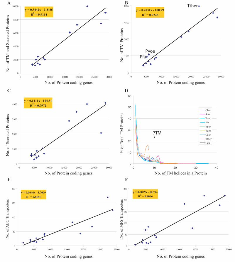
y = 0.2031x - 100.99R2 = 0.9228
0
1500
3000
4500
6000
0 5000 10000 15000 20000 25000 30000
Tther
PfaPyoe
0
10
20
30
40
50
60
0 10 20 30 40
GlamScerTcruPfaTparTgonCparTtherCele
7TM
Ay = 0.3442x - 215.85
R2 = 0.9114
0
2000
4000
6000
8000
10000
0 5000 10000 15000 20000 25000 30000
No.
of T
M a
nd S
ecre
ted
Prot
eins
No. of Protein coding genes No. of Protein coding genes
No.
of T
M P
rote
ins
B
y = 0.1411x - 114.31R2 = 0.7972
0
1000
2000
3000
4000
0 5000 10000 15000 20000 25000 30000
No. of Protein coding genes
No.
of S
ecre
ted
Prot
eins
No. of TM helices in a Protein
% o
f Tot
alTM
Pro
tein
s
No. of Protein coding genes
y = 0.0079x - 10.794R2 = 0.8066
0
50
100
150
200
250
0 5000 10000 15000 20000 25000 30000
No. of Protein coding genes
C D
E F
y = 0.0046x - 5.7009R2 = 0.8181
0
50
100
150
200
0 5000 10000 15000 20000 25000 30000
No.
of A
BC
Tra
nspo
rters
No.
of M
FS T
rans
porte
rs
lakshmin
Text Box
Figure 2

MFS DNAJ
R45 kinase
Rhoptry kinase
Insulinase
T.parva
T.gondii
P.falciparum
P.falciparum
C.parvum
T.thermophilaCpar
Pcha
Pcha
Pyoe
Pyoe
Pber
Pfa
Pfa
Pfa
PfaPfa
Pfa
Cpar
Cpar
CparCpar
Cpar
Cpar
Cpar
Cpar
AtAt
At At
Tgon
Tgon
Tgon
Tthe
Tthe
TtheTthe
Tthe
Tpar Tthe
Tthe
y = 149.31x-1.7983
R2 = 0.8081
1
10
100
1000
1 10 100 1000
Size of LSE cluster (Log scale)
Num
ber o
f LSE
clu
ster
s (Lo
g sc
ale)
P.falciparum
A B
lakshmin
Text Box
Figure 3

lakshmin
Text Box
Figure 4

lakshmin
Text Box
Figure 5

lakshmin
Text Box
Figure 6

3' C
hr10
5' C
hr14
3' C
hr4
Ring
Trophozoite
Schizont
LateSchizont/Merozoite
Plasmodium falciparum
Chr 1 Chr 2 Chr 3 Chr 4 Chr 5
Chr 6 Chr 7 Chr 8 Chr 9 Chr 10
Chr 11 Chr 12 Chr 13 Chr 14
0
0.1
0.2
0.3
0.4
0.5
0
0.1
0.2
0.3
0.4
0
0.1
0.2
0.3
0.4
0.5
0
0.15
0.3
0.45
0.6
0
0.1
0.2
0.3
0.4
0.5
0
0.15
0.3
0.45
0.6
0
0.1
0.2
0.3
0.4
0.5
0
0.1
0.2
0.3
0.4
0.5
0
0.15
0.3
0.45
0.6
0
0.15
0.3
0.45
0.6
0
0.15
0.3
0.45
0.6
0
0.15
0.3
0.45
0.6
0
0.1
0.2
0.3
0
0.15
0.3
0.45
0.6
Cryptosporidium parvum
0
0.1
0.2
0.3
0.4
Chr 1 Chr 20
0.2
0.4
0.6
0.8
Chr 30
0.2
0.4
0.6
0.8
Chr 40
0.2
0.4
0.6
0.8
Chr 50
0.2
0.4
0.6
0.8
Chr 60
0.2
0.4
0.6
Chr 70
0.1
0.2
0.3
0.4
Chr 80
0.2
0.4
0.6
0.8
Theileria parva
Chr 1 Chr 2 Chr 3 Chr 40
0.2
0.4
0.6
0
0.15
0.3
0.45
0.6
0
0.1
0.2
0.3
0.4
0.5
0
0.15
0.3
0.45
0.6
PFI0005w PFI0010c PFI0045c PFI0010c PFI0010cPFI0060cPfemp1 RIFIN RIFIN RIFINSTEVOR
PRESANFragment?
Cgd1_10
Cgd2_70
Cgd3_4170 Cgd3_4240Cgd3_4230
Cgd1_110
Cgd1_20
Cgd1_120
Cgd1_30
Cgd1_130
Cgd1_40
Cgd1_140 Cgd1_150 Cgd1_160
11TM
ABC Transporter (2 ABC Atpase and Tms)Sp with conserved C
12TMSecreted
InsulinaseSecretedInsulinase
SP with C
SPSP/9TMTransporter
SP SKSR SP SKSR SP SKSR
CHR1
CHR1 CHR2
CHR3X6 X5
11TM
[]
TP02_0001Faint Dom
X13[]TP02_0015SP
TP02_0016
ABC Transporter
X2 TP02_0018Faint Dom
X2CHR2
TP03_0888Faint Dom
X6 TP03_0894
TP03_0339-like MFS
TP03_0895Transporter
TP02_0018
SPTP03_0463-like
X7CHR3 TP03_0905TPR Rpts
X24[]TP03_0930FAINT Dom
[]
PFI0095c
PFI0045c PFI0085c
PFI0090c PFI0130c PFI0135c PFI0140w PFI0145w PFI0150c PFI0160w
STEVOR
TM TM SP+Seraprotease SP+TM TM RER1 3TM 4TMsSP+R45
CHR9
X7
CHR14
[]
PF14_0747 PF14_0748 PF14_0751 PF14_0752 PF14_0754PF14_0753
TM+VGD+Pfemp1-C TMPfFACS
SP+TM+TM
TM+PRESAN
[]
TM+PRESAN
PF14_0755 PF14_0756
PF14_0757 PF14_0758 PF14_0760PF14_0759
SP+TM+TMPFB0995w-like
SP
SP+TMPF14_0759-like
TM+TM
SP+TM+TMPFB0995w-like
TM+PRESAN
SP+TMPF14_0759-like
PF14_0751PFACS
PF14_0762
SP PFL2575c-like
PF14_0763X2
TM+PRESAN
PF14_0765
PRESANFragment?
PF14_0766RIFIN/STEVOR
X7
B
A
Cgd2_80 Cgd2_90
5' C
hr10
3' C
hr14
3' C
hr2
5' C
hr8
5' C
hr10
3' C
hr10
5' C
hr11
3' C
hr11
5' C
hr14
3' C
hr14
3' C
hr2
5' C
hr5
3' C
hr5
5' C
hr9
3' C
hr13
3' C
hr10
3' C
hr11
5' C
hr11
5' C
hr14
3' C
hr14
5' C
hr2
3' C
hr2
3' C
hr5
5' C
hr9
5' C
hr5
lakshmin
Text Box
Figure 7