1 2 DNA FRAGMENTATION IN MICROORGANISMS...
35
1 1 DNA FRAGMENTATION IN MICROORGANISMS ASSESSED IN SITU 2 3 4 Running title: DNA fragmentation in microorganisms 5 6 7 José Luis Fernández a, b * , Mónica Cartelle a , Lourdes Muriel a , Rebeca Santiso a, b , 8 María Tamayo a, b , Vicente Goyanes a , Jaime Gosálvez c , Germán Bou d 9 10 a INIBIC-Genética, Complejo Hospitalario Universitario Juan Canalejo, As Xubias 84, 11 15006- A Coruña, Spain. 12 13 b Laboratorio de Genética Molecular y Radiobiología, Centro Oncológico de Galicia, 14 Avda. de Montserrat s/n, 15009-A Coruña, Spain. 15 16 c Unidad de Genética, Facultad de Biología, Universidad Autónoma de Madrid, 28049- 17 Madrid, Spain. 18 19 d INIBIC-Microbiología, Complejo Hospitalario Universitario Juan Canalejo, As 20 Xubias, 84, 15006-A Coruña, Spain 21 22 23 24 25 26 27 * Correspondence 28 Dr. José Luis Fernández 29 Sección de Genética y Unidad de Investigación, 30 Complejo Hospitalario Universitario Juan Canalejo, 31 As Xubias, 84, 32 15006- A Coruña, Spain 33 Tel: 34 981 287499 34 Fax: 34 981 287122 35 E-mail: [email protected] ; [email protected] 36 ACCEPTED Copyright © 2008, American Society for Microbiology and/or the Listed Authors/Institutions. All Rights Reserved. Appl. Environ. Microbiol. doi:10.1128/AEM.00318-08 AEM Accepts, published online ahead of print on 8 August 2008 on April 19, 2018 by guest http://aem.asm.org/ Downloaded from
Transcript of 1 2 DNA FRAGMENTATION IN MICROORGANISMS...

1
1
DNA FRAGMENTATION IN MICROORGANISMS ASSESSED IN SITU 2
3
4
Running title: DNA fragmentation in microorganisms 5
6
7
José Luis Fernández a, b *
, Mónica Cartelle a
, Lourdes Muriel a
, Rebeca Santiso a, b
, 8
María Tamayo a, b
, Vicente Goyanes a
, Jaime Gosálvez c, Germán Bou
d 9
10
a INIBIC-Genética, Complejo Hospitalario Universitario Juan Canalejo, As Xubias 84, 11
15006- A Coruña, Spain. 12
13 b
Laboratorio de Genética Molecular y Radiobiología, Centro Oncológico de Galicia, 14
Avda. de Montserrat s/n, 15009-A Coruña, Spain. 15
16 c Unidad de Genética, Facultad de Biología, Universidad Autónoma de Madrid, 28049-17
Madrid, Spain. 18
19 d INIBIC-Microbiología, Complejo Hospitalario Universitario Juan Canalejo, As 20
Xubias, 84, 15006-A Coruña, Spain 21
22
23
24
25
26
27 * Correspondence 28
Dr. José Luis Fernández 29
Sección de Genética y Unidad de Investigación, 30
Complejo Hospitalario Universitario Juan Canalejo, 31
As Xubias, 84, 32
15006- A Coruña, Spain 33
Tel: 34 981 287499 34
Fax: 34 981 287122 35
E-mail: [email protected] ; [email protected] 36
ACCEPTED
Copyright © 2008, American Society for Microbiology and/or the Listed Authors/Institutions. All Rights Reserved.Appl. Environ. Microbiol. doi:10.1128/AEM.00318-08 AEM Accepts, published online ahead of print on 8 August 2008
on April 19, 2018 by guest
http://aem.asm
.org/D
ownloaded from

2
ABSTRACT 37
38
Chromosomal DNA fragmentation may be a direct or indirect outcome of cell death. 39
Unlike research in higher eukaryotic cells, DNA fragmentation in microorganisms is 40
rarely studied. We report an adaptation of a diffusion-based assay, developed as a kit, 41
which allows for simple and rapid discrimination of bacteria with fragmented DNA. 42
Intact cells were embedded in an agarose microgel on a slide, incubated in a lysis buffer 43
to partially remove the cell walls, membranes, and proteins, and then stained with a 44
DNA fluorochrome, SYBR Gold. Identifying cells with fragmented DNA uses 45
peripheral diffusion of DNA fragments. Cells without DNA fragmentation only show 46
limited spreading of DNA fiber loops. These results have been seen in several Gram-47
negative and Gram-positive bacteria, as well as in yeast. Detection of DNA 48
fragmentation was confirmed by fluoroquinolone treatment and by DNA Breakage 49
Detection-Fluorescence In situ Hybridization (DBD-FISH). Proteus mirabilis with 50
DNA spontaneously fragmented during exponentially and stationary growth, or 51
Escherichia coli with DNA damaged after exposure to hydrogen peroxide or antibiotics 52
such us ciprofloxacin or ampicillin, were clearly detected. Similarly, fragmented DNA 53
was detected in Saccharomyces cerevisiae after amphotericin B treatment. Our assay 54
may be useful for a simple and rapid evaluation of DNA damage and repair as well as 55
cell death, either spontaneous or induced by exogenous stimuli, including antimicrobial 56
agents, or environmental conditions. 57
58
Key words: DNA fragmentation; DNA damage; cell death; quinolones; DBD-FISH59
ACCEPTED
on April 19, 2018 by guest
http://aem.asm
.org/D
ownloaded from

3
INTRODUCTION 60
61
Chromosomal DNA fragmentation, resulting from massive DNA double-strand breaks, 62
is a hallmark of cell death. In higher eukaryotic cells, this may be a consequence of 63
active programmed cell death (PCD), i. e. apoptosis, where DNA is cleaved by an 64
activated endonuclease (26). Otherwise, DNA fragmentation may occur passively 65
through necrotic cell death. Passive DNA fragmentation is likely in microorganisms 66
killed by various causes. Recent studies suggest, however, a possible PCD in unicellular 67
bacteria and yeast (38). In fact, bactericidal antibiotics may trigger a PCD response. It 68
has been reported that bactericidal antibiotics can stimulate the production of hydroxyl 69
radicals that contribute to cell death (20). Tolerant bacterial cells, that are resistant to the 70
bactericidal action of some antibiotics, may be cells with a disabled PCD. Ecological 71
pressure, differentiation processes, starvation and certain DNA damaging agents, may 72
activate PCD involving DNA fragmentation (1, 6, 18, 22, 35). DNA fragments released 73
during autolysis may be absorbed by other bacteria, contributing to antigenic variation 74
and the spread of antibiotic resistance (12). Bacterial autolytic mechanisms have been 75
described primarily with reference to the cell wall level. Nevertheless, active DNA 76
fragmentation has only partially been addressed. 77
78
The presence of DNA breakage is usually evaluated using biochemical or molecular 79
procedures such us alkaline unwinding, DNA elution, gel electrophoresis, sucrose 80
gradient sedimentation, melting curve analysis, viscoelastometry, or light-scattering (2, 81
37). Unfortunately, evaluating DNA fragmentation within an individual cell is not 82
possible nor is it possible to identify important low level intercellular variations. In situ 83
procedures allow for cells with fragmented DNA to be discriminated from those without 84
ACCEPTED
on April 19, 2018 by guest
http://aem.asm
.org/D
ownloaded from

4
DNA fragmentation. However, in situ procedures have been developed primarily for use 85
in higher eukaryotic cells. DNA breaks can be enzymatically labelled by attaching 86
modified nucleotides that can be visualized by direct and indirect methods (10). Usually 87
this process involves the E. coli DNA polymerase I in the In Situ Nick Translation 88
(ISNT) assay or end-labelling with the Klenow fragment of DNA polymerase I, or the 89
terminal deoxynucleotidyl transferase (TdT) in the TUNEL procedure (10). In each of 90
these, a free and accessible 3’-OH group at the end of the break is necessary as substrate 91
for extension, but the ends of DNA breaks may be chemically modified by many DNA-92
damaging agents. 93
94
In situ detection of DNA breaks in higher eukaryotes can also be seen through the 95
single-cell gel electrophoresis (SCGE) or comet assay (27). In this technique, the cells 96
are trapped in an inert agarose microgel on microscope slide, deproteinized by 97
incubation with a lysing solution, and then electrophoresed. DNA staining with a 98
fluorochrome reveals a comet, with a head and a tail of chromatin in the direction of the 99
positive pole of the electric field. Cells with DNA breaks have a higher tail and/or 100
greater DNA concentration in the tail. In contrast to enzymatic labelling, cells in the 101
SCGE assay are unfixed, fully accessible to lysis, and the DNA migration is not 102
dependent on the chemical nature of the breaks. An image analysis system is habitually 103
used for evaluation. Nevertheless, electrophoresis is not necessary to identify cells with 104
fragmented DNA. After lysis in the agarose microgel, cells with fragmented DNA can 105
be identified because they produce a peripheral halo of diffused DNA fragments in the 106
agarose matrix. This is the underlying principle of the diffusion assay to detect cells 107
with DNA fragmentation (33, 36). 108
109
ACCEPTED
on April 19, 2018 by guest
http://aem.asm
.org/D
ownloaded from
5
Regarding microorganisms, the TUNEL assay has been used to determine DNA 110
fragmentation in spheroplasts from yeast, but only one paper has described its use in 111
bacteria, specifically in E. coli and in the archaeon Haloferax volcanii (31). The 112
procedure was time-consuming, requiring fixation, centrifugation and permeabilization 113
steps. Moreover, the evaluation used a flow cytometer, requiring a high number of cells. 114
These technical factors make this procedure impractical for routine assessment of DNA 115
fragmentation in the standard microbiology laboratory. For the comet assay, only one 116
paper has described its use for E. coli (34). The procedure was also lengthy, requiring 117
lysozyme digestion of the cell wall prior to incubation in the lysis solution and in 118
conjunction with prolonged proteinase K digestion. The resulting images are difficult to 119
interpret. 120
121
Despite its importance from both a basic research and a clinical point of view, DNA 122
fragmentation has not been assessed in the microbiology laboratory. This may be due to 123
the lack of a simple and rapid evaluation procedure. Most current techniques are 124
complex, technically demanding, and are not specifically adapted for the different 125
microorganisms. Here we present a diffusion-based assay to identify DNA 126
fragmentation in bacteria and yeast, using fluorescence microscopy. This assay is 127
assembled as a kit in order to implement a simple, fast, reproducible and accurate 128
method for studying DNA fragmentation in microorganisms. 129
ACCEPTED
on April 19, 2018 by guest
http://aem.asm
.org/D
ownloaded from

6
MATERIALS AND METHODS 130
131
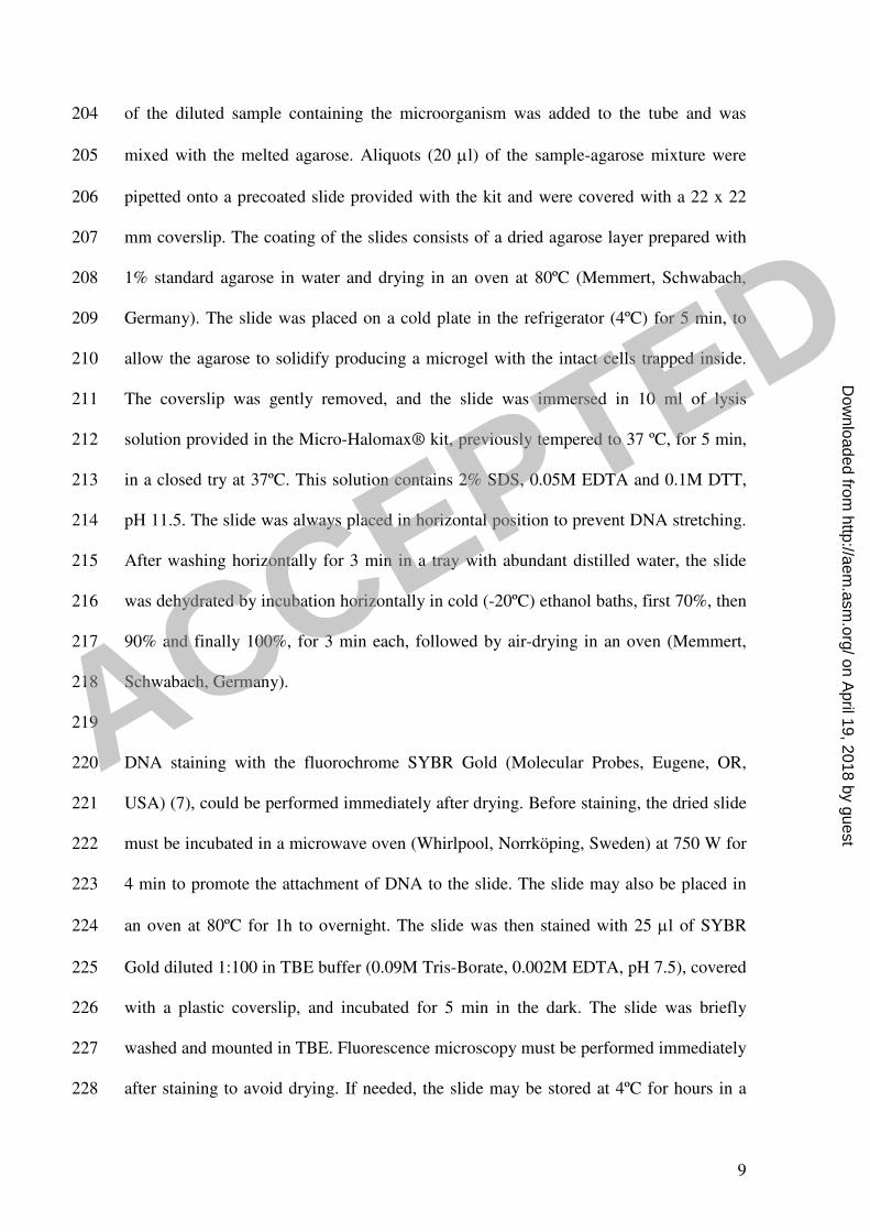
Microorganisms and cultures. Chromosomal DNA fragmentation was assayed in 132
Gram-negative and Gram-positive bacteria (Table 1). Gram-negative bacteria were 133
grown in Luria Bertani (LB) broth (1% Bacto Tryptone, 0.5% yeast extract, 0.5% NaCl) 134
or on LB agar, at 37ºC in aerobic conditions. Gram-positive bacteria were grown on 135
Trypticase Soy Agar (TSA) plates (Diagnostics Systems, Sparks, MD, USA). Candida 136
albicans and S. cerevisiae yeast were grown in yeast extract/peptone/dextrose (YPD) 137
broth and plates. Cell growth in liquid cultures was monitored with the 138
spectrophotometer (Unicam 8625, Cambridge, UK). In amphotericin B experiments, 139
viability was determined by colony-counting after sequential dilutions and plating. 140
141
Experiments. Five different experiments illustrated the use of the procedure to 142
determine chromosomal DNA fragmentation. In the first experiment, aliquots of 143
exponentially growing cultures of the E. coli strain TG1, were exposed to ciprofloxacin 144
(5 µg/ml and 0.012 µg/ml), for 40 min in LB broth, at 37ºC. Fluoroquinolone is an 145
inducer of DNA fragmentation; this experiment validated the assay. In the second 146
experiment, P. mirabilis was incubated in LB medium at 37ºC in aerobic conditions. 147
The initial optical density in the spectrophotometer, measured at 600 nm, was 0.05. P. 148
mirabilis in exponentially growing phase and in stationary phase were analyzed for 149
frequency of cells with fragmented DNA and for membrane permeability, in aliquots 150
that were removed periodically along 106 h. These were batch cultures, where growth 151
uses an unsupplemented amount of nutrients, so the nutrients will decrease in time, 152
together with an increase in metabolites. In a third experiment, TG1, either 153
exponentially growing or in stationary phase, was incubated with 10mM hydrogen 154
ACCEPTED
on April 19, 2018 by guest
http://aem.asm
.org/D
ownloaded from

7
peroxide for 10 min at room temperature in LB broth. Immediately the cells were 155
processed with the DNA fragmentation kit, as described later. The purpose of this 156
experiment was two-fold: 1) to assess the DNA damage induced by hydroxyl radicals, 157
and 2) to explore the influence of the growth phase on the effect of hydrogen peroxide 158
on DNA. In the fourth experiment, exponentially growing cultures of TG1 were 159
exposed to ampicillin (300 µg/ml) for 40 min in LB broth, at 37ºC. This compared the 160
effect on DNA of incubation with an antibiotic with a different mechanism of action to 161
that of quinolones. Finally, exponentially growing cultures of S. cerevisiae were 162
incubated with increasing doses of amphotericin B (0, 0.5, 1, 2, 4, 8 and 16 µg/ml), at 163
30ºC, for 3 and 24 h, in suspension, in YPD broth. After incubation, the cells were 164
processed with the DNA fragmentation kit, as described later. 1000-5000 165
microorganisms were scored per treatment. 166
167
Membrane permeability. A mixture of SYBR Green II (21) and propidium iodide 168
(Molecular Probes, Eugene, OR, USA) was prepared in PBS, at 333X and 0.17 µg/ml, 169
respectively. A 4µl aliquot of microorganisms growing in liquid medium was diluted in 170
16 µl of culture medium and was incubated for 5 min with 4 µl of dye mixture in the 171
dark. If the microorganisms were at a very low density, the dye mixture was added to a 172
20 µl aliquot of cell culture without dilution. 5 µl of the stained cell suspension were 173
dropped onto a glass slide, covered with a coverslip, and examined by fluorescence 174
microscopy. Permeable cells that did not exclude the propidium iodide appeared red, 175
whereas those “alive” only fluoresced green. In S. cerevisiae, non-permeable cells 176
appeared unstained. 177
178
ACCEPTED
on April 19, 2018 by guest
http://aem.asm
.org/D
ownloaded from

8
Determination of DNA fragmentation. The commercial sperm-Halomax® kits 179
(Halotech DNA SL, Madrid, Spain) used to determine DNA fragmentation in different 180
mammalian spermatozoa were evaluated (13, 17, 30). Using sperm-Halomax® as a 181
reference, a prototype Micro-Halomax® kit was developed for microorganisms 182
(Halotech DNA SL, Madrid, Spain). While Gram-negative bacteria could be directly 183
processed, a previous brief cell wall digestion step was necessary for Gram-positive 184
bacteria. S. agalactiae was incubated with mutanolysin (0.1 mg/ml), E. faecalis with a 185
mix of mutanolysin (0.1 mg/ml) and lysozyme (4 mg/ml), and S. aureus with a mix of 186
lysostaphin (0.05 mg/ml) and lysozyme (0.25 mg/ml). S. pyogenes was incubated with 187
lysozyme (1 mg/ml). To this purpose, bacteria were scrapped from the culture plate and 188
resuspended in 0.25 ml of LB medium or phosphate buffer saline (PBS) pH 6.88, in 0.5 189
ml snap cap micro-centrifuge tubes. The enzyme was added at the desired final 190
concentration and was incubated for 15 min at 37ºC. C. albicans and S. cerevisiae were 191
digested with lyticase, 2.5 U/ml for 15 min and 0.07 U/ml for 10 min, respectively, in 192
1M sorbitol, 1M EDTA and 15mM betamercaptoethanol, pH 7.5, at 30ºC. All enzymes 193
were purchased from Sigma (St Louis, MN, USA). 194
195
An aliquot of each sample was diluted to a concentration of 5-10x106 196
microorganisms/ml in the broth culture medium specific for each microorganism. The 197
yeast was centrifuged and resuspended in the lyticase buffer without the enzyme. 0.5 ml 198
snap cap micro-centrifuge tubes containing gelled aliquots of 60 µl of low-melting-point 199
agarose (Pronadisa, Laboratorios Conda, Madrid, Spain) in distilled water are provided 200
with the Micro-Halomax® kit. The tube was placed in a water bath at 90-100ºC for 5 201
min to melt the agarose and then transferred to a water bath at 37ºC (Memmert, 202
Schwabach, Germany). After 5 min incubation, to allow for equilibration to 37ºC, 25 µl 203
ACCEPTED
on April 19, 2018 by guest
http://aem.asm
.org/D
ownloaded from
9
of the diluted sample containing the microorganism was added to the tube and was 204
mixed with the melted agarose. Aliquots (20 µl) of the sample-agarose mixture were 205
pipetted onto a precoated slide provided with the kit and were covered with a 22 x 22 206
mm coverslip. The coating of the slides consists of a dried agarose layer prepared with 207
1% standard agarose in water and drying in an oven at 80ºC (Memmert, Schwabach, 208
Germany). The slide was placed on a cold plate in the refrigerator (4ºC) for 5 min, to 209
allow the agarose to solidify producing a microgel with the intact cells trapped inside. 210
The coverslip was gently removed, and the slide was immersed in 10 ml of lysis 211
solution provided in the Micro-Halomax® kit, previously tempered to 37 ºC, for 5 min, 212
in a closed try at 37ºC. This solution contains 2% SDS, 0.05M EDTA and 0.1M DTT, 213
pH 11.5. The slide was always placed in horizontal position to prevent DNA stretching. 214
After washing horizontally for 3 min in a tray with abundant distilled water, the slide 215
was dehydrated by incubation horizontally in cold (-20ºC) ethanol baths, first 70%, then 216
90% and finally 100%, for 3 min each, followed by air-drying in an oven (Memmert, 217
Schwabach, Germany). 218
219
DNA staining with the fluorochrome SYBR Gold (Molecular Probes, Eugene, OR, 220
USA) (7), could be performed immediately after drying. Before staining, the dried slide 221
must be incubated in a microwave oven (Whirlpool, Norrköping, Sweden) at 750 W for 222
4 min to promote the attachment of DNA to the slide. The slide may also be placed in 223
an oven at 80ºC for 1h to overnight. The slide was then stained with 25 µl of SYBR 224
Gold diluted 1:100 in TBE buffer (0.09M Tris-Borate, 0.002M EDTA, pH 7.5), covered 225
with a plastic coverslip, and incubated for 5 min in the dark. The slide was briefly 226
washed and mounted in TBE. Fluorescence microscopy must be performed immediately 227
after staining to avoid drying. If needed, the slide may be stored at 4ºC for hours in a 228
ACCEPTED
on April 19, 2018 by guest
http://aem.asm
.org/D
ownloaded from

10
self-made humid box in the dark to prevent drying. If dried, the coverslip may be 229
removed by incubation in PBS and, after a brief wash, may be re-stained again. If 230
immediate evaluation is not necessary, dried slides may be left overnight or a couple of 231
days in a high temperature oven (80ºC) and then stored in a tightly closed box, in the 232
dark, at room temperature, for several months, before staining. 233
234
Fluorescence microscopy allows for 10x to 100x magnification, but 100X is necessary 235
for a precise visualization of the small spots from nucleoids with fragmented DNA. 236
Three microgels can be placed on a same slide, including a control sample if required, 237
so that all microgels are simultaneously processed under the same conditions. 238
239
DNA Breakage Detection-Fluorescence In Situ Hybridization (DBD-FISH) (14, 240
15). To confirm the presence of DNA breakage in cells with diffused DNA spots, the 241
DBD-FISH procedure was used in E. coli, P. mirabilis, Acinetobacter haemolyticus, 242
Staphylococcus aureus and S. cerevisiae nucleoids. The cells were immersed in agarose 243
microgels and were lysed as described. They were then washed in 0.9%NaCl and were 244
incubated in an alkaline unwinding solution (0.03M NaOH) for 2.5 min at 22ºC. The 245
gels were neutralized in 0.4M TrisHCl, pH 7.5, washed in distilled water, dehydrated in 246
increasing ethanol baths (70-90-100%) for 2 min each, and air-dried. 247
248
A DNA probe to label the total DNA from the microorganism was prepared. DNA from 249
each microorganism was isolated using standard procedures, and was labeled with 250
biotin-16-dUTP, using a nick translation kit, according to the manufacturer’s 251
instructions (Roche Applied Science, San Cugat del Vallés, Spain). The DNA probe 252
was mixed at 4.3 ng/µl in the hybridization buffer (50% formamide / 2xSSC, 10% 253
ACCEPTED
on April 19, 2018 by guest
http://aem.asm
.org/D
ownloaded from

11
dextran sulfate, 100mM calcium phosphate, pH 7.0) (1xSSC is 0.015M NaCitrate, 254
0.15M NaCl, pH 7.0). The probe in hybridization buffer was denatured by incubation at 255
80ºC for 8 min and was then incubated on ice. This solution (30 µl) was pipetted onto 256
the dried slide, covered with a glass coverslip (22x60mm) and incubated overnight at 257
room temperature, in the dark, in a humid chamber. The coverslip was removed, and the 258
slides were washed twice in 50% formamide/2xSSC, pH 7.0, for 5 min, and twice in 259
2xSSC pH 7.0, for 3 min, at room temperature. The slides were incubated with 80 µl of 260
blocking solution (4XSSC, 0.1% Triton X-100, 5%BSA) for 5 min, covered with a 261
plastic coverslip, in a humid chamber, at 37ºC. This solution was decanted, and the 262
bound probe was detected by incubation with 80 µl of streptavidin-Cy3 (Sigma Chem, 263
St Louis, MN, USA) in 4XSSC, 0.1% Triton X-100, 1%BSA (1:200), covered with a 264
plastic coverslip, in a humid chamber at 37ºC. After washing in 4XSSC, 0.1% Triton X-265
100, three times, 2 min each, slides were counterstained with 20 µl of DAPI (2 µg/ml) 266
in Vectashield (Vector, Burlingame, CA). 267
Fluorescence Microscopy and Digital Image Analysis. Images were viewed with an
epifluorescence microscope (Nikon E800), with a 100x objective and appropriate
fluorescence filters for FITC-SYBR Gold (excitation 465-495 nm, emission 515-555
nm), PI-Cy3 (excitation 540/25 nm, emission 605/55 nm) and DAPI (excitation 340-380
nm, emission 435-485 nm). The images were captured with a high-sensitivity CCD
camera (KX32ME, Apogee Instruments, Roseville, CA). Groups of 16 bit digital
images were obtained and stored as .tiff files. Image analysis used a macro in Visilog
5.1 software (Noesis, Gif sur Yvette, France). This allowed for thresholding,
background subtraction, and measurement of the total fluorescence intensity (surface
area, in pixels x mean fluorescence intensity, in grey level) of the signals. In the
ACCEPTED
on April 19, 2018 by guest
http://aem.asm
.org/D
ownloaded from

12
experiment concerning ciprofloxacin exposure, the surface area of diffusion of the DNA
fragments from nucleoids, in number of pixels, was established, for control, 0.012
µg/ml, and 5 µg/ml. Since the data were not normally distributed, as ascertained by the
Kolmogorov-Smirnov test, a non parametric Mann-Whitney U test was performed to
compare between doses.
ACCEPTED
on April 19, 2018 by guest
http://aem.asm
.org/D
ownloaded from

13
RESULTS AND DISCUSSION 268
269
Technical implications. Here we present an adapted single-cell diffusion assay, 270
initially used to identify DNA fragmentation in mammalian sperm cells (13, 17, 30), for 271
assessing chromosomal DNA integrity in microorganisms, with relatively small 272
genomes. Intact unfixed microorganisms were immersed in an agarose microgel on a 273
slide, lysed, and stained with a DNA fluorochrome. In higher eukaryotic cells, cells 274
without DNA fragmentation only release DNA loops around a central core, but cells 275
with fragmented DNA produce a large halo of diffusion of DNA spots or fragments. 276
277
Given the relatively small genome size of microorganisms, classical fluorochromes such 278
us propidium iodide, DAPI, Hoechst, etc., are not suitable for staining. In order to 279
visualize DNA fragments, it is necessary to use a highly sensitive fluorochrome such us 280
one from the SYBR family. SYBR Gold provides excellent sensitivity and 281
photostability in comparison with other fluorochromes from the same family, giving an 282
accurate visual assessment under the fluorescence microscope (7). Antifading solution 283
was not used since it diminishes the contrast between the small DNA dots and the 284
background. 285
286
Validation of the assay: ciprofloxacin treatment and DBD-FISH. Ciprofloxacin is a 287
fluoroquinone that induces DNA double-strand breaks by trapping DNA gyrase and 288
topoisomerase IV on DNA (19). DNA breaks have been shown by several 289
methodologies including viscosity measurements of cell lysates (24, 25). After 290
processing with the Micro-Halomax® kit and SYBR Gold staining, bacteria from 291
untreated control cultures showed nucleoids with DNA loops spreading from a central 292
ACCEPTED
on April 19, 2018 by guest
http://aem.asm
.org/D
ownloaded from

14
core, which corresponds to the residual bacterium, with a compact, microgranular 293
surface, extended peripherally to may branches (Fig. 1a, b). Remarkably, a few 294
bacteria, 0.4%, spontaneously had a very big halo of DNA spots radiating from the 295
residual central core (Fig. 1a, 5a). These images were similar to those visualized in 296
higher eukaryotic cells with fragmented DNA, with the diffusion-based assay (13, 17, 297
30, 36). After treatment with 5 µg/ml ciprofloxacin, all the nucleoids appeared similar 298
to nucleoids with extensive diffusion of DNA spots as observed occasionally in the 299
control cultures. Thus, ciprofloxacin demonstrates that 1) our procedure confidently 300
detects DNA fragmentation, and that 2) images of nucleoids with a big halo of diffusion 301
of DNA spots indicate extremely fragmented DNA. Quantitative analysis of digital 302
images revealed that the mean surface of the nucleoids was 11.5-fold higher than of the 303
untreated control bacteria (Fig. 1d; Table 2). 304
305
Interestingly, the effect of ciprofloxacin on DNA integrity was increased with respect to 306
untreated control cultures, at the minimum inhibitory concentration (MIC), i.e., 0.012 307
µg/ml (Fig. 1c). After this low dose, the DNA damage was less and was constant among 308
the bacteria. In fact, the nucleoids appeared more spread, with their average surface 309
being 3.9-fold higher than the untreated control cells (Table 2) and having larger 310
peripheral DNA fragments than after the high dose. As postulated by Drlica et al. (11), 311
ciprofloxacin at low doses like MIC and short incubation times may block growth 312
without killing the cells, suggesting the formation of reversible complexes. At higher 313
doses, like 5 µg/ml, the DNA is extremely fragmented, as here observed, perhaps 314
causing rapid death. The experiment demonstrates the sensitivity and potential value of 315
our procedure for determining the activity of quinolones, both in basic and in clinical 316
research. This approach is currently under investigation in our laboratory. 317
ACCEPTED
on April 19, 2018 by guest
http://aem.asm
.org/D
ownloaded from

15
318
To further confirm the presence of DNA breaks in nucleoids with diffused DNA 319
fragments in control E. coli cultures, the DBD-FISH technique was employed (14, 15). 320
This procedure uses the same microgel as the diffusion assay, allowing simultaneous or 321
sequential visualization of the nucleoids with or without fragmented DNA and labelling 322
of DNA breaks. DBD-FISH is a powerful procedure that involves microgel-embedding, 323
lysis, and incubation with a limited alkaline DNA unwinding step (3, 32). This final step 324
transforms DNA breaks into limited single-stranded DNA (ssDNA) segments generated 325
from the ends of the breaks, which hybridize to fluorescent DNA probes. As DNA 326
breaks increase, more ssDNA is produced, increasing probe hybridization and 327
fluorescence intensity. Fluorescence may be quantified using image analysis software. 328
When hybridizing with a whole genome probe, DNA breaks in the entire genome are 329
assessed. Damage within specific DNA sequence areas may be evaluated by hybridizing 330
specific DNA probes. 331
332
The DBD-FISH procedure was applied to E. coli (Fig. 2) and other microorganisms 333
lysed in the microgel. Nucleoids with diffused spots were strongly labelled, further 334
confirming massive DNA breaks. 335
336
Three kinds of experiments demonstrated the potential of the procedure to determine 337
chromosomal DNA fragmentation: 1) analysis of cells with spontaneous fragmented 338
DNA in culture, 2) reactive oxygen species (ROS) induced DNA damage, and 3) 339
analysis of antibiotic and antifungal agent effects. 340
341
ACCEPTED
on April 19, 2018 by guest
http://aem.asm
.org/D
ownloaded from

16
Batch cultures. P. mirabilis was incubated in liquid LB medium for 106 h, monitoring 342
turbidity and removing aliquots periodically to determine membrane permeability and 343
DNA fragmentation. Bacteria with fragmented DNA were easily separated from 344
nucleoids without fragmented DNA (Fig. 3a). The frequency of bacteria with DNA 345
fragmentation was established using the Micro-Halomax® kit. This evaluated nucleoids 346
with diffused DNA fragments in 1000-5000 cells per experimental point. Membrane 347
permeability was determined by SYBR Green II and propidium iodide staining. All 348
cells were permeable to SYBR Green II, but propidium iodide is a vital dye, so only 349
cells with membrane permeability, which do not exclude the dye, appear red under the 350
propidium iodide filter set of the microscope. The frequency of propidium iodide 351
permeable cells was established in 1000-5000 microorganisms, per experimental point 352
(Fig. 4). 353
354
The culture changed from exponentially growing to stationary phase after 9 h. The 355
percentage of cells permeable to propidium iodide increased at 48 h from 0.5% to 5%, 356
and then progressively rose through the end of the experiment to 88%. The proportion 357
of bacteria with fragmented DNA significantly increased after 81 h from 0.5-1% to 358
9.5%. It remained constant at 35% from 99 to 103 h and then rose to 52 % 3 h later. 359
360
These results suggest that membrane permeability does not indicate the presence of 361
fragmented DNA, being independent parameters related to different initial targets and 362
not correlated in time. Moreover, the stationary phase seems not to be steady in the 363
frequency of bacteria with fragmented DNA. In the initial period of the stationary 364
phase, the proportion of bacteria with DNA fragmentation did not increase over the 365
exponential phase. The percentage increased later, probably reflecting a progressive 366
ACCEPTED
on April 19, 2018 by guest
http://aem.asm
.org/D
ownloaded from

17
change in the turnover rate between dead and dividing cells. Perhaps with the 367
accumulation of metabolites and the depletion of nutrients, the fraction of cells with 368
fragmented DNA should further increase. 369
370
Hydrogen peroxide treatment. The effect of ROS on DNA integrity was evaluated. 371
Hydrogen peroxide decomposes into hydroxyl radicals (.OH) through catalysis by low 372
valence transition metal ions in a Fenton-Haber-Weiss reaction. These oxidizing agents 373
strongly reacted with macromolecules. .OH attack on DNA results in a variety of base 374
damages and DNA breaks (8, 9). In the only report using the TUNEL assay in bacteria 375
(31), labelling of DNA breaks was detected in exponentially growing cultures of E. coli, 376
after exposure to extremely high doses of H2O2 (86 mM for 30 min). Surprisingly, in 377
stationary phase cultures, even doubling the H2O2 dose (172 mM) did not result in DNA 378
breakage. This suggested that H2O2 does not directly cause DNA breaks, which could be 379
transient intermediates in DNA repair produced by the DNA repair enzymes (31). 380
381
We tested this hypothesis using our DNA fragmentation assay. The percentage of E. 382
coli cells with fragmented DNA was 0.4% and 37.6% in untreated control cells, 383
growing exponentially or in stationary phase, respectively (Fig. 5a, c). Using a lower 384
dose for a shorter time than in the previous report (10mM, 10 min), 100% of nucleoids 385
showed extensively fragmented DNA, either in exponential or in stationary growth 386
phase (Fig 5b, d). This result suggests a higher sensitivity in the microgel-based assay 387
compared with TUNEL in bacteria and illustrates a difficulty with enzymatic 388
procedures for labelling DNA breaks. In the case of TdT, a free 3’-OH group at the 389
terminus of the DNA break is essential as substrate in order to polymerize the 390
nucleotides (10). Attack by agents like H2O2 does not produce “clean” DNA termini, but 391
ACCEPTED
on April 19, 2018 by guest
http://aem.asm
.org/D
ownloaded from

18
rather chemically modified ends, such that direct DNA breaks could be undetectable to 392
enzymes (9). To allow for DNA repair, Exonuclease III removes blocking groups at the 393
3’ terminus (9). Labelling by TdT should therefore be possible. The absence of 394
enzymatic labelling in stationary phase cultures could be explained if end-processing is 395
impaired at this stage. Nevertheless, H2O2-induced DNA breaks are visible with our 396
assay, since it is independent on the chemical nature of the DNA break. Overall, our 397
diffusion assay identifies DNA damage by .OH, both in exponentially and stationary 398
growth phases. 399
400
Ampicillin incubation. To evaluate the influence of ampicillin treatment on 401
chromosomal DNA, exponentially growing cultures of TG1 were exposed to 300 µg/ml 402
ampicillin for 40 min or 24 h. This dose was much higher than the MIC of 3 µg/ml. In 403
contrast to ciprofloxacin, that affects DNA, the cell wall is the primary target for 404
ampicillin; it inhibits peptidoglycan synthesis after binding to penicillin binding 405
proteins and activating autolysins (5, 16). 406
407
Contrary to ciprofloxacin, 40 min treatment with ampicillin barely increased the 408
frequency of cells with fragmented DNA or with appearance of DNA damage. The 409
nucleoids were similar to those from untreated control cells. When incubated for 24 h, 410
the density of bacteria, and correspondent nucleoids, was reduced, but had a uniform 411
background of DNA spots that were probably from spontaneously lysed cells that 412
released the DNA fragments to the medium (Fig. 6). This suggests that cell death 413
initially appears to be independent on DNA damage, but may evolve late in DNA 414
degradation. 415
416
ACCEPTED
on April 19, 2018 by guest
http://aem.asm
.org/D
ownloaded from

19
Amphotericin B incubation in yeast. A presumed PCD has been described in S. 417
cerevisiae, following acidic, oxidative, or osmotic stress, and after ultraviolet exposure 418
(23). This has also been reported for Candida albicans after acetic acid, H2O2, or 419
amphotericin B treatment (28, 29). Apoptotic cells were very significantly increased 420
after 200 min incubation with 4 µg/ml amphotericin B (28). Apoptotic cells were 421
considered those not growing and not permeable to propidium iodide. DNA 422
fragmentation was not assessed. After a dose of 16 µg/ml, practically all Candida cells 423
did not grow, appearing 10% propidium iodide impermeable that were assumed 424
apoptotic, whereas the propidium iodide permeable cells were presumed necrotic. 425
426
An image of C. albicans showing DNA fragmentation is presented in figure 3b. 427
Nevertheless, we assessed the possible induction of DNA fragmentation by 428
amphotericin B in S. cerevisiae. In untreated control cultures, no cells with fragmented 429
DNA were detected in 6000 yeasts. There was no evidence of fragmented DNA with 430
any dose of the antifungal agent when incubated for 3 h. After 24 h incubation with 431
amphotericin B, yeast cells with fragmented DNA were recorded in a dose-dependence 432
manner (Fig. 7). The frequency of cells with fragmented DNA was lower than that with 433
propidium iodide permeable membrane. In fact, with the highest dose assayed, 5% of 434
the cells contained fragmented DNA, whereas 85% were propidium iodide permeable. 435
This decouples membrane permeability from DNA fragmentation as being indicative of 436
death in these microorganisms, at least after treatment with antifungal agents that target 437
to yeast cell membrane, like amphotericin B (4). A substantial and proportional decrease 438
in viability with dose, assayed 48 h after treatment, was demonstrated (Fig. 7), 439
suggesting that DNA fragmentation after amphotericin B treatment is either a rare 440
phenomenon or a late response. 441
ACCEPTED
on April 19, 2018 by guest
http://aem.asm
.org/D
ownloaded from

20
442
Conclusion. The experiments presented here illustrate the ability of the technique, 443
assembled as a kit, to determine the presence of fragmented DNA in microorganisms. 444
Its simplicity, short assay time (50 min) and efficacy, makes this technique useful for 445
the routine determination of DNA fragmentation and intercellular variation. 446
Applications may be extensive in both basic and clinical research. Only a fluorescence 447
microscope is required. Though direct visual identification is quite sharp, the scoring 448
process may be partially automated by adapted image analysis software. This 449
automation could be more complete by integrating a microscope with a motorized plate 450
and focus, a CCD camera for image capture, and image analysis software. This could be 451
useful when scoring many thousands of microorganisms, resembling the flow-cytometer 452
facilities. 453
454
Acknowledgements. Dr. J.L. Fernández , Dr. V. Goyanes and Dr. J. Gosálvez 455
collaborate as scientific advisers of Halotech DNA SL. We are grateful to prof. 456
Christopher de Jonge, from Minnesota University, for the critical reading of the 457
manuscript. This work has been supported by a public grant from the Xunta de Galicia 458
07CSA050916PR and INCITE07PXI916201ES, and by FIS PI061368. 459
ACCEPTED
on April 19, 2018 by guest
http://aem.asm
.org/D
ownloaded from

21
FIGURES 460
461
Figure 1. Images after application of the Micro-Halomax® kit to E. coli cultures. Cells 462
were embedded in an agarose microgel, lysed, and stained with SYBR Gold. a: 463
Nucleoids from control untreated cells, showing spread of DNA loops from a central 464
core. One has highly diffused DNA spots (asterisk). b: A detailed image of an 465
undamaged nucleoid, showing an uneven microgranular surface and multibranched 466
appearance. c: Ciprofloxacin treatment at the MIC dose, 0.012 µg/ml, for 40 min, 467
resulted in DNA fragments. d: These fragments increased after 5 µg/ml treatment; 468
nucleoids showing a big halo of diffusion of DNA spots, similar to the image marked by 469
asterisk in (a). Bar: 5 µm in a, c, d; 2.5 µm in b. 470
471
Figure 2. DBD-FISH detected DNA breaks in nucleoids from control cultures of E. 472
coli. Cells in an agarose microgel were lysed and treated with an alkaline unwinding 473
treatment to transform DNA breaks into restricted single-stranded DNA motifs detected 474
by hybridization with a whole genome probe, Cy-3 labeled (red). The central nucleoid 475
with diffused DNA spots appears intensely labelled, confirming the presence of 476
spontaneous massive DNA breaks (asterisk). Nucleoids without fragmented DNA were 477
visible with DAPI (blue). Bar: 5 µm. 478
479
Figure 3. Images after the application of Micro-Halomax® to cultures of P. mirabilis 480
(a) and C. albicans (b), processed as indicated in the Materials and Methods. Nucleoids 481
without DNA fragmentation released DNA loops around a central core from the residual 482
cell. Otherwise, nucleoids with fragmented DNA were clearly identified by a big halo of 483
DNA spots (asterisks). Bar: 5 µm. 484
ACCEPTED
on April 19, 2018 by guest
http://aem.asm
.org/D
ownloaded from

22
485
Figure 4. Kinetics of the frequency of P. mirabilis cells with fragmented DNA and with 486
propidium iodide permeable membrane. Identification of bacteria with fragmented DNA 487
was performed using the Micro-Halomax® kit, as indicated in the Materials and 488
Methods. 489
490
Figure 5. Exponentially growing cultures from E. coli control cells (a) and exposed to 491
10mM hydrogen peroxide for 10 min (b), evaluated with the diffusion-based assay, 492
using the Micro-Halomax® kit. a: bacterial nucleoids from control cultures only show 493
spreading of DNA loops. Some nucleoids had a big halo of diffused DNA spots, as 494
indicated in the image (asterisk). b: all nucleoids observed after H2O2 treatment reveal a 495
halo of DNA spots. In stationary phase cultures, untreated cells (c) showed similar 496
images to (a), with a higher proportion of background nucleoids with diffused DNA 497
spots (asterisks), whereas those treated with H2O2 (d) were similar to (b). Bar: 5 µm. 498
499
Figure 6. E. coli cultures processed with the Micro-Halomax® kit after ampicillin 500
treatment, 300 µg/ml, 24 h. Nucleoids from residual cells appear more relaxed, 501
accompanied by a dense background of DNA fragments. Bar: 5 µm. 502
503
Figure 7. S. cerevisiae cells with fragmented DNA (right scale), propidium iodide (PI) 504
permeable membrane, and viability (left scale), after incubation for 24 h with increasing 505
doses of amphotericin B.506
ACCEPTED
on April 19, 2018 by guest
http://aem.asm
.org/D
ownloaded from

23
REFERENCES 507
508
1. Aersten A., and C.W. Michiels. 2004. Stress and how bacteria cope with death and 509
supervivence. Crit. Rev. Microbiol. 30:263-273. 510
2. Ahnström, G. 1988. Techniques to measure DNA strand breaks in cells: a review. 511
Int. J. Radiat. Biol. 54:695-707. 512
3. Ahnström, G., and K. Erixon. 1973. Radiation-induced strand breakage in DNA 513
from mammalian cells. Strand separation in alkaline solution. Int. J. Radiat. Biol. 514
23:285-289. 515
4. Baginski, M., J. Czub, K. and K. Sternal. 2006. Interaction of amphotericin B and 516
its selected derivatives with membranes: molecular modeling studies. Chem. Rec. 517
6:320-332. 518
5. Bayles, K.W. 2000. The bactericidal action of penicillin: new clues to an unsolved 519
mystery. Trends Microbiol. 8:274-278. 520
6. Beppu, T., and K. Arima. 1971. Properties of the Colicin E2-induced Degradation of 521
Deoxyribonucleic Acid in Escherichia coli. J. Biochem. 70:263-271. 522
7. Chen, F., J-R. Lu, B. J. Binder, Y-C. Liu, and R.E. Hodson. 2001. Application of 523
digital image analysis and flow cytometry to enumerate marine viruses stained with 524
SYBR Gold. Appl. Env. Microbiol. 67:539-545. 525
8. Dahm-Daphi, J., C. Sab, and W. Alberti. 2000. Comparison of biological effects of 526
DNA damage induced by ionizing radiation and hydrogen peroxide in CHO cells. Int. J. 527
Radiat. Biol. 76:67-75. 528
9. Demple, D., A. Johnson, and D. Fung. 1986. Exonuclease III and endonuclease IV 529
remove 3’ blocks from DNA synthesis primers in H2O2-damaged Escherichia coli. 530
Proc. Natl. Acad. Sci. USA. 83:7731-7735. 531
ACCEPTED
on April 19, 2018 by guest
http://aem.asm
.org/D
ownloaded from

24
10. Didenko, V., ed. In situ detection of DNA damage. Humana Press, Totowa, New 532
Yersey, 2002. 533
11. Drlica, K., M. Malik, R.J. Kerns, and X. Zhao. 2008. Quinolone-mediated 534
bacterial death. Antimicrob. Agents Chemother. 52:385-392. 535
12. Dubnau, D. 1999. DNA Uptake in bacteria. Annu. Rev. Microbiol. 53:217-224. 536
13. Enciso, M., C. López-Fernández , J.L. Fernández
, P. García
, A. Gosálbez
, and 537
J. Gosálvez. 2006. A new method to analyze boar sperm DNA fragmentation under 538
bright-field or fluorescence microscopy. Theriogenology 65:308-316. 539
14. Fernández, J.L., and J. Gosálvez. 2002. DNA Breakage Detection-FISH (DBD-540
FISH) Methods Mol. Biol. 203:203-216. 541
15. Fernández, J.L., V. Goyanes, and J. Gosálvez. 2002. DNA Breakage Detection-542
FISH (DBD-FISH), p. 282-290. In B. Rautenstrauss, and T. Liehr (ed.), FISH 543
technology-Springer lab manual. Springer-Verlag, Heidelberg. 544
16. Fuda, C., D. Hesek, M. Lee, W. Heilmayer, R. Novak, S.B. Vakulenko, and S. 545
Mobashery. 2006. Mechanistic basis for the action of new cephalosporin antibiotics 546
effective against methicillin-and vancomycin-resitant Stapylococcus aureus. J. Biol. 547
Chem. 281:10035-10041. 548
17. García-Macías, V., P. de Paz, F. Martínez-Pastor, M. Alvarez, S. Gomes-Alves, 549
J. Bernardo, E. Anel, and L. Anel. 2007. DNA fragmentation assessment by flow 550
cytometry and Sperm-Bos-Halomax (bright-field microscopy and fluorescence 551
microscopy) in bull sperm. Int. J. Androl. 30:88-98. 552
18. Herrero, M., and F. Moreno. 1986. Microcin B17 blocks DNA replication and 553
induces SOS system in Escherichia coli. J. Gen. Microbiol. 132:393-402. 554
555
ACCEPTED
on April 19, 2018 by guest
http://aem.asm
.org/D
ownloaded from

25
19. Hooper, D.C. 2001. Mechanisms of action of antimicrobials: focus on 556
fluoroquinolones. Clin. Inf. Dis. 32:9-15. 557
20. Kohanski M.A., D.J. Dwyer, B. Hayete, C.A. Lawrence, and J.J. Collins. 2007. 558
A common mechanism of cellular death induced by bactericidal antibiotics. Cell 559
130:797-810. 560
21. Lebaron, P., N. Parthuisot, and P. Catala. 1998. Comparison of blue nucleic acid 561
dyes for flow cytometric enumeration of bacteria in aquatic systems. Appl. Env. 562
Microbiol. 64:1725-1730. 563
22. Lewis, K. 2000. Programmed death in bacteria. Microbiol. Mol. Biol. Rev. 64:503-564
514. 565
23. Madeo F., S. Engelhardt, E. Herker, N. Lehmann, C. Maldener, A. Proksch, S. 566
Wissing, and K.U. Frohlich. 2002. Apoptosis in yeast: a new model system with 567
applications in cell biology and medicine. Curr. Genet. 41:208-216. 568
24. Malik, M., X. Zhao, and K. Drlica. 2006. Lethal fragmentation of bacterial 569
chromosomes mediated by DNA gyrase and quinolones. Mol. Microbiol. 61:810-825. 570
25. Malik, M., X. Zhao, and K. Drlica. 2007. Effect of anaerobic growth on quinolone 571
lethality with Escherichia coli. Antimicrob. Agents Chemother. 51:28-34. 572
26. Nagata, S. 2000. Apoptotic DNA fragmentation. Exp. Cell Res. 256:12-18. 573
27. Olive, P.L., and R.E. Durand. 2005. Heterogeneity in DNA damage using the 574
comet assay. Cytometry 66:1-8. 575
28. Philips, A.J., I. Sudbery, and M. Ramsdale. 2003. Apoptosis induced by 576
environmental stresses and amphotericin B in Candida albicans. Proc. Nat. Acad. Sci. 577
USA. 100:14327-14332. 578
ACCEPTED
on April 19, 2018 by guest
http://aem.asm
.org/D
ownloaded from

26
29. Philips, A.J., J.D. Crowe, and M. Ramsdale. 2006. Ras pathway signalling 579
accelerates programmed cell death in the pathogenic fungus Candida albicans. Proc. 580
Nat. Acad. Sci. USA. 103:726-731. 581
30. Rodríguez, S., V. Goyanes, E. Segrelles, M. Blasco, J. Gosálvez, and J.L. 582
Fernández. 2005. Critically short telomeres are associated with sperm DNA 583
fragmentation. Fertil. Steril. 84:843-845. 584
31. Rohwer, F., and F. Azam. 2000. Detection of DNA damage in prokaryotes by 585
terminal deoxyribonucleotyde transferase-mediated dUTP nick end labeling. Appl. Env. 586
Microbiol. 66:1001-1006. 587
32. Rydberg, B. 1975. The rate of strand separation in alkali of DNA of irradiated 588
mammalian cells. Radiat. Res. 61:274-287. 589
33. Singh, N.P. 2000. A simple method for accurate estimation of apoptotic cells. Exp. 590
Cell Res. 256:328-337. 591
34. Singh, N.P., R.E. Stephens, H. Singh, and H. Lai. 1999. Visual quantification of 592
DNA double-strand breaks in bacteria. Mutat. Res. 429:159-168. 593
35. Valenti, A., A. Napoli, M. C. Ferrara, M. Nadal, M. Rossi, and M. Ciaramella. 594
2006. Selective degradation of reverse gyrase and DNA fragmentation induced by 595
alkylating agent in the archaeon Sulfolobus solfataricus. Nucleic Acids Res. 34:2098-596
2108. 597
36. Vasquez, M., and R.R. Tice. 1997. Comparative analysis of apoptosis versus 598
necrosis using the single cell gel (SCG) assay. Environ. Mol. Mutagen. 29:53. 599
37. von Sonntag, C. The chemical basis of radiation biology. Taylor and Francis Ltd., 600
London, 1987. 601
38. Yarmolinsky, M.B. 1995. Programmed cell death in bacterial populations. Science 602
267:836-837. 603
ACCEPTED
on April 19, 2018 by guest
http://aem.asm
.org/D
ownloaded from
Gram Negative Bacteria Gram Positive Bacteria
Escherichia coli Enterococcus faecalis
Enterobacter cloacae Staphylococcus aureus
Pseudomonas aeruginosa Streptococcus agalactiae
Proteus mirabilis Streptococcus pyogenes
Salmonella spp.
Stenotrophomonas maltophilia
Acinetobacter haemolyticus
Acinetobacter baumannii
Klebsiella oxytoca
Klebsiella pneumoniae
Citrobacter freundii
Table 1. Bacteria assessed with the Micro-Halomax® kit.
ACCEPTED
on April 19, 2018 by guest
http://aem.asm
.org/D
ownloaded from

Dose
(µg/ml) n
Surface Area
(mean ± SD)
Relative Increase
in Surface P
0 150 7411.9 ± 2173.8 - -
0.012 145 29198.8 ± 12747.9 3.9 < 0.0001
5.0 146 85156.8 ± 19301.1 11.5 <0.0001
Table 2. Data of digital image analysis of nucleoids from E. coli treated with
ciprofloxacin, 40 min, in LB broth, and processed with the Micro-Halomax® kit
(n = number of nucleoids examined; surface area is in number of pixels; P =
significance level obtained with Mann-Whitney U test).
ACCEPTED
on April 19, 2018 by guest
http://aem.asm
.org/D
ownloaded from

0
20
40
60
80
100
0 20 40 60 80 100 120
Time (h)
(%)
0
1
2
3
4
5
OD
600
DNA Fragmentation
Membrane Permeability
OD600
ACCEPTED
on April 19, 2018 by guest
http://aem.asm
.org/D
ownloaded from

0,00
10,00
20,00
30,00
40,00
50,00
60,00
70,00
80,00
90,00
100,00
0 0.5 1 2 4 8 16
[Amphotericin B µµµµg/ml]
% PI Permeable Cells
% Viability
0,00
0,50
1,00
1,50
2,00
2,50
3,00
3,50
4,00
4,50
5,00
% Cells with Fragmented
DNA
% PI Permeable Cells
% Viability
% Cells with Fragmented DNA
ACCEPTED on A
pril 19, 2018 by guesthttp://aem
.asm.org/
Dow
nloaded from


![Sperm DNA Fragmentation is Significantly Increased in ... · Sperm DNA fragmentation assessment The sperm DNA damage was evaluated by Sperm Chromatin Dispersion (SCD) test [23] using](https://static.fdocuments.us/doc/165x107/5f3a6b0098469b5f937b3512/sperm-dna-fragmentation-is-significantly-increased-in-sperm-dna-fragmentation.jpg)