1, 2, 2 1,4 1 3 - Developmentdev.biologists.org/content/develop/139/4/793.full.pdf · This movement...
12
RESEARCH ARTICLE 793 Development 139, 793-804 (2012) doi:10.1242/dev.071720 © 2012. Published by The Company of Biologists Ltd INTRODUCTION The evolution of animal complexity has fascinated scientists ever since Darwin presented his ideas on embryology and ‘descent with modification’ (Darwin, 1890). Species’ adaptation to new environments required specific organ system modifications. The adaptation of fish from hyperosmotic saltwater to hypoosmotic freshwater environments is an extreme example of organ system adaptation. This movement required drastic modification of salt and water homeostasis mechanisms. Two organ systems essential for maintaining osmotic balance are the gills and kidney. In aquatic animals, the gills are gatekeepers for maintaining salt balance whereas the kidney is needed for maintaining both salt and water concentrations. Thus, maintaining homeostasis requires cooperation between these two systems. How differential adaptation of the gills and kidney to new environments is encoded in the genomes of diverse animals is not well understood. Gross anatomical observations readily demonstrate kidney diversity across species. For over a century and a half, scientists have compared adult mammalian kidneys, cataloging both similarities and differences (Wagner and Tulk, 1845). These differences are illustrated when comparing the horse (Fig. 1A) and human (Fig. 1B) kidney. Despite such outward structural diversity among mammals, the building blocks of their kidneys, the nephrons, are nearly identical. However, comparing nephrons among chordates reveals clear differences (Dantzler, 1989; Smith, 1937). As early as 1937, key variances in nephron structure were noted to track with broad evolutionary changes in vertebrates (Smith, 1937). When examining the earliest developing kidney, the pronephros, variations are observed when comparing glomerular absence (aglomerular; Fig. 1C) or presence (non-integrated or integrated glomerulus; Fig. 1D,E), and by examining the complexity of tubules and ducts extending from the glomerulus. Aglomerular kidneys (Fig. 1C), found in a subset of teleosts such as Opsanus tau and Lophius piscatorius (Marshall, 1930), remove waste by tubule lumen secretion (Beyenbach, 2004). In organisms containing a glomerulus, two examples of glomerular pronephric evolution are the non-integrated and the integrated glomerulus. The non-integrated form of the pronephros is found in Xenopus, in which the coelom separates the glomerulus from the tubules and ducts (Fig. 1D, arrow) (Dressler, 2006). By contrast, the zebrafish pronephros (Fig. 1E) represents the most common teleost kidney type – a glomerulus integrated with the pronephric tubules and duct – and is similar to the nephron found in adult mammalian kidneys (Fig. 1F). The molecular basis underlying these developmental variations of the vertebrate pronephros is unknown. Broadly, three related but molecularly distinct models have been proposed as the genetic basis of organ diversity. The first involves conservation of master regulatory genes (Carroll, 2005); the second outlines gene regulatory networks (GRNs) (Davidson and Erwin, 2006); and the third focuses on dynamic gene families (Demuth et al., 2006). First, the ‘master regulation’ model – whereby a handful of key conserved genes are necessary and sufficient for organ development – is the best characterized. Changes in number and regulation of key highly conserved genes is an important mechanism underlying evolutionary diversity (Carroll, 2005). For example, Hox genes specify variation in body segments, including 1 Department of Biochemistry and Molecular Biology, Mayo Clinic, Rochester, MN 55905, USA. 2 Department of Genetics, Cell Biology and Development, University of Minnesota, Minneapolis, MN 55455, USA. 3 STEM Squad, Mayo Clinic, Rochester, MN 55905, USA. 4 Department of Biology, Temple University, Philadelphia, PA 19122, USA. *These authors contributed equally to this work ‡ Author for correspondence ([email protected]) Accepted 15 December 2011 SUMMARY The Homeobox (Hox) and Paired box (Pax) gene families are key determinants of animal body plans and organ structure. In particular, they function within regulatory networks that control organogenesis. How these conserved genes elicit differences in organ form and function in response to evolutionary pressures is incompletely understood. We molecularly and functionally characterized one member of an evolutionarily dynamic gene family, plac8 onzin related protein 1 (ponzr1), in the zebrafish. ponzr1 mRNA is expressed early in the developing kidney and pharyngeal arches. Using ponzr1-targeting morpholinos, we show that ponzr1 is required for formation of the glomerulus. Loss of ponzr1 results in a nonfunctional glomerulus but retention of a functional pronephros, an arrangement similar to the aglomerular kidneys found in a subset of marine fish. ponzr1 is integrated into the pax2a pathway, with ponzr1 expression requiring pax2a gene function, and proper pax2a expression requiring normal ponzr1 expression. In addition to pronephric function, ponzr1 is required for pharyngeal arch formation. We functionally demonstrate that ponzr1 can act as a transcription factor or co-factor, providing the first molecular mode of action for this newly described gene family. Together, this work provides experimental evidence of an additional mechanism that incorporates evolutionarily dynamic, lineage-specific gene families into conserved regulatory gene networks to create functional organ diversity. KEY WORDS: Kidney development, Kidney evolution, Pharyngeal arch development, Zebrafish, Plac8 The lineage-specific gene ponzr1 is essential for zebrafish pronephric and pharyngeal arch development Victoria M. Bedell 1, *, Anthony D. Person 2, *, Jon D. Larson 2 , Anna McLoon 2 , Darius Balciunas 1,4 , Karl J. Clark 1 , Kevin I. Neff 1 , Katie E. Nelson 3 , Brent R. Bill 2 , Lisa A. Schimmenti 2 , Soraya Beiraghi 2 and Stephen C. Ekker 1,2,‡ DEVELOPMENT DEVELOPMENT
Transcript of 1, 2, 2 1,4 1 3 - Developmentdev.biologists.org/content/develop/139/4/793.full.pdf · This movement...

RESEARCH ARTICLE 793
Development 139, 793-804 (2012) doi:10.1242/dev.071720© 2012. Published by The Company of Biologists Ltd
INTRODUCTIONThe evolution of animal complexity has fascinated scientists eversince Darwin presented his ideas on embryology and ‘descent withmodification’ (Darwin, 1890). Species’ adaptation to newenvironments required specific organ system modifications. Theadaptation of fish from hyperosmotic saltwater to hypoosmoticfreshwater environments is an extreme example of organ systemadaptation. This movement required drastic modification of saltand water homeostasis mechanisms. Two organ systems essentialfor maintaining osmotic balance are the gills and kidney. In aquaticanimals, the gills are gatekeepers for maintaining salt balancewhereas the kidney is needed for maintaining both salt and waterconcentrations. Thus, maintaining homeostasis requirescooperation between these two systems. How differentialadaptation of the gills and kidney to new environments is encodedin the genomes of diverse animals is not well understood.
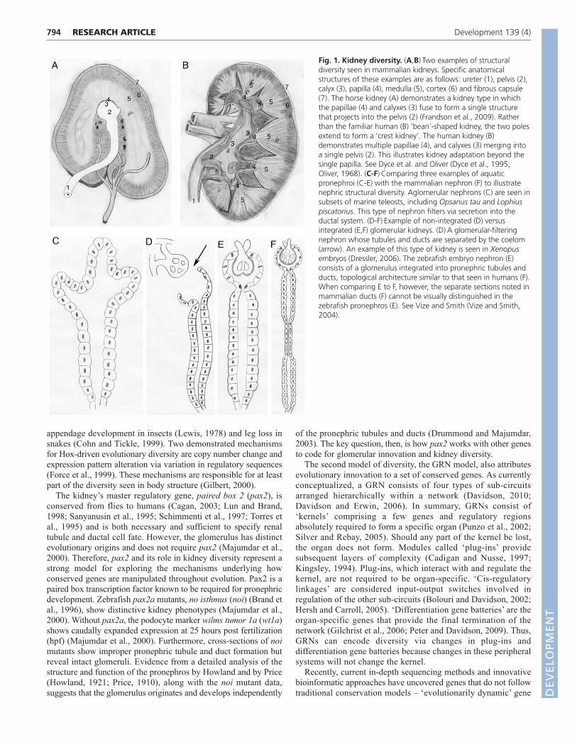
Gross anatomical observations readily demonstrate kidneydiversity across species. For over a century and a half, scientistshave compared adult mammalian kidneys, cataloging bothsimilarities and differences (Wagner and Tulk, 1845). Thesedifferences are illustrated when comparing the horse (Fig. 1A) andhuman (Fig. 1B) kidney. Despite such outward structural diversityamong mammals, the building blocks of their kidneys, the
nephrons, are nearly identical. However, comparing nephronsamong chordates reveals clear differences (Dantzler, 1989; Smith,1937). As early as 1937, key variances in nephron structure werenoted to track with broad evolutionary changes in vertebrates(Smith, 1937). When examining the earliest developing kidney, thepronephros, variations are observed when comparing glomerularabsence (aglomerular; Fig. 1C) or presence (non-integrated orintegrated glomerulus; Fig. 1D,E), and by examining thecomplexity of tubules and ducts extending from the glomerulus.Aglomerular kidneys (Fig. 1C), found in a subset of teleosts suchas Opsanus tau and Lophius piscatorius (Marshall, 1930), removewaste by tubule lumen secretion (Beyenbach, 2004). In organismscontaining a glomerulus, two examples of glomerular pronephricevolution are the non-integrated and the integrated glomerulus. Thenon-integrated form of the pronephros is found in Xenopus, inwhich the coelom separates the glomerulus from the tubules andducts (Fig. 1D, arrow) (Dressler, 2006). By contrast, the zebrafishpronephros (Fig. 1E) represents the most common teleost kidneytype – a glomerulus integrated with the pronephric tubules and duct– and is similar to the nephron found in adult mammalian kidneys(Fig. 1F). The molecular basis underlying these developmentalvariations of the vertebrate pronephros is unknown.
Broadly, three related but molecularly distinct models have beenproposed as the genetic basis of organ diversity. The first involvesconservation of master regulatory genes (Carroll, 2005); the secondoutlines gene regulatory networks (GRNs) (Davidson and Erwin,2006); and the third focuses on dynamic gene families (Demuth etal., 2006). First, the ‘master regulation’ model – whereby a handfulof key conserved genes are necessary and sufficient for organdevelopment – is the best characterized. Changes in number andregulation of key highly conserved genes is an importantmechanism underlying evolutionary diversity (Carroll, 2005). Forexample, Hox genes specify variation in body segments, including
1Department of Biochemistry and Molecular Biology, Mayo Clinic, Rochester, MN 55905, USA. 2Department of Genetics, Cell Biology and Development,University of Minnesota, Minneapolis, MN 55455, USA. 3STEM Squad, Mayo Clinic,Rochester, MN 55905, USA. 4Department of Biology, Temple University, Philadelphia,PA 19122, USA.
*These authors contributed equally to this work‡Author for correspondence ([email protected])
Accepted 15 December 2011
SUMMARYThe Homeobox (Hox) and Paired box (Pax) gene families are key determinants of animal body plans and organ structure. Inparticular, they function within regulatory networks that control organogenesis. How these conserved genes elicit differences inorgan form and function in response to evolutionary pressures is incompletely understood. We molecularly and functionallycharacterized one member of an evolutionarily dynamic gene family, plac8 onzin related protein 1 (ponzr1), in the zebrafish.ponzr1 mRNA is expressed early in the developing kidney and pharyngeal arches. Using ponzr1-targeting morpholinos, we showthat ponzr1 is required for formation of the glomerulus. Loss of ponzr1 results in a nonfunctional glomerulus but retention of afunctional pronephros, an arrangement similar to the aglomerular kidneys found in a subset of marine fish. ponzr1 is integratedinto the pax2a pathway, with ponzr1 expression requiring pax2a gene function, and proper pax2a expression requiring normalponzr1 expression. In addition to pronephric function, ponzr1 is required for pharyngeal arch formation. We functionallydemonstrate that ponzr1 can act as a transcription factor or co-factor, providing the first molecular mode of action for this newlydescribed gene family. Together, this work provides experimental evidence of an additional mechanism that incorporatesevolutionarily dynamic, lineage-specific gene families into conserved regulatory gene networks to create functional organdiversity.
KEY WORDS: Kidney development, Kidney evolution, Pharyngeal arch development, Zebrafish, Plac8
The lineage-specific gene ponzr1 is essential for zebrafishpronephric and pharyngeal arch developmentVictoria M. Bedell1,*, Anthony D. Person2,*, Jon D. Larson2, Anna McLoon2, Darius Balciunas1,4, Karl J. Clark1,Kevin I. Neff1, Katie E. Nelson3, Brent R. Bill2, Lisa A. Schimmenti2, Soraya Beiraghi2 and Stephen C. Ekker1,2,‡
DEVELO
PMENT
DEVELO
PMENT
794
appendage development in insects (Lewis, 1978) and leg loss insnakes (Cohn and Tickle, 1999). Two demonstrated mechanismsfor Hox-driven evolutionary diversity are copy number change andexpression pattern alteration via variation in regulatory sequences(Force et al., 1999). These mechanisms are responsible for at leastpart of the diversity seen in body structure (Gilbert, 2000).
The kidney’s master regulatory gene, paired box 2 (pax2), isconserved from flies to humans (Cagan, 2003; Lun and Brand,1998; Sanyanusin et al., 1995; Schimmenti et al., 1997; Torres etal., 1995) and is both necessary and sufficient to specify renaltubule and ductal cell fate. However, the glomerulus has distinctevolutionary origins and does not require pax2 (Majumdar et al.,2000). Therefore, pax2 and its role in kidney diversity represent astrong model for exploring the mechanisms underlying howconserved genes are manipulated throughout evolution. Pax2 is apaired box transcription factor known to be required for pronephricdevelopment. Zebrafish pax2a mutants, no isthmus (noi) (Brand etal., 1996), show distinctive kidney phenotypes (Majumdar et al.,2000). Without pax2a, the podocyte marker wilms tumor 1a (wt1a)shows caudally expanded expression at 25 hours post fertilization(hpf) (Majumdar et al., 2000). Furthermore, cross-sections of noimutants show improper pronephric tubule and duct formation butreveal intact glomeruli. Evidence from a detailed analysis of thestructure and function of the pronephros by Howland and by Price(Howland, 1921; Price, 1910), along with the noi mutant data,suggests that the glomerulus originates and develops independently
of the pronephric tubules and ducts (Drummond and Majumdar,2003). The key question, then, is how pax2 works with other genesto code for glomerular innovation and kidney diversity.
The second model of diversity, the GRN model, also attributesevolutionary innovation to a set of conserved genes. As currentlyconceptualized, a GRN consists of four types of sub-circuitsarranged hierarchically within a network (Davidson, 2010;Davidson and Erwin, 2006). In summary, GRNs consist of‘kernels’ comprising a few genes and regulatory regionsabsolutely required to form a specific organ (Punzo et al., 2002;Silver and Rebay, 2005). Should any part of the kernel be lost,the organ does not form. Modules called ‘plug-ins’ providesubsequent layers of complexity (Cadigan and Nusse, 1997;Kingsley, 1994). Plug-ins, which interact with and regulate thekernel, are not required to be organ-specific. ‘Cis-regulatorylinkages’ are considered input-output switches involved inregulation of the other sub-circuits (Bolouri and Davidson, 2002;Hersh and Carroll, 2005). ‘Differentiation gene batteries’ are theorgan-specific genes that provide the final termination of thenetwork (Gilchrist et al., 2006; Peter and Davidson, 2009). Thus,GRNs can encode diversity via changes in plug-ins anddifferentiation gene batteries because changes in these peripheralsystems will not change the kernel.
Recently, current in-depth sequencing methods and innovativebioinformatic approaches have uncovered genes that do not followtraditional conservation models – ‘evolutionarily dynamic’ gene
RESEARCH ARTICLE Development 139 (4)
Fig. 1. Kidney diversity. (A,B)Two examples of structuraldiversity seen in mammalian kidneys. Specific anatomicalstructures of these examples are as follows: ureter (1), pelvis (2),calyx (3), papilla (4), medulla (5), cortex (6) and fibrous capsule(7). The horse kidney (A) demonstrates a kidney type in whichthe papillae (4) and calyxes (3) fuse to form a single structurethat projects into the pelvis (2) (Frandson et al., 2009). Ratherthan the familiar human (B) ‘bean’-shaped kidney, the two polesextend to form a ‘crest kidney’. The human kidney (B)demonstrates multiple papillae (4), and calyxes (3) merging intoa single pelvis (2). This illustrates kidney adaptation beyond thesingle papilla. See Dyce et al. and Oliver (Dyce et al., 1995;Oliver, 1968). (C-F)Comparing three examples of aquaticpronephroi (C-E) with the mammalian nephron (F) to illustratenephric structural diversity. Aglomerular nephrons (C) are seen insubsets of marine teleosts, including Opsanus tau and Lophiuspiscatorius. This type of nephron filters via secretion into theductal system. (D-F)Example of non-integrated (D) versusintegrated (E,F) glomerular kidneys. (D)A glomerular-filteringnephron whose tubules and ducts are separated by the coelom(arrow). An example of this type of kidney is seen in Xenopusembryos (Dressler, 2006). The zebrafish embryo nephron (E)consists of a glomerulus integrated into pronephric tubules andducts, topological architecture similar to that seen in humans (F).When comparing E to F, however, the separate sections noted inmammalian ducts (F) cannot be visually distinguished in thezebrafish pronephros (E). See Vize and Smith (Vize and Smith,2004).
DEVELO
PMENT
DEVELO
PMENT

families (Obbard et al., 2009). In particular, vertebrate-specificgene families have been discovered (Boutet et al., 2010; Katsubeet al., 2009). One such example is the vertebrate-specific Ccnfamily of small, reactive, cysteine-rich proteins that are crucial forsignaling of many vertebrate traits, including vasculogenesis andchondrogenesis (Katsube et al., 2009).
The master regulatory gene and GRN models exhibit substantialconceptual overlap, while each offers unique insight intomechanisms that can ultimately cooperate to encode evolutionarydiversity. However, how these newly described, lineage-restrictedgene families are specifically utilized by core, highly conservedgenes and their corresponding signaling networks throughevolution is still an open question.
Here, we molecularly and functionally characterize one memberof an evolutionarily dynamic gene family, plac8 onzin relatedprotein 1 (ponzr1). Throughout development, ponzr1 expressionlocalizes to the pronephros and pharyngeal arches. We show thatponzr1 functions downstream of pax2a and forms a feedback loopthat also modifies pax2a expression. Morpholino knockdownreveals ectopic midline expression of pax2a and wt1a at 24 hpf. At3 days post fertilization (dpf), ponzr1 knockdown results in amodified zebrafish kidney with loss of the glomerulus anddisrupted podocytes. Despite the loss of the glomerulus, theresulting kidney in ponzr1 morphants unexpectedly reveals afunctioning structure reminiscent of the simpler kidney found inaglomerular fish (Cagan, 2003; Vize and Smith, 2004). These datalay the foundation for a new model of kidney development inwhich pax2a signals for kidney differentiation in the pronephricducts and tubules, and ponzr1 serves as a switch to signal for amore complex kidney that filters with an integrated glomerulus.Examining the second organ system involved in osmotichomeostasis, we find that the wilms tumor 1b (wt1b)-expressingpharyngeal arches, which will eventually develop into the gills, donot form in ponzr1 morphants. Finally, we show ponzr1 canfunction as a transcription factor or co-factor, providing the firstmechanistic insight for this gene family. This protein gives us anadditional mechanism of control for conserved genes using amember of a dynamic gene family to generate diversity.
MATERIALS AND METHODSponzr1 isolation and constructsponzr1 was isolated by RT-PCR from total RNA isolated from 2 dpfembryos. The reverse transcription reaction was performed with 5 mg totalRNA, 100 ng of random hexamers (Invitrogen), and Superscript II(Invitrogen). PCR was with the following ponzr1 primers: 5�-CGCGGTAAAACACATTTGCTG-3� and 5�-TATCAGCGATCACAA -AGTTAACT-3� with cDNA from 2 dpf embryos and High FidelityPolymerase (Roche). ponzr1 PCR amplicon was gel extracted with the GelExtraction Kit (Qiagen) and inserted into pCR4TOPO (Invitrogen) togenerate pCR4TOPO-ponzr1.
To generate pT3TS-ponzr1, ponzr1 was PCR amplified with SpeI sitesengineered onto each primer: 5�-GGACTAGTGCCACCATGGCA -GCAATTTCTACTACG-3�, 5�-GGACTAGTCTAATATCTGATCTGCT -GGGT-3�. After PCR amplification the band was gel extracted and cut withSpeI restriction enzyme (New England Biolabs) and ligated with T4 ligase(Roche) into pT3TS (Hyatt and Ekker, 1999).
pT3TSpax2a vector was generated by PCR amplifying pax2a Isoform2 (Lun and Brand, 1998) from a pCMV-S6Pax2-Z vector (OpenBiosystems) using the following primers with BglII and SpeI sitesengineered onto the 5� and 3� primers respectively: 5�-GGAAGATC -TGCCACCATGGATATTCACTGCAAAGCA, 3�-GGACTAGTCTAG -TGGCGGTCATAGGCAG. ponzr1 amplicon was gel extracted (QiagenGel Extraction Kit) and cut with BglII and SpeI (New England Biolabs)and inserted into pT3TS (Hyatt and Ekker, 1999).
pT3TSdsRed was made by cutting the dsRed coding sequence out ofpFRM2.1dsRed with BamHI and XbaI followed by ligation into BglII/SpeIsites in pT3TS to generate pT3TSdsRed.
pT3TSponzr1, pT3TSpax2a, pT3TSdsRed were linearized with XbaIand 5� (7-methyl guanosine) capped mRNA was transcribed with T3 RNApolymerase (T3 mMESSAGE mMACHINE, Ambion).
The ponzr1 5� untranslated region (UTR) was amplified engineeringa BamH1 and EcoR1 site onto the 5� and 3� primers, respectively: 5�-GGATCCCAGCTTGCTCCACACAAGACAAG, 3�-GAATTCCTCA -CAACCGTAGTAGAAATTGCTGC. The PCR product and pCS2+-GFPwas digested with BamH1 and EcoR1 and ligated. pCS2+ponzr1 5�UTR-GFP was digested using Not1 and capped mRNA was made usingSP6 RNA polymerase (SP6 mMESSAGE mMACHINE, Ambion). ThismRNA was co-injected with ponzr1 morpholino oligonucleotides (MOs)to check efficacy of MO download as previously described (Chen et al.,2004).
Whole-mount in situ hybridizationpCR4TOPOponzr1 (described above) was cut with NotI (New EnglandBiolabs) and DIG-labeled riboprobe generated by in vitro transcription withT3 RNA polymerase (Roche) according to the manufacturer’s protocol.pGEM-3Zf(+)pax2a cut with EcoRI and DIG-labeled riboprobe wastranscribed with SP6 as described (Krauss et al., 1991). For double in situhybridizations, pax2a riboprobes were made by incorporating fluorescein-labeled UTPs instead of DIG. pBluescript KS+ lim-1 (Toyama et al., 1995)was cut with SpeI and riboprobe transcribed with T7 RNA polymerase(Roche). cdh17, wt1a and podocin cDNAs were all isolated by RT-PCRfrom total RNA from 24 hpf embryos. cdh17 primers (5�-ACAG -CTGGAGACCCTCAGAA-3�, 5�-GTCCTGAAGGCAGATGAAGC-3�),wt-1 primers (5�-TGGCTGTCACACTCCTTCTG-3�, 5�-TAGGGTTT -CTCCCCTGTGTG-3�), podocin primers (5�-CAAAGCAGCCAAAT -CTGTGA-3�, 5�-GTCTGGAATGCTAGCGAAGG-3�). cdh17, wt1a andpodocin cDNAs were all inserted into pCRIITOPO (Invitrogen).pCRIITOPOcdh17, pCRIITOPOwt1a and pCRIITOPOpodocin were alllinearized with Not1 and transcribed with SP6 to generate DIG-labeledriboprobes. Whole-mount in situ hybridizations were performed asdescribed (Thisse and Thisse, 2008).
Morpholino experimentsThe following MOs were used in this study: ponzr1 MO1 5�-GAAG -TCCTTGTCTTGTGTGGAGCAA-3�, ponzr1 MO2 5�-CCGTAGTA -GAAATTGCTGCCATGAC-3�, Control MO 5�-CAAGACCTTGTG -TTGTGTGCAGCAT-3�. Morpholino injections were performed aspreviously described (Nasevicius and Ekker, 2000).
Glomerular capillary experimentsDouble transgenic larvae were generated by crossing the Tg(wt1b:EGFP)to Tg(gata1:dsRed) line. Embryos were imaged in fluorinated ethylenepropylene tubing (Cole Parmer, USA, P/N EW-00244-ZU) as described by(Petzold et al., 2010) held in a custom-built capillary stage that allowedrotation of the capillary and rotation in the lateral plane about the focalpoint. Images and movies were taken using a Zeiss Examiner D1 stand,equipped with the W Plan-Aprochromat 20�/1.0 objective (Zeiss,Germany, P/N 421452-9800) and a digital videocamera (Sony, Japan,HDR-HC9) capable of HD 1080i videorecording.
Alcian Blue stainingAlcian Blue cartilage staining was performed as previously described(Walker and Kimmel, 2007) with two modifications. First, we used a0.01% Alcian Blue concentration to stain the larvae. Second, the larvaewere stained for 6 hours then washed overnight in 20% glycerol in 0.25%KOH.
Zebrafish workAll zebrafish work was conducted under full animal care and useguidelines with prior approval by the local institutional animal care and usecommittee. Danio rerio pax2a null noitu29–/– and pax2a hypomorphicstrains noitb21–/– used in our studies were described previously (Lun andBrand, 1998). Danio rerio Tg(wt1b:EGFP)-expressing strain was
795RESEARCH ARTICLEZebrafish kidney requires ponzr1
DEVELO
PMENT
DEVELO
PMENT
796
previously described (Perner et al., 2007), as was the mRFP-expressingline, GBT0046, used as a filtration assay (Clark et al., 2011; Petzold et al.,2009). Danio rerio transgenic lines were described previously:Tg(atp1a1a.4:GFP) (Liu et al., 2007), Tg(enpep:GFP) (Seiler and Pack,2011) Tg(fli1:EGFP) (Traver et al., 2003) and Tg(gata1:dsRed) (Lawsonand Weinstein, 2002).
StatisticsAll histograms are the mean of two or three experiments. The error barswere calculated using the mean variance. Statistics was run using a one-way ANOVA and the Newman-Keuls multiple comparison test to look forstatistical significance while still accounting for multiple comparisons. Allstatistical analysis was run on RCF Prism v50a.
RESULTSThe evolutionarily dynamic, chordate-specificPonzr gene familyponzr1 was isolated as a part of a forward genetic screen for novelgenes required for zebrafish organogenesis (Pickart et al., 2006).ponzr1 encodes a 124-amino acid predicted protein with a plac8domain, a protein motif of unknown function. The human genomeencodes three known proteins containing this same motif: placenta-specific 8 (PLAC8), PLAC8-like 1 (PLAC8L1), and Cornifelin(CNFN). The signature family motif contains two conserved,cysteine-rich domains (domains 1 and 2) separated by a variableregion (supplementary material Fig. S1A). Using the conservedsequence obtained from the human and zebrafish Ponzr1alignment, we mined numerous animal genomes for Ponzr familymembers (supplementary material Fig. S1B). Ponzr familymembers are found in the extended vertebrate lineage(supplementary material Fig. S1A) and do not appear in thegenomes of Drosophila melanogaster or Caenorhabditis elegans(data not shown). The Ponzr gene family is evolutionarily diverse.For example, several branches of the Ponzr family tree have nohuman or mouse orthologs (supplementary material Fig. S1B);other branches contain two human genes that have been truncatedand are thought to be no longer functional (PLAC8 pseudogenes)that align to a Ponzr member from the invertebrate chordate Cionaintestinalis (supplementary material Fig. S1B). Furthermore, evenin animals with the same number of Ponzr family members, suchas Ciona and Xenopus, the identified Ponzr genes are notorthologous. Zebrafish ponzr1 represents one of a dozen Ponzrgenes in this teleost.
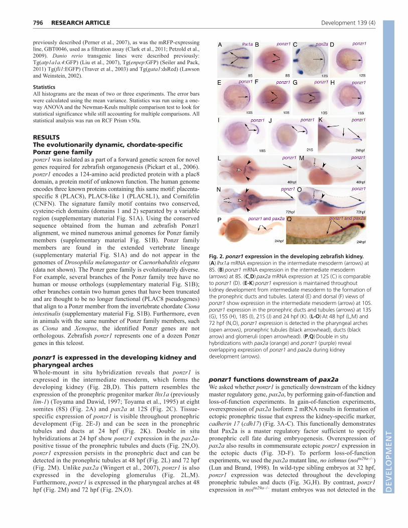
ponzr1 is expressed in the developing kidney andpharyngeal archesWhole-mount in situ hybridization reveals that ponzr1 isexpressed in the intermediate mesoderm, which forms thedeveloping kidney (Fig. 2B,D). This pattern resembles theexpression of the pronephric progenitor marker lhx1a (previouslylim-1) (Toyama and Dawid, 1997; Toyama et al., 1995) at eightsomites (8S) (Fig. 2A) and pax2a at 12S (Fig. 2C). Tissue-specific expression of ponzr1 is visible throughout pronephricdevelopment (Fig. 2E-J) and can be seen in the pronephrictubules and ducts at 24 hpf (Fig. 2K). Double in situhybridizations at 24 hpf show ponzr1 expression in the pax2a-positive tissue of the pronephric tubules and ducts (Fig. 2N,O).ponzr1 expression persists in the pronephric duct and can bedetected in the pronephric tubules at 48 hpf (Fig. 2L) and 72 hpf(Fig. 2M). Unlike pax2a (Wingert et al., 2007), ponzr1 is alsoexpressed in the developing glomerulus (Fig. 2L,M).Furthermore, ponzr1 is expressed in the pharyngeal arches at 48hpf (Fig. 2M) and 72 hpf (Fig. 2N,O).
ponzr1 functions downstream of pax2aWe asked whether ponzr1 is genetically downstream of the kidneymaster regulatory gene, pax2a, by performing gain-of-function andloss-of-function experiments. In gain-of-function experiments,overexpression of pax2a Isoform 2 mRNA results in formation ofectopic pronephric tissue that express the kidney-specific marker,cadherin 17 (cdh17) (Fig. 3A-C). This functionally demonstratesthat Pax2a is a master regulatory factor sufficient to specifypronephric cell fate during embryogenesis. Overexpression ofpax2a also results in commensurate ectopic ponzr1 expression inthe ectopic ducts (Fig. 3D-F). To perform loss-of-functionexperiments, we used the pax2a mutant line, no isthmus (noitu29a–/–)(Lun and Brand, 1998). In wild-type sibling embryos at 32 hpf,ponzr1 expression was detected throughout the developingpronephric tubules and ducts (Fig. 3G,H). By contrast, ponzr1expression in noitu29a–/– mutant embryos was not detected in the
RESEARCH ARTICLE Development 139 (4)
Fig. 2. ponzr1 expression in the developing zebrafish kidney.(A)lhx1a mRNA expression in the intermediate mesoderm (arrows) at8S. (B)ponzr1 mRNA expression in the intermediate mesoderm(arrows) at 8S. (C,D)pax2a mRNA expression at 12S (C) is comparableto ponzr1 (D). (E-K)ponzr1 expression is maintained throughoutkidney development from intermediate mesoderm to the formation ofthe pronephric ducts and tubules. Lateral (E) and dorsal (F) views ofponzr1 show expression in the intermediate mesoderm (arrow) at 10S.ponzr1 expression in the pronephric ducts and tubules (arrows) at 13S(G), 15S (H), 18S (I), 21S (J) and 24 hpf (K). (L-O)At 48 hpf (L,M) and72 hpf (N,O), ponzr1 expression is detected in the pharyngeal arches(open arrows), pronephric tubules (black arrowhead), ducts (blackarrow) and glomeruli (open arrowhead). (P,Q)Double in situhybridizations with pax2a (orange) and ponzr1 (purple) revealoverlapping expression of ponzr1 and pax2a during kidneydevelopment (arrows).
DEVELO
PMENT
DEVELO
PMENT
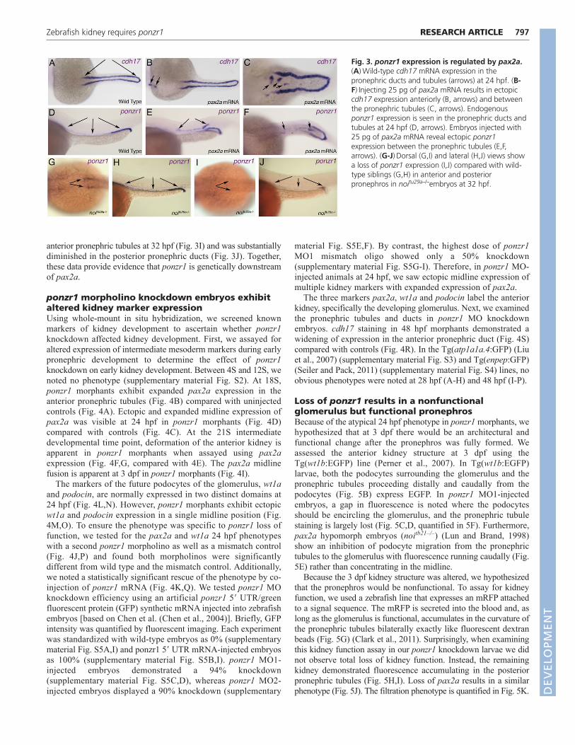
anterior pronephric tubules at 32 hpf (Fig. 3I) and was substantiallydiminished in the posterior pronephric ducts (Fig. 3J). Together,these data provide evidence that ponzr1 is genetically downstreamof pax2a.
ponzr1 morpholino knockdown embryos exhibitaltered kidney marker expressionUsing whole-mount in situ hybridization, we screened knownmarkers of kidney development to ascertain whether ponzr1knockdown affected kidney development. First, we assayed foraltered expression of intermediate mesoderm markers during earlypronephric development to determine the effect of ponzr1knockdown on early kidney development. Between 4S and 12S, wenoted no phenotype (supplementary material Fig. S2). At 18S,ponzr1 morphants exhibit expanded pax2a expression in theanterior pronephric tubules (Fig. 4B) compared with uninjectedcontrols (Fig. 4A). Ectopic and expanded midline expression ofpax2a was visible at 24 hpf in ponzr1 morphants (Fig. 4D)compared with controls (Fig. 4C). At the 21S intermediatedevelopmental time point, deformation of the anterior kidney isapparent in ponzr1 morphants when assayed using pax2aexpression (Fig. 4F,G, compared with 4E). The pax2a midlinefusion is apparent at 3 dpf in ponzr1 morphants (Fig. 4I).
The markers of the future podocytes of the glomerulus, wt1aand podocin, are normally expressed in two distinct domains at24 hpf (Fig. 4L,N). However, ponzr1 morphants exhibit ectopicwt1a and podocin expression in a single midline position (Fig.4M,O). To ensure the phenotype was specific to ponzr1 loss offunction, we tested for the pax2a and wt1a 24 hpf phenotypeswith a second ponzr1 morpholino as well as a mismatch control(Fig. 4J,P) and found both morpholinos were significantlydifferent from wild type and the mismatch control. Additionally,we noted a statistically significant rescue of the phenotype by co-injection of ponzr1 mRNA (Fig. 4K,Q). We tested ponzr1 MOknockdown efficiency using an artificial ponzr1 5� UTR/greenfluorescent protein (GFP) synthetic mRNA injected into zebrafishembryos [based on Chen et al. (Chen et al., 2004)]. Briefly, GFPintensity was quantified by fluorescent imaging. Each experimentwas standardized with wild-type embryos as 0% (supplementarymaterial Fig. S5A,I) and ponzr1 5� UTR mRNA-injected embryosas 100% (supplementary material Fig. S5B,I). ponzr1 MO1-injected embryos demonstrated a 94% knockdown(supplementary material Fig. S5C,D), whereas ponzr1 MO2-injected embryos displayed a 90% knockdown (supplementary
material Fig. S5E,F). By contrast, the highest dose of ponzr1MO1 mismatch oligo showed only a 50% knockdown(supplementary material Fig. S5G-I). Therefore, in ponzr1 MO-injected animals at 24 hpf, we saw ectopic midline expression ofmultiple kidney markers with expanded expression of pax2a.
The three markers pax2a, wt1a and podocin label the anteriorkidney, specifically the developing glomerulus. Next, we examinedthe pronephric tubules and ducts in ponzr1 MO knockdownembryos. cdh17 staining in 48 hpf morphants demonstrated awidening of expression in the anterior pronephric duct (Fig. 4S)compared with controls (Fig. 4R). In the Tg(atp1a1a.4:GFP) (Liuet al., 2007) (supplementary material Fig. S3) and Tg(enpep:GFP)(Seiler and Pack, 2011) (supplementary material Fig. S4) lines, noobvious phenotypes were noted at 28 hpf (A-H) and 48 hpf (I-P).
Loss of ponzr1 results in a nonfunctionalglomerulus but functional pronephrosBecause of the atypical 24 hpf phenotype in ponzr1 morphants, wehypothesized that at 3 dpf there would be an architectural andfunctional change after the pronephros was fully formed. Weassessed the anterior kidney structure at 3 dpf using theTg(wt1b:EGFP) line (Perner et al., 2007). In Tg(wt1b:EGFP)larvae, both the podocytes surrounding the glomerulus and thepronephric tubules proceeding distally and caudally from thepodocytes (Fig. 5B) express EGFP. In ponzr1 MO1-injectedembryos, a gap in fluorescence is noted where the podocytesshould be encircling the glomerulus, and the pronephric tubulestaining is largely lost (Fig. 5C,D, quantified in 5F). Furthermore,pax2a hypomorph embryos (noitb21–/–) (Lun and Brand, 1998)show an inhibition of podocyte migration from the pronephrictubules to the glomerulus with fluorescence running caudally (Fig.5E) rather than concentrating in the midline.
Because the 3 dpf kidney structure was altered, we hypothesizedthat the pronephros would be nonfunctional. To assay for kidneyfunction, we used a zebrafish line that expresses an mRFP attachedto a signal sequence. The mRFP is secreted into the blood and, aslong as the glomerulus is functional, accumulates in the curvature ofthe pronephric tubules bilaterally exactly like fluorescent dextranbeads (Fig. 5G) (Clark et al., 2011). Surprisingly, when examiningthis kidney function assay in our ponzr1 knockdown larvae we didnot observe total loss of kidney function. Instead, the remainingkidney demonstrated fluorescence accumulating in the posteriorpronephric tubules (Fig. 5H,I). Loss of pax2a results in a similarphenotype (Fig. 5J). The filtration phenotype is quantified in Fig. 5K.
797RESEARCH ARTICLEZebrafish kidney requires ponzr1
Fig. 3. ponzr1 expression is regulated by pax2a.(A)Wild-type cdh17 mRNA expression in thepronephric ducts and tubules (arrows) at 24 hpf. (B-F)Injecting 25 pg of pax2a mRNA results in ectopiccdh17 expression anteriorly (B, arrows) and betweenthe pronephric tubules (C, arrows). Endogenousponzr1 expression is seen in the pronephric ducts andtubules at 24 hpf (D, arrows). Embryos injected with25 pg of pax2a mRNA reveal ectopic ponzr1expression between the pronephric tubules (E,F,arrows). (G-J)Dorsal (G,I) and lateral (H,J) views showa loss of ponzr1 expression (I,J) compared with wild-type siblings (G,H) in anterior and posteriorpronephros in noitu29a–/–embryos at 32 hpf.
DEVELO
PMENT
DEVELO
PMENT

798
As the wt1b fluorescence showed altered architecture of thepodocytes and pronephric tubules, we examined the glomerulususing serial sections stained with haematoxylin and eosin (H&E).In wild-type embryos, the glomerulus (arrowhead, Fig. 5L) isclearly visible, as are the flanking tubules (arrows, Fig. 5L) andpronephric ducts (Fig. 5O). In noitb21–/– mutant embryos, theglomerulus is unaltered (Fig. 5M), but the pronephric ducts areovertly dilated (Fig. 5P). This phenotype is comparable to thatnoted in noitb29a–/– mutant embryos (Majumdar et al., 2000). Bycontrast, ponzr1 morphants show a loss of the central glomerulusand a dilation of the pronephric tubules (Fig. 5N) as well as the
ducts (Fig. 5Q). These data confirm that ponzr1 morphants lack acentralized glomerulus but are still able to retain some kidneyfiltration function.
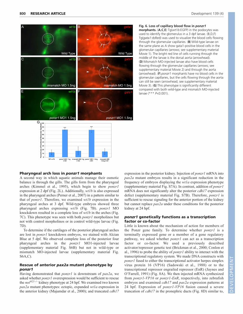
To further assess the defect in the glomerulus, in ponzr1morphant embryos we examined the glomerular capillaries, whichmake up the bulk of the cells within the podocytes. Because thevasculature is so abundant in the head, we were unable to visualizethe capilliaries using Tg(fli1:EGFP) (Lawson and Weinstein, 2002)(data not shown). Moreover, confidence in focal plane alignmentwas low in that line. Therefore, we used a double transgenicTg(wt1b:EGFP) and Tg(gata1:dsRed) (Traver et al., 2003) to ask
RESEARCH ARTICLE Development 139 (4)
Fig. 4. ponzr1 is required for correct kidneypatterning by pax2a. (A)pax2a mRNA expression inanterior pronephric tubules (arrows) at 18S.(B)Expanded expression of pax2a at 18S (arrows) inponzr1 morphants. (C)Wild-type expression of pax2ain the anterior pronephric tubules (arrows) at 24 hpf.(D)ponzr1 morphants show expanded/ectopic pax2aexpression toward the midline at 24 hpf (arrow).(E)Wild-type pax2a at 21S (arrows). (F,G)ponzr1morphants show a pax2a phenotype intermediate 18Sand 24 hpf where the invagination (F) of pax2a-expressing cells and the start of the midline fusion (G)can be seen. (H)Wild-type pax2a at 3 dpf. (I)ponzr1morphants show a maintenance of the ectopicmidline expression of pax2a (arrow). (J)Ectopic pax2aphenotype is seen using two independent ponzr1morpholinos but not in ponzr1 mismatch control.(K)Ectopic pax2a phenotype is rescued using ponzr1mRNA (P<0.001) but not dsRed mRNA. (L-O)wt1a (L)and podocin (N) mark future podocytes with twodistinct expression domains at 24 hpf in controls(arrows). A single midline wt1a (M) and podocin (O)expression domain is seen in ponzr1 morphants at 24hpf. (P,Q)wt1a phenotype is seen with two ponzr1morpholinos (P) and can be rescued using ponzr1mRNA (Q, P<0.001). (R,S)cdh17 expression isdetected in the pronephric ducts (R, arrows) andtubules at 48 hpf in controls but is expanded in thepronephric ducts (S, arrows) in ponzr1 morphants at48 hpf. ***P<0.001
DEVELO
PMENT
DEVELO
PMENT

whether the glomerulus had functional capillaries. We used anupdated Specimen in a Corrected Optical Rotational Enclosure(SCORE) protocol (Petzold et al., 2010) combined with a 20Xwater-emersion, long working-distance lens to visualize thesamples and a high-definition camera to record the data. TheTg(wt1b:EGFP) was used to focus on the glomerulus; then wemonitored RFP fluorescence to visualize blood flow through thecapillaries. In both uninjected embryos (supplementary material
Movie 1; Fig. 6A,B) as well as mismatch MO controls(supplementary material Movie 2; Fig. 6C,D), blood cells could beseen moving through the capillaries. However, in ponzr1 MO1-injected larvae, no blood cells could be seen flowing through thearea where the glomerular capillaries should be located(supplementary material Movie 3; Fig. 6E,F). The loss of bloodflow in the ponzr1 MO-injected larval glomeruli is significantlydifferent from both wild-type and mismatch MO controls (Fig. 6G).
799RESEARCH ARTICLEZebrafish kidney requires ponzr1
Fig. 5. ponzr1 is required for anterior kidney form and function. (A)A drawing of a larval zebrafish at 3-4 dpf. The green box is the area ofthe larva seen in images B-E, and the red box is the area seen in images G-J. (B)Tg(wt1b:EGFP) larvae (Perner et al., 2007) mark podocytessurrounding the glomerulus (arrowhead) and the pronephric tubules (arrows). (C,D)ponzr1 morphants show a loss of fluorescence in the areasurrounding the glomerulus, except at the midline (brackets). The pronephric tubular staining is lost (arrows) in a dose-dependent manner (D: 1 ngMO1; E: 1.5 ng MO1). (E)noitb21–/– mutant embryos exhibit a loss of fluorescence around the glomerulus (brackets) and the pronephric tubulefluorescence runs posteriorly instead of laterally (arrows). (F)Two ponzr1 MOs induce a similar phenotype that is ameliorated in mismatch controlMO injections. (G)mRFP appears bilaterally in the curvature of the pronephric tubules (arrows) in control embryos due to glomerular filtration. (H-J)In ponzr1 morphants, mRFP is collected in a more posterior part of the tubules – sometimes only one pronephric tubule collects fluorescenceand sometimes fluorescence is seen in both (H: 1 ng MO1; I: 1.5 ng MO1). A similar phenotype is seen in noitb21–/– mutant embryos (J).(K)Percentage of embryos with the filtration phenotype. (L-Q)Larvae at 4dpf that have been sectioned and H&E stained. (L)Uninjected embryosshow a glomerulus (arrowhead) with two pronephric tubules (arrows). (M)noitb21–/– mutant embryo has a glomerulus (arrowhead) but showsdilated tubules (arrow). (N)ponzr1 morphants show dilated pronephric tubules (arrows) with no glomerulus. (O)The posterior pronephric tubules inwild-type embryos (arrows). noitb21–/– mutant (P) and ponzr1 morphant embryos (Q) both show dilated tubules (arrows). **P<0.01, ***P<0.001
DEVELO
PMENT
DEVELO
PMENT
800
Pharyngeal arch loss in ponzr1 morphantsA second way in which aquatic animals manage their osmoticbalance is through the gills. The gills form from the pharyngealarches (Kimmel et al., 1995), which begin to show ponzr1expression at 2 dpf (Fig. 2L). Additionally, wt1b is also expressedin the pharyngeal arches (Perner et al., 2007) in a pattern similar tothat of ponzr1. Therefore, we examined wt1b expression in thepharyngeal arches at 3 dpf. Wild-type embryos showed threepharyngeal arches expressing wt1b (Fig. 7B). ponzr1 MOknockdown resulted in a complete loss of wt1b in the arches (Fig.7C). This phenotype was seen with both ponzr1 morpholinos butnot with control morpholinos or in control wild-type larvae (Fig.7D).
To determine if the cartilages of the posterior pharyngeal archesare lost in ponzr1 knockdown embryos, we stained with AlcianBlue at 5 dpf. We observed complete loss of the posterior fourpharyngeal arches in the ponzr1 MO1-injected larvae(supplementary material Fig. S6B) but not in wild-type ormismatch MO-injected larvae (supplementary material Fig.S6A,C).
Rescue of anterior pax2a mutant phenotype byponzr1Having demonstrated that ponzr1 is downstream of pax2a, weasked whether ponzr1 overexpression would be sufficient to rescuethe noitb21–/– kidney phenotype at 24 hpf. We examined two knownpax2a mutant phenotypes: ectopic, expanded wt1a expression inthe anterior kidney (Majumdar et al., 2000); and truncated cdh17
expression in the posterior kidney. Injection of ponzr1 mRNA intopax2a mutant embryos results in a significant reduction in thefrequency of embryos displaying the wt1a expression phenotype(supplementary material Fig. S7A). In contrast, addition of ponzr1mRNA does not significantly alter the posterior cdh17 expressiondefect (supplementary material Fig. S7B). Therefore, ponzr1 issufficient to rescue signaling for the anterior portion of the kidneybut cannot replace pax2a under these conditions for the posteriorkidney at 24 hpf.
ponzr1 genetically functions as a transcriptionfactor or co-factorLittle is known about the mechanism of action for members ofthe Ponzr gene family. To determine whether ponzr1 is aterminally expressed gene or a member of a gene regulatorypathway, we asked whether ponzr1 can act as a transcriptionfactor or co-factor. We used a previously describedactivator/repressor genetic test (Brickman et al., 2000; Conlon etal., 1996) to probe the ability of ponzr1 ability to interact with thetranscriptional regulatory system. We made DNA constructs withponzr1 fused to either the transcriptional activator herpes simplexvirus protein 16 (VP16) (Sadowski et al., 1988) or to thetranscriptional repressor engrailed repressor (EnR) (Jaynes andO’Farrell, 1991) (Fig. 8A). We then injected mRNA synthesizedfrom ponzr1-VP16 or ponzr1-EnR, respectively, into zebrafishembryos and examined cdh17 and pax2a expression patterns at24 hpf. Expression of ponzr1-VP16 fusion caused a severetruncation of cdh17 in the pronephric ducts (Fig. 8D) similar to,
RESEARCH ARTICLE Development 139 (4)
Fig. 6. Loss of capillary blood flow in ponzr1morphants. (A,C,E) Tg(wt1b:EGFP) in the podocytes wasused to identify the glomerulus in a 3 dpf larvae. (B,D,F)Tg(gata1:dsRed) was used to visualize the blood cells flowingthrough the glomerular capillaries. (B)Wild-type larvae onthe same plane as A show gata1-positive blood cells in theglomerular capillaries (arrows; see supplementary materialMovie 1). The bright red line of cells running through themiddle of the larvae is the dorsal aorta (arrowhead).(D)Mismatch MO-injected larvae also have blood cellsflowing through the glomerular capillaries (arrows; seesupplementary material Movie 2) and through the aorta(arrowhead). (F)ponzr1 morphants have no blood cells in theglomerular capillaries, but the cells flowing through the aortacan still be seen (arrowhead; see supplementary materialMovie 3). (G)This phenotype is significantly differentcompared with both wild-type and mismatch MO-injectedlarvae (*** P<0.001).
DEVELO
PMENT
DEVELO
PMENT

but more extreme than, the overexpression of ponzr1 mRNA (Fig.8C). The truncation phenotype is significantly increasedcompared with wild-type embryos (Fig. 8E). Injections ofponzr1-VP16 mRNA also resulted in a reduction in pax2aexpression in the anterior pronephric tubules (Fig. 8G,J)compared with controls (Fig. 8F,I). By contrast, expression of theponzr1-EnR mRNA resulted in enhanced pax2a expression (Fig.8H,K), resembling the change of pax2a noted in ponzr1 morphantembryos (Fig. 4B). However, we did not observe a cdh17phenotype in ponzr1-EnR injected embryos (Fig. 8E). The pax2aphenotypes seen in the ponzr1-VP16 and ponzr1-EnR injections
are significantly different from wild type (Fig. 8L). Together,these data strongly suggest that ponzr1 normally functions as atranscription factor or co-factor during kidney development.
DISCUSSIONThe mysterious glomerulusThe conserved regulatory gene, pax2, plays a conserved role inkidney biology from flies to humans. However, because pax2 is akey kidney regulatory gene, it is found in both glomerular andaglomerular vertebrates, presenting a conundrum regarding themechanism underlying differences in kidney biology between
801RESEARCH ARTICLEZebrafish kidney requires ponzr1
Fig. 7. ponzr1 is required for wt1b-expressing pharyngeal arches. (A)A drawing of a 3 dpf larval zebrafish showing where the pharyngealarches are located. The green box is the area seen in images B and C. (B)Tg(wt1b:EGFP) larvae 3 dpf lateral view shows three pharyngeal archesexpress wt1b. (C)When ponzr1 MO is injected, the pharyngeal arches are lost. (D)The loss of pharyngeal arches phenotype is significantly differentin the two ponzr1 MOs compared with both the wild type and mismatch controls. **P<0.01, ***P<0.001
Fig. 8. ponzr1 can function as a transcription factoror co-factor. (A)Diagrams showing the transcriptionalactivator and repressor mRNAs injected into zebrafish. TheNLS was included to facilitate nuclear access of the fusionproteins. (B)Dorsal view of wild-type cdh17 expression inthe pronephric ducts at 24 hpf. (C)ponzr1 mRNA-injectedembryos reveal cdh17 expression loss at the cloaca(arrow). (D)ponzr1-VP16 mRNA-injected embryos show aseverely truncated cdh17 expression in the pronephricducts (arrows). (E)cdh17 phenotype in ponzr1- andponzr1-VP16-injected embryos are significantly differentfrom control, whereas ponzr1-EnR-injected embryos arenot. (F)Wild-type pax2a expression in the anteriorpronephric ducts at 24 hpf (magnified I). (G-K)ponzr1-VP16 mRNA-injected embryos (G) show reduced pax2aexpression (magnified J) whereas ponzr1-EnR mRNA-injected embryos (H) demonstrate expanded pax2aexpression in the anterior pronephric ducts (arrows)(magnified K) at 24 hpf. (L)Quantification of pax2aphenotypes standardized to wild-type pax2a at 100%.Percentage of embryos with reduced pax2a is seen asbelow 100% (as seen in ponzr1VP16-injected embryos)and expanded pax2a expression is seen above 100% (asseen in ponzr1EnR-injected embryos). Both ponzr1-VP16and ponzr1-EnR are significantly different from wild type.***P<0.001
DEVELO
PMENT
DEVELO
PMENT
802
vertebrate lineages. The glomerulus is a complex organ structurewhose development requires multiple cell types of diverse origin(Ditrich, 2005; Kramer-Zucker et al., 2005). Over the past few years,great strides have been made in discovering the molecular signals fortubular and ductal development (Drummond, 2004; Drummond etal., 1998; Vize et al., 1997) as well as identifying signals directingindividual cell types, such as the vasculature (Eremina and Quaggin,2004; Kitamoto et al., 1997; Majumdar and Drummond, 1999) andpodocytes (Majumdar and Drummond, 2000; Quaggin andKreidberg, 2008), to migrate into the glomerulus. However,localized, cell-type-specific development of a centralized glomerulusrequires coordination through global signals that remain largelyunidentified. For example, VEGF has been implicated in glomerularvascularization, but what regulates VEGF in this developmentalsetting is unknown (Eremina and Quaggin, 2004; Kitamoto et al.,1997). We show that in zebrafish, ponzr1 is needed to organize thefiltration apparatus into a single, central glomerular structure. Howponzr1, a likely transcription factor or co-factor, regulates these cellmovements and other processes underlying zebrafish kidney biologyis a key next step to furthering our understanding of the role ofponzr1 in organogenesis.
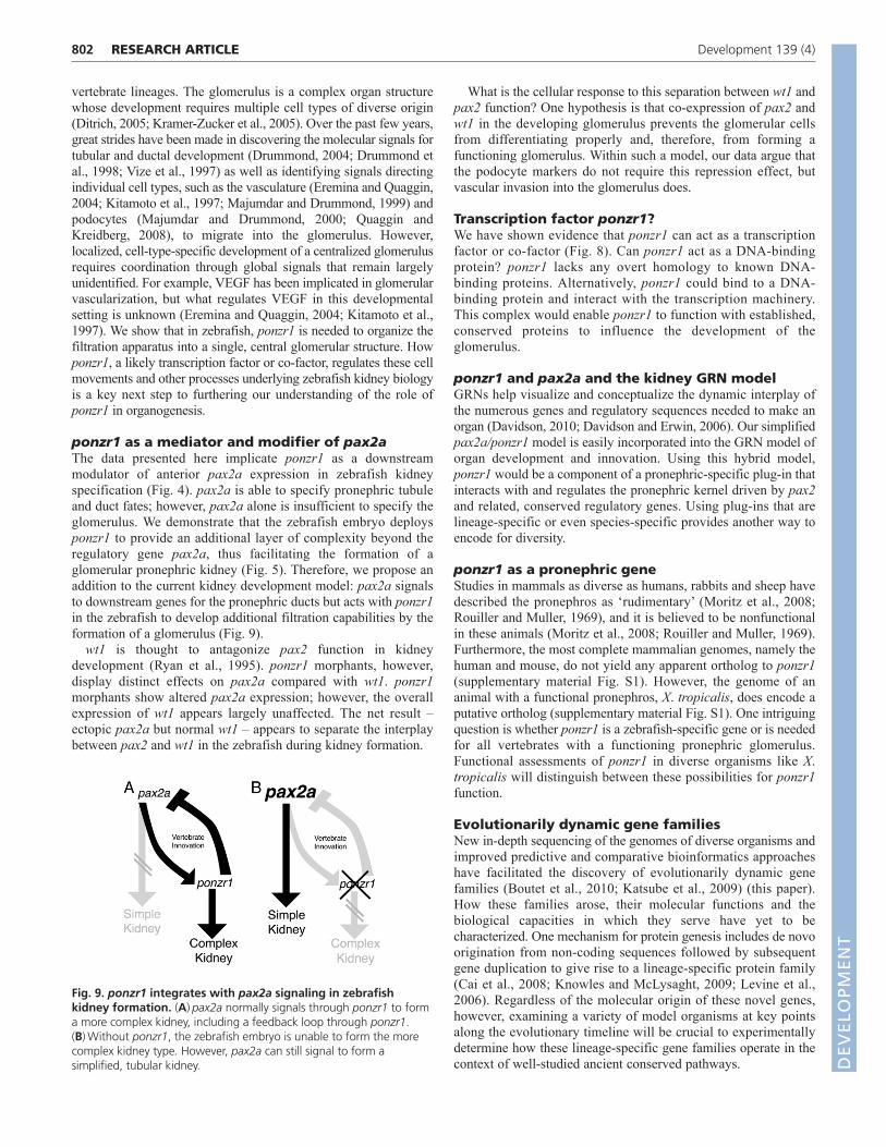
ponzr1 as a mediator and modifier of pax2aThe data presented here implicate ponzr1 as a downstreammodulator of anterior pax2a expression in zebrafish kidneyspecification (Fig. 4). pax2a is able to specify pronephric tubuleand duct fates; however, pax2a alone is insufficient to specify theglomerulus. We demonstrate that the zebrafish embryo deploysponzr1 to provide an additional layer of complexity beyond theregulatory gene pax2a, thus facilitating the formation of aglomerular pronephric kidney (Fig. 5). Therefore, we propose anaddition to the current kidney development model: pax2a signalsto downstream genes for the pronephric ducts but acts with ponzr1in the zebrafish to develop additional filtration capabilities by theformation of a glomerulus (Fig. 9).
wt1 is thought to antagonize pax2 function in kidneydevelopment (Ryan et al., 1995). ponzr1 morphants, however,display distinct effects on pax2a compared with wt1. ponzr1morphants show altered pax2a expression; however, the overallexpression of wt1 appears largely unaffected. The net result –ectopic pax2a but normal wt1 – appears to separate the interplaybetween pax2 and wt1 in the zebrafish during kidney formation.
What is the cellular response to this separation between wt1 andpax2 function? One hypothesis is that co-expression of pax2 andwt1 in the developing glomerulus prevents the glomerular cellsfrom differentiating properly and, therefore, from forming afunctioning glomerulus. Within such a model, our data argue thatthe podocyte markers do not require this repression effect, butvascular invasion into the glomerulus does.
Transcription factor ponzr1?We have shown evidence that ponzr1 can act as a transcriptionfactor or co-factor (Fig. 8). Can ponzr1 act as a DNA-bindingprotein? ponzr1 lacks any overt homology to known DNA-binding proteins. Alternatively, ponzr1 could bind to a DNA-binding protein and interact with the transcription machinery.This complex would enable ponzr1 to function with established,conserved proteins to influence the development of theglomerulus.
ponzr1 and pax2a and the kidney GRN modelGRNs help visualize and conceptualize the dynamic interplay ofthe numerous genes and regulatory sequences needed to make anorgan (Davidson, 2010; Davidson and Erwin, 2006). Our simplifiedpax2a/ponzr1 model is easily incorporated into the GRN model oforgan development and innovation. Using this hybrid model,ponzr1 would be a component of a pronephric-specific plug-in thatinteracts with and regulates the pronephric kernel driven by pax2and related, conserved regulatory genes. Using plug-ins that arelineage-specific or even species-specific provides another way toencode for diversity.
ponzr1 as a pronephric geneStudies in mammals as diverse as humans, rabbits and sheep havedescribed the pronephros as ‘rudimentary’ (Moritz et al., 2008;Rouiller and Muller, 1969), and it is believed to be nonfunctionalin these animals (Moritz et al., 2008; Rouiller and Muller, 1969).Furthermore, the most complete mammalian genomes, namely thehuman and mouse, do not yield any apparent ortholog to ponzr1(supplementary material Fig. S1). However, the genome of ananimal with a functional pronephros, X. tropicalis, does encode aputative ortholog (supplementary material Fig. S1). One intriguingquestion is whether ponzr1 is a zebrafish-specific gene or is neededfor all vertebrates with a functioning pronephric glomerulus.Functional assessments of ponzr1 in diverse organisms like X.tropicalis will distinguish between these possibilities for ponzr1function.
Evolutionarily dynamic gene familiesNew in-depth sequencing of the genomes of diverse organisms andimproved predictive and comparative bioinformatics approacheshave facilitated the discovery of evolutionarily dynamic genefamilies (Boutet et al., 2010; Katsube et al., 2009) (this paper).How these families arose, their molecular functions and thebiological capacities in which they serve have yet to becharacterized. One mechanism for protein genesis includes de novoorigination from non-coding sequences followed by subsequentgene duplication to give rise to a lineage-specific protein family(Cai et al., 2008; Knowles and McLysaght, 2009; Levine et al.,2006). Regardless of the molecular origin of these novel genes,however, examining a variety of model organisms at key pointsalong the evolutionary timeline will be crucial to experimentallydetermine how these lineage-specific gene families operate in thecontext of well-studied ancient conserved pathways.
RESEARCH ARTICLE Development 139 (4)
Fig. 9. ponzr1 integrates with pax2a signaling in zebrafishkidney formation. (A)pax2a normally signals through ponzr1 to forma more complex kidney, including a feedback loop through ponzr1.(B)Without ponzr1, the zebrafish embryo is unable to form the morecomplex kidney type. However, pax2a can still signal to form asimplified, tubular kidney. D
EVELO
PMENT
DEVELO
PMENT

Do ancient pathways and lineage-specific geneslead to new structures?The data presented in this study represent functional evidence thatmembers of evolutionarily dynamic gene families can provide anadditional pathway to innovation. We find that one member of adynamic family, ponzr1, is integrated into the ancient pax2pathway, facilitating innovation in the pronephros through theformation of the glomerulus. Whether other Ponzr family membersor additional dynamic gene families – only now being discoveredthrough the extensive new comparative genomics projects – areresponsible for diversity in additional organ systems is unknown.Do other lineage-specific genome additions function in ancientpathways to encode for innovation?
AcknowledgementsWe thank Dr Englert for the Tg(wt1b:EGFP) fish line, Dr Drummond for theTg(atp1a1a.4:GFP) fish line, Dr Pack for the Tg(enpep.4:GFP) fish line, andStephanie Westcot and Dr Keith Cheng for key manuscript revisions.
FundingThis work was funded by National Institute of General Medical Sciences(NIGMS) [GM63904]; National Institute of Diabetes and Digestive and KidneyDiseases (NIDDK) [1F30DK083219-01]; and National Institute on Drug Abuse(NIDA) [DA14546]. Deposited in PMC for release after 12 months.
Competing interests statementThe authors declare no competing financial interests.
Supplementary materialSupplementary material available online athttp://dev.biologists.org/lookup/suppl/doi:10.1242/dev.071720/-/DC1
ReferencesBeyenbach, K. W. (2004). Kidneys sans glomeruli. Am. J. Physiol. Renal Physiol.
286, F811-F827.Bolouri, H. and Davidson, E. H. (2002). Modeling DNA sequence-based cis-
regulatory gene networks. Dev. Biol. 246, 2-13.Boutet, A., Comai, G. and Schedl, A. (2010). The WTX/AMER1 gene family:
evolution, signature and function. BMC Evol. Biol. 10, 280.Brand, M., Heisenberg, C. P., Jiang, Y. J., Beuchle, D., Lun, K., Furutani-Seiki,
M., Granato, M., Haffter, P., Hammerschmidt, M., Kane, D. A. et al. (1996).Mutations in zebrafish genes affecting the formation of the boundary betweenmidbrain and hindbrain. Development 123, 179-190.
Brickman, J. M., Jones, C. M., Clements, M., Smith, J. C. and Beddington, R.S. (2000). Hex is a transcriptional repressor that contributes to anterior identityand suppresses Spemann organiser function. Development 127, 2303-2315.
Cadigan, K. M. and Nusse, R. (1997). Wnt signaling: a common theme in animaldevelopment. Genes Dev. 11, 3286-3305.
Cagan, R. (2003). The signals that drive kidney development: a view from the flyeye. Curr. Opin. Nephrol. Hypertens. 12, 11-17.
Cai, J., Zhao, R., Jiang, H. and Wang, W. (2008). De novo origination of a newprotein-coding gene in Saccharomyces cerevisiae. Genetics 179, 487-496.
Carroll, S. B. (2005). Endless Forms Most Beautiful: the New Science of Evo Devoand the Making of the Animal Kingdom. New York: Norton.
Chen, E., Hermanson, S. and Ekker, S. C. (2004). Syndecan-2 is essential forangiogenic sprouting during zebrafish development. Blood 103, 1710-1719.
Clark, K. J., Balciunas, D., Pogoda, H. M., Ding, Y., Westcot, S. E., Bedell, V.M., Greenwood, T. M., Urban, M. D., Skuster, K. J., Petzold, A. M. et al.(2011). In vivo protein trapping produces a functional expression codex of thevertebrate proteome. Nat. Methods 8, 506-515.
Cohn, M. J. and Tickle, C. (1999). Developmental basis of limblessness and axialpatterning in snakes. Nature 399, 474-479.
Conlon, F. L., Sedgwick, S. G., Weston, K. M. and Smith, J. C. (1996).Inhibition of Xbra transcription activation causes defects in mesodermalpatterning and reveals autoregulation of Xbra in dorsal mesoderm. Development122, 2427-2435.
Dantzler, W. H. (1989). Comparative Physiology of the Vertebrate Kidney. Berlin:Springer-Verlag.
Darwin, C. (1890). The Origin of Species by Means of Natural Selection. NewYork: D. Appleton and Company.
Davidson, E. H. (2010). Emerging properties of animal gene regulatory networks.Nature 468, 911-920.
Davidson, E. H. and Erwin, D. H. (2006). Gene regulatory networks and theevolution of animal body plans. Science 311, 796-800.
Demuth, J. P., De Bie, T., Stajich, J. E., Cristianini, N. and Hahn, M. W. (2006).The evolution of mammalian gene families. PLoS ONE 1, e85.
Ditrich, H. (2005). Renal Structure and Function in Vertebrates. Enfield, NH:Science Publishers.
Dressler, G. R. (2006). The cellular basis of kidney development. Annu. Rev. CellDev. Biol. 22, 509-529.
Drummond, I. A. (2004). Zebrafish kidney development. Methods Cell Biol. 76,501-530.
Drummond, I. A. and Majumdar, A. (2003). The pronephric glomerulus. In TheKidney From Normal Development to Congenital Disease (ed. P. D. Vize, A. S.Woolf and J. B. L. Bard), pp. 61-73. San Diego: Academic Press.
Drummond, I. A., Majumdar, A., Hentschel, H., Elger, M., Solnica-Krezel, L.,Schier, A. F., Neuhauss, S. C., Stemple, D. L., Zwartkruis, F., Rangini, Z. etal. (1998). Early development of the zebrafish pronephros and analysis ofmutations affecting pronephric function. Development 125, 4655-4667.
Dyce, K. M., Sack, W. O. and Wensing, C. J. G. (1995). Textbook of VeterinaryAnatomy. Philadelphia: Saunders.
Eremina, V. and Quaggin, S. E. (2004). The role of VEGF-A in glomerulardevelopment and function. Curr. Opin. Nephrol. Hypertens. 13, 9-15.
Force, A., Lynch, M., Pickett, F. B., Amores, A., Yan, Y. L. and Postlethwait, J.(1999). Preservation of duplicate genes by complementary, degenerativemutations. Genetics 151, 1531-1545.
Frandson, R. D., Wilke, W. L. and Fails, A. D. (2009). Anatomy and Physiologyof Farm Animals. Ames, Iowa: Wiley-Blackwell.
Gilbert, S. F. (2000). Developmental Biology. Sunderland, MA: Sinauer Associates.Gilchrist, M., Thorsson, V., Li, B., Rust, A. G., Korb, M., Roach, J. C., Kennedy,
K., Hai, T., Bolouri, H. and Aderem, A. (2006). Systems biology approachesidentify ATF3 as a negative regulator of Toll-like receptor 4. Nature 441, 173-178.
Hersh, B. M. and Carroll, S. B. (2005). Direct regulation of knot gene expressionby Ultrabithorax and the evolution of cis-regulatory elements in Drosophila.Development 132, 1567-1577.
Howland, R. B. (1921). Experiments on the effect of removal of the pronephros ofAmblystoma punctatum. J. Exp. Zool. 32, 355-396.
Hyatt, T. M. and Ekker, S. C. (1999). Vectors and techniques for ectopic geneexpression in zebrafish. Methods Cell Biol. 59, 117-126.
Jaynes, J. B. and O’Farrell, P. H. (1991). Active repression of transcription by theengrailed homeodomain protein. EMBO J. 10, 1427-1433.
Katsube, K., Sakamoto, K., Tamamura, Y. and Yamaguchi, A. (2009). Role ofCCN, a vertebrate specific gene family, in development. Dev. Growth Differ. 51,55-67.
Kimmel, C. B., Ballard, W. W., Kimmel, S. R., Ullmann, B. and Schilling, T. F.(1995). Stages of embryonic development of the zebrafish. Dev. Dyn. 203, 253-310.
Kingsley, D. M. (1994). The TGF-beta superfamily: new members, new receptors,and new genetic tests of function in different organisms. Genes Dev. 8, 133-146.
Kitamoto, Y., Tokunaga, H. and Tomita, K. (1997). Vascular endothelial growthfactor is an essential molecule for mouse kidney development: glomerulogenesisand nephrogenesis. J. Clin. Invest. 99, 2351-2357.
Knowles, D. G. and McLysaght, A. (2009). Recent de novo origin of humanprotein-coding genes. Genome Res. 19, 1752-1759.
Kramer-Zucker, A. G., Wiessner, S., Jensen, A. M. and Drummond, I. A.(2005). Organization of the pronephric filtration apparatus in zebrafish requiresNephrin, Podocin and the FERM domain protein Mosaic eyes. Dev. Biol. 285,316-329.
Krauss, S., Johansen, T., Korzh, V. and Fjose, A. (1991). Expression of thezebrafish paired box gene pax[zf-b] during early neurogenesis. Development113, 1193-1206.
Lawson, N. D. and Weinstein, B. M. (2002). In vivo imaging of embryonicvascular development using transgenic zebrafish. Dev. Biol. 248, 307-318.
Levine, M. T., Jones, C. D., Kern, A. D., Lindfors, H. A. and Begun, D. J.(2006). Novel genes derived from noncoding DNA in Drosophila melanogasterare frequently X-linked and exhibit testis-biased expression. Proc. Natl. Acad. Sci.USA 103, 9935-9939.
Lewis, E. B. (1978). A gene complex controlling segmentation in Drosophila.Nature 276, 565-570.
Liu, Y., Pathak, N., Kramer-Zucker, A. and Drummond, I. A. (2007). Notchsignaling controls the differentiation of transporting epithelia and multiciliatedcells in the zebrafish pronephros. Development 134, 1111-1122.
Lun, K. and Brand, M. (1998). A series of no isthmus (noi) alleles of the zebrafishpax2.1 gene reveals multiple signaling events in development of the midbrain-hindbrain boundary. Development 125, 3049-3062.
Majumdar, A. and Drummond, I. A. (1999). Podocyte differentiation in theabsence of endothelial cells as revealed in the zebrafish avascular mutant,cloche. Dev. Genet. 24, 220-229.
Majumdar, A. and Drummond, I. A. (2000). The zebrafish floating head mutantdemonstrates podocytes play an important role in directing glomerulardifferentiation. Dev. Biol. 222, 147-157.
803RESEARCH ARTICLEZebrafish kidney requires ponzr1
DEVELO
PMENT
DEVELO
PMENT
804
Majumdar, A., Lun, K., Brand, M. and Drummond, I. A. (2000). Zebrafish noisthmus reveals a role for pax2.1 in tubule differentiation and patterning eventsin the pronephric primordia. Development 127, 2089-2098.
Marshall, E. K. J. (1930). A comparison of the function of the glomerular andaglomerular kidney. Am. J. Physiol. 94, 1-10.
Moritz, K. M., Wintour-Coghlan, M., Bertram, J. F., Black, M. J. and Caruana,G. (2008). Factors Influencing Mammalian Kidney Development: Implications forHealth in Adult Life. Berlin: Springer-Verlag.
Nasevicius, A. and Ekker, S. C. (2000). Effective targeted gene ‘knockdown’ inzebrafish. Nat. Genet. 26, 216-220.
Obbard, D. J., Welch, J. J. and Little, T. J. (2009). Inferring selection in theAnopheles gambiae species complex: an example from immune-related serineprotease inhibitors. Malar. J. 8, 117.
Oliver, J. (1968). Nephrons and Kidneys; a Quantitative Study of Developmentaland Evolutionary Mammalian Renal Architectonics. New York: Hoeber MedicalDivision.
Perner, B., Englert, C. and Bollig, F. (2007). The Wilms tumor genes wt1a andwt1b control different steps during formation of the zebrafish pronephros. Dev.Biol. 309, 87-96.
Peter, I. S. and Davidson, E. H. (2009). Modularity and design principles in thesea urchin embryo gene regulatory network. FEBS Lett. 583, 3948-3958.
Petzold, A. M., Balciunas, D., Sivasubbu, S., Clark, K. J., Bedell, V. M.,Westcot, S. E., Myers, S. R., Moulder, G. L., Thomas, M. J. and Ekker, S. C.(2009). Nicotine response genetics in the zebrafish. Proc. Natl. Acad. Sci. USA106, 18662-18667.
Petzold, A. M., Bedell, V. M., Boczek, N. J., Essner, J. J., Balciunas, D., Clark,K. J. and Ekker, S. C. (2010). SCORE imaging: specimen in a corrected opticalrotational enclosure. Zebrafish 7, 149-154.
Pickart, M. A., Klee, E. W., Nielsen, A. L., Sivasubbu, S., Mendenhall, E. M.,Bill, B. R., Chen, E., Eckfeldt, C. E., Knowlton, M., Robu, M. E. et al. (2006).Genome-wide reverse genetics framework to identify novel functions of thevertebrate secretome. PLoS ONE 1, e104.
Price, G. C. (1910). The structure and function of the adult head kidney ofBdellostoma stouti. J. Exp. Zool. 9, 849-864.
Punzo, C., Seimiya, M., Flister, S., Gehring, W. J. and Plaza, S. (2002).Differential interactions of eyeless and twin of eyeless with the sine oculisenhancer. Development 129, 625-634.
Quaggin, S. E. and Kreidberg, J. A. (2008). Development of the renalglomerulus: good neighbors and good fences. Development 135, 609-620.
Rouiller, C. and Muller, A. F. (1969). The Kidney: Morphology, Biochemistry,Physiology. New York: Academic Press.
Ryan, G., Steele-Perkins, V., Morris, J. F., Rauscher, F. J., 3rd and Dressler, G.R. (1995). Repression of Pax-2 by WT1 during normal kidney development.Development 121, 867-875.
Sadowski, I., Ma, J., Triezenberg, S. and Ptashne, M. (1988). GAL4-VP16 is anunusually potent transcriptional activator. Nature 335, 563-564.
Sanyanusin, P., Schimmenti, L. A., McNoe, L. A., Ward, T. A., Pierpont, M. E.,Sullivan, M. J., Dobyns, W. B. and Eccles, M. R. (1995). Mutation of thePAX2 gene in a family with optic nerve colobomas, renal anomalies andvesicoureteral reflux. Nat. Genet. 9, 358-364.
Schimmenti, L. A., Cunliffe, H. E., McNoe, L. A., Ward, T. A., French, M. C.,Shim, H. H., Zhang, Y. H., Proesmans, W., Leys, A., Byerly, K. A. et al.(1997). Further delineation of renal-coloboma syndrome in patients withextreme variability of phenotype and identical PAX2 mutations. Am. J. Hum.Genet. 60, 869-878.
Seiler, C. and Pack, M. (2011). Transgenic labeling of the zebrafish pronephricduct and tubules using a promoter from the enpep gene. Gene Expr. Patterns11, 118-121.
Silver, S. J. and Rebay, I. (2005). Signaling circuitries in development: insightsfrom the retinal determination gene network. Development 132, 3-13.
Smith, H. W. (1937). The Physiology of the Kidney. New York: Oxford UniversityPress.
Thisse, C. and Thisse, B. (2008). High-resolution in situ hybridization to whole-mount zebrafish embryos. Nat. Protoc. 3, 59-69.
Torres, M., Gomez-Pardo, E., Dressler, G. R. and Gruss, P. (1995). Pax-2 controlsmultiple steps of urogenital development. Development 121, 4057-4065.
Toyama, R. and Dawid, I. B. (1997). lim6, a novel LIM homeobox gene in thezebrafish: comparison of its expression pattern with lim1. Dev. Dyn. 209, 406-417.
Toyama, R., O’Connell, M. L., Wright, C. V., Kuehn, M. R. and Dawid, I. B.(1995). Nodal induces ectopic goosecoid and lim1 expression and axisduplication in zebrafish. Development 121, 383-391.
Traver, D., Paw, B. H., Poss, K. D., Penberthy, W. T., Lin, S. and Zon, L. I.(2003). Transplantation and in vivo imaging of multilineage engraftment inzebrafish bloodless mutants. Nat. Immunol. 4, 1238-1246.
Vize, P. D. and Smith, H. W. (2004). A Homeric view of kidney evolution: Areprint of H.W. Smith’s classic essay with a new introduction. Evolution of thekidney. 1943. Anat. Rec. A Discov. Mol. Cell. Evol. Biol. 277, 344-354.
Vize, P. D., Seufert, D. W., Carroll, T. J. and Wallingford, J. B. (1997). Modelsystems for the study of kidney development: use of the pronephros in theanalysis of organ induction and patterning. Dev. Biol. 188, 189-204.
Wagner, R. and Tulk, A. (1845). Elements of the Comparative Anatomy of theVertebrate Animals, Designed Especially for the Use of Students. London:Longman, Brown, Green and Longmans.
Walker, M. B. and Kimmel, C. B. (2007). A two-color acid-free cartilage andbone stain for zebrafish larvae. Biotech. Histochem. 82, 23-28.
Wingert, R. A., Selleck, R., Yu, J., Song, H. D., Chen, Z., Song, A., Zhou, Y.,Thisse, B., Thisse, C., McMahon, A. P. et al. (2007). The cdx genes andretinoic acid control the positioning and segmentation of the zebrafishpronephros. PLoS Genet. 3, 1922-1938.
RESEARCH ARTICLE Development 139 (4)
DEVELO
PMENT
DEVELO
PMENT