1 1 2 3 4 5 6 7 8 9 10 11 The hypothalamo-pituitary-gonadal axis 12 ...
31
1 1 2 3 4 5 6 7 8 9 10 11 The hypothalamo-pituitary-gonadal axis 12 13 Tony M. Plant 14 15 Department of Obstetrics, Gynecology and Reproductive Sciences, University of 16 Pittsburgh School of Medicine, and the Magee-Womens Research Institute, 17 Pittsburgh, USA. 18 19 Postal address: 20 Magee-Womens Research Institute 21 204 Craft Avenue; Room B311 22 Pittsburgh, PA 15213 USA 23 [email protected] 24 25 26 Keywords: GnRH, pulsatility, LH surge, kisspeptin, neurokinin B, KNDy neurons, 27 GnIH, arcuate nucleus, puberty, sexual differentiation, ovarian cycles, negative 28 feedback, inhibin, positive feedback, leptin, season, thyroid hormone, melatonin, 29 therapeutics 30 31 32 Word count: 33 34 35 36 37 38 39 40 41 42 43 44 45 46 Page 1 of 31 Accepted Preprint first posted on 21 April 2015 as Manuscript JOE-15-0113 Copyright © 2015 by the Society for Endocrinology.
Transcript of 1 1 2 3 4 5 6 7 8 9 10 11 The hypothalamo-pituitary-gonadal axis 12 ...

1
1
2
3
4
5
6
7
8
9
10
11
The hypothalamo-pituitary-gonadal axis 12
13
Tony M. Plant 14
15
Department of Obstetrics, Gynecology and Reproductive Sciences, University of 16
Pittsburgh School of Medicine, and the Magee-Womens Research Institute, 17
Pittsburgh, USA. 18
19
Postal address: 20
Magee-Womens Research Institute 21
204 Craft Avenue; Room B311 22
Pittsburgh, PA 15213 USA 23
25
26
Keywords: GnRH, pulsatility, LH surge, kisspeptin, neurokinin B, KNDy neurons, 27
GnIH, arcuate nucleus, puberty, sexual differentiation, ovarian cycles, negative 28
feedback, inhibin, positive feedback, leptin, season, thyroid hormone, melatonin, 29
therapeutics 30
31
32
Word count: 33
34
35
36
37
38
39
40
41
42
43
44
45
46
Page 1 of 31 Accepted Preprint first posted on 21 April 2015 as Manuscript JOE-15-0113
Copyright © 2015 by the Society for Endocrinology.

2
47
Introduction 48
49
In the fourth chapter of Geoffrey Harris’ widely acclaimed 1955 monograph 50
(Harris 1955), which now 60 years later provides the corner stone of this special 51
issue of the journal, he succinctly and systematically presented his views, and the 52
evidence upon which they were based, on the neural mechanisms controlling the 53
pituitary-gonadal axis. That gonadal function was under control by the central 54
nervous system was well established at the time of Harris’ monograph, as was the 55
recognition of the gonad-stimulating properties of pituitary gonadotropin, the 56
relative insignificance of gonadal nerves to gonadal function and the concept of 57
neurosecretion. The problem for Harris and his fellow neuroendocrinologists was 58
how did the hypothalamus regulate the secretion of the anterior pituitary hormones, 59
specifically gonadotropin in the context of this review, and what was the role of the 60
hypopophysial portal system in this regard. After elegantly interpreting and 61
summarizing the extant data, Harris proposed that of the hypotheses that were 62
being debated at the time “the most likely seems to be that nerve fibres from the 63
hypothalmus liberate some humoral substance(s) into the capillaries of the primary 64
plexus in the median eminence and that this substance is carried by the portal 65
vessels to excite or inhibit the cells of the pars distalis.” It is to be recalled, that this 66
idea had been proposed by Harris and Green and Harris on several occasions prior 67
to publication of the monograph (see Harris, 1955). It is also worth noting that the 68
evidence upon which Harris’ hypothesis was based had been obtained primarily 69
from studies of the female, most likely because ovulation was a discrete and readily 70
detected event and, at the time, the only reliable surrogate marker of acute 71
hypothalamic activation. 72
73
The main purpose of the present chapter is to describe the essential 74
refinements and additional complexities that have been added to the fundamental 75
model Harris put forward in 1955 for the neuroendocrine control of gonadal 76
function (Figure 1). The major additions to the neuroendocrinologist’s 77
armamentarium that have facilitated the development of the ideas of Harris are, 78
according to an historical timeline, the introduction of radioimmunoassay to 79
measure concentrations of pituitary and gonadal hormones in the peripheral 80
circulation and GnRH in portal blood, the development of immunohistochemical 81
techniques to localize neuropeptides and neurotransmitters in the hypothalamus, 82
the application of techniques in molecular endocrinology to the study of gene 83
expression in the hypothalamus and pituitary, the introduction of transgenic models, 84
the advances in molecular genetics, and the arrival of the “omics” era. After 85
discussing refinements to the Harris model resulting from the application of the 86
foregoing technologies, the review will close with a brief outline of ways in which 87
knowledge of the operation of this neuroendocrine axis has been applied to clinical 88
practice, and a personal glimpse into the future of this field. 89
90
91
92
Page 2 of 31

3
93
Harris’ “humoral” substance (gonadotropin releasing hormone, GnRH) and its 94
mode of release. 95
96
After many years of herculean effort, a struggle between two rival laboratories 97
and many setbacks1, the isolation of the humoral substance of hypothalamic origin 98
that is secreted into the hypophysial portal circulation to regulate the synthesis and 99
secretion of the gonadotopins, luteinizing hormone (LH) and follicle stimulating 100
hormone (FSH), was independently achieved from bovine and ovine brain by the 101
groups of Schally and Guilleman, respectively; ironically this work was published in 102
1971, the year of Harris’ death (Amoss, et al. 1971; Matsuo, et al. 1971). It was a 103
decapeptide that was initially termed luteinizing hormone releasing hormone 104
(LHRH) or luteinizing hormone releasing factor (LRF), but the molecule is now 105
generally referred to as GnRH12. For their labors, Schally and Guilleman were 106
awarded the Nobel Prize in Physiology or Medicine in 1977. Had it not been for 107
Harris’ untimely death at an age of 58 he surely would have shared this most 108
prestigious of awards for laying the conceptual underpinnings for the work of 109
Schally and Guilleman! 110
111
In 1969, two years before Schally and Guilleman reported the isolation of 112
GnRH, Knobil’s laboratory studying the ovariectomized rhesus monkey had found 113
that LH was secreted in a pulsatile or episodic manner with a frequency in the 114
agonadal condition of approximately 1 pulse per hour: they proposed that this 115
pulsatile mode of LH secretion may be due to intermittent signals from the central 116
nervous system that are relayed to the pituitary by an LH releasing factor 117
(Dierschke, et al. 1970; Knobil 1992). Knobil’s laboratory went on to demonstrate 118
in 1978 that intermittent GnRH stimulation of the pituitary was essential for 119
sustained secretion of both LH and FSH (Belchetz, et al. 1978) (Figure 2). 120
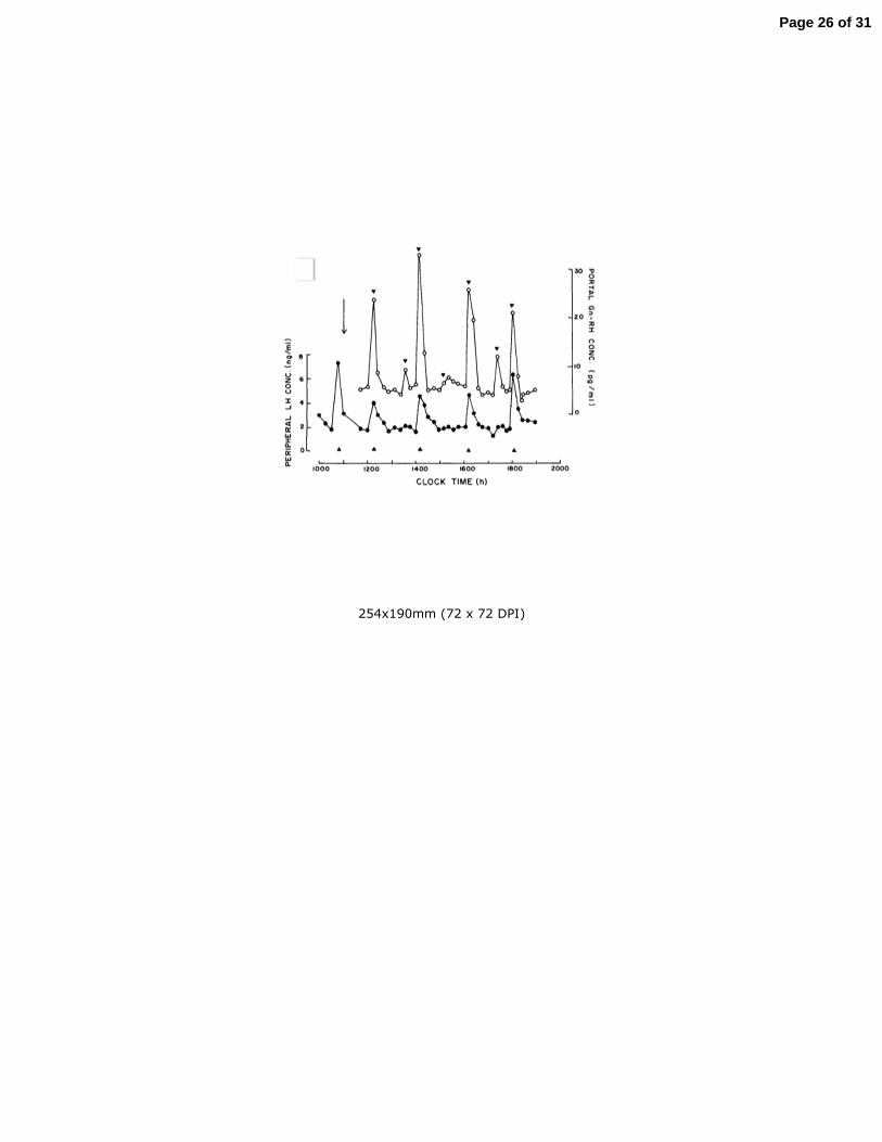
Interestingly, it was not until 1982, after Clarke and Cummins had developed a 121
technique to sample portal blood in the un-sedated ewe, that the episodic nature of 122
hypothalamic GnRH release was empirically demonstrated by these investigators 123
(Clarke and Cummins 1982) (Figure 3). Together, the foregoing findings provided 124
the foundation for the concept that pulsatile GnRH release was driven by a neural 125
timing mechanism resident in the hypothalamus (Karsch 1980) (Pohl and Knobil 126
1982) that subsequently came to be known as the hypothalamic GnRH pulse 127
generator. However, it was the later observation that the episodes of LH release in 128
ovariectomized monkeys were tightly coupled to brief increases in multiunit 129
electrophysiological activity (MUA) recorded in the mediobasal hypothalamus 130
(MBH) (Wilson, et al. 1984), that led to the general acceptance of the notion of a 131
GnRH pulse generator. This hypothalamic mechanisms drives “basal” or “tonic” 132
gonadotropin secretion that, in the female, is responsible for folliculogenesis, 133
maintenance of the corpus luteum and the synthesis of ovarian hormones and, in the 134
male, for maintaining spermatogenesis and testosterone secretion (Plant and 135
Marshall 2001; Zeleznik 2015). 136 1 Recounted by Nicholas Wade in The Nobel Duel (Anchor Press/DoubleDay, 1981) and in a series of 137 three articles published in Science in April/May, 1978. 138
Page 3 of 31

4
2 In this review GnRH1 (mammalian GnRH) will be referred to as GnRH. 139
Ovulation at the end of the follicular phase of the ovarian cycle is triggered by 140
a massive and relatively prolonged discharge of gonadotropin, a mode of release 141
termed “surge” secretion. The view held by Harris that ovulation was under neural 142
control was, in part, based on the classical studies in 1950 by Everett and Sawyer 143
(Everett and Sawyer 1950) indicating that, in the rat, a recurring daily neural signal 144
was generated by the hypothalamus during a brief critical period in the light phase 145
of the 24 h cycle, and which, on the day of proestrus, was responsible for eliciting 146
the pre-ovulatory gonadotropin surge from the pituitary. The prediction that this 147
neural signal was relayed to the pituitary by Harris’ humoral substance was 148
confirmed when Fink and his colleagues in 1976 demonstrated a large surge in the 149
concentration of GnRH in portal blood during the critical period of the proestrus rat 150
(Sarkar, et al. 1976). The neural mechanism responsible for the preovulatory surge 151
of GnRH that occurs over a period of several hours is now conceptualized as the 152
GnRH surge generator. The precise relationship between the GnRH neurons 153
mediating surge and pulsatile release of the decapeptide, however, remains to be 154
determined. 155
156
Other “humoral” substances regulating gonadotroph function. 157
158
The notion that regulation of anterior pituitary hormone secretion by 159
humoral substances from the hypothalamus might be achieved by inhibiting the 160
cells of the pars distalis was built into the Harris’ hypothesis (see above). In the 161
context of the hypothalamo-pituitary-gonadal axis, evidence for such an inhibitory 162
control system was obtained in studies of Japanese quail in 2000 reporting that a 163
neuropeptide belonging to the RFamide related peptide (RFRP)3 family was found 164
to be concentrated in the external zone of the median eminence of quail and, in vitro, 165
inhibited LH secretion from the pituitary of these birds (Tsutsui, et al. 2000). RFRPs 166
were known to exhibit a wide range of biological actions and Tsutsui and colleagues 167
termed the peptide they had discovered in the quail brain, gonadotropin inhibitory 168
hormone (GnIH). Subsequent studies have demonstrated that GnIH not only 169
regulates gonadotropin secretion in avian species by a direct action on the pituitary 170
but also by an indirect action at the hypothalamic level to modulate GnRH release 171
(Tsutsui, et al. 2012). GnIH is not expressed in mammalian brain but two related 172
peptides, RFRP-1 and RFRP-3, are encoded by a gene orthologous to that encoding 173
avian GnIH (Tsutsui et al. 2012). Evidence that RFRPs serve a hypophysiotropic role 174
in the control of the pituitary-gonadal axis of mammalian species in general, 175
however, is tenuous (see Herbison, 2015). 176
177
178
179
180
181 182 3 The carboxyl terminal of these peptides is preceded by the amino acid sequence, LP(LorQ)RFamide 183 (Tsutsui et al. 2012). 184
Page 4 of 31

5
The GnRH Neuron (birth, location, morphology, electrophysiology). 185
186
Two years after the structure of GnRH was announced, the first antibody to the 187
peptide was reported (Barry, et al. 1973) and since then numerous studies of the 188
immunohistochemical distribution of GnRH neurons in the developing and mature 189
hypothalamus have been conducted (Herbison 2015). Based on careful examination 190
of the embryonic mouse brain the groups of Pfaff and Wray in 1989 independently 191
proposed that GnRH neurons are born in the olfactory placode and after entering 192
the forebrain during early embryonic development migrate to the hypothalamus 193
where several hundred of these cells are found diffusely distributed in the preoptic 194
area (POA) and more caudal areas in the MBH (Schwanzel-Fukuda, et al. 1989) 195
(Wray, et al. 1989). As may be expected from such an amazing journey through the 196
brain, the neurobiology of GnRH neuron migration is complex and involves the 197
interplay of guidance cues, adhesion molecules, growth factors and 198
neurotransmitters (Wierman, et al. 2011). 199
200
In the adult hypothalamus, the typical GnRH neuron has two dendritic 201
projections, which, as revealed by studies conducted with contemporary 202
morphological techniques over the last 10 years, may extend remarkable distances 203
(2-3 mm) from the cell body or perikaryon (Herbison 2015) (Figure 4). Not 204
surprisingly, a principal target of hypothalamic GnRH neurons is the primary plexus 205
of the hypophysial portal system in the median eminence. Interestingly, the latter 206
projections combine characteristics of both dendrites and axons and have been 207
termed “dendrons” by Herbison and his colleagues (Herde, et al. 2013). While 208
considerable attention has been paid to the electrophysiology of the GnRH neuron 209
and the underlying ion channels in the membrane of the cell body, a meaningful 210
correlation between electrical activity and intermittent terminal release of GnRH, as 211
classically demonstrated for oxytocin neurons, has yet to be obtained. 212
213
GnRH biosynthesis and action. 214
215
GnRH is encoded by the GNRH1 gene, which was cloned by Seeburg and 216
Adelman in 1984 (Seeburg and Adelman 1984). As pointed out by Herbison (2015), 217
GnRH neurons maintain a high content of their peptide, and it is therefore unlikely 218
that regulation of GNRH1 expression plays a critical role in moment to moment 219
control of either GnRH surges or pulses. GnRH action on the pituitary gonadotroph 220
is mediated by G-protein coupled receptors that signal via Gq and or G11 to activate 221
phospholipase-C that leads to mobilization of Ca++ by inositol phosphate3 (McArdle 222
2015). 223
224
Neurobiology of the hypothalamic GnRH pulse generator 225
226
The concept of the hypothalamic GnRH pulse generator that emerged in the 1980s 227
remained for more than 20 years in the realm of a “black box”. One school of 228
thought proposed that pulsatility was intrinsic to the GnRH neurons themselves and 229
that extensive inter-cellular mechanisms orchestrated synchrony within the 230
Page 5 of 31
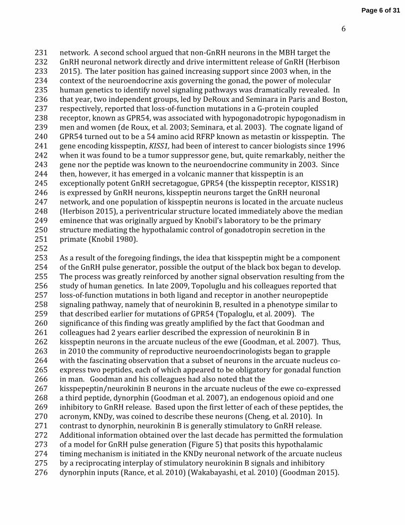
6
network. A second school argued that non-GnRH neurons in the MBH target the 231
GnRH neuronal network directly and drive intermittent release of GnRH (Herbison 232
2015). The later position has gained increasing support since 2003 when, in the 233
context of the neuroendocrine axis governing the gonad, the power of molecular 234
human genetics to identify novel signaling pathways was dramatically revealed. In 235
that year, two independent groups, led by DeRoux and Seminara in Paris and Boston, 236
respectively, reported that loss-of-function mutations in a G-protein coupled 237
receptor, known as GPR54, was associated with hypogonadotropic hypogonadism in 238
men and women (de Roux, et al. 2003; Seminara, et al. 2003). The cognate ligand of 239
GPR54 turned out to be a 54 amino acid RFRP known as metastin or kisspeptin. The 240
gene encoding kisspeptin, KISS1, had been of interest to cancer biologists since 1996 241
when it was found to be a tumor suppressor gene, but, quite remarkably, neither the 242
gene nor the peptide was known to the neuroendocrine community in 2003. Since 243
then, however, it has emerged in a volcanic manner that kisspeptin is an 244
exceptionally potent GnRH secretagogue, GPR54 (the kisspeptin receptor, KISS1R) 245
is expressed by GnRH neurons, kisspeptin neurons target the GnRH neuronal 246
network, and one population of kisspeptin neurons is located in the arcuate nucleus 247
(Herbison 2015), a periventricular structure located immediately above the median 248
eminence that was originally argued by Knobil’s laboratory to be the primary 249
structure mediating the hypothalamic control of gonadotropin secretion in the 250
primate (Knobil 1980). 251
252
As a result of the foregoing findings, the idea that kisspeptin might be a component 253
of the GnRH pulse generator, possible the output of the black box began to develop. 254
The process was greatly reinforced by another signal observation resulting from the 255
study of human genetics. In late 2009, Topoluglu and his colleagues reported that 256
loss-of-function mutations in both ligand and receptor in another neuropeptide 257
signaling pathway, namely that of neurokinin B, resulted in a phenotype similar to 258
that described earlier for mutations of GPR54 (Topaloglu, et al. 2009). The 259
significance of this finding was greatly amplified by the fact that Goodman and 260
colleagues had 2 years earlier described the expression of neurokinin B in 261
kisspeptin neurons in the arcuate nucleus of the ewe (Goodman, et al. 2007). Thus, 262
in 2010 the community of reproductive neuroendocrinologists began to grapple 263
with the fascinating observation that a subset of neurons in the arcuate nucleus co-264
express two peptides, each of which appeared to be obligatory for gonadal function 265
in man. Goodman and his colleagues had also noted that the 266
kisspepeptin/neurokinin B neurons in the arcuate nucleus of the ewe co-expressed 267
a third peptide, dynorphin (Goodman et al. 2007), an endogenous opioid and one 268
inhibitory to GnRH release. Based upon the first letter of each of these peptides, the 269
acronym, KNDy, was coined to describe these neurons (Cheng, et al. 2010). In 270
contrast to dynorphin, neurokinin B is generally stimulatory to GnRH release. 271
Additional information obtained over the last decade has permitted the formulation 272
of a model for GnRH pulse generation (Figure 5) that posits this hypothalamic 273
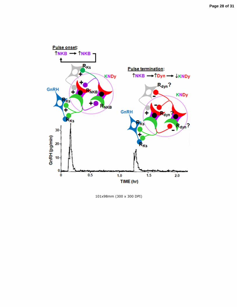
timing mechanism is initiated in the KNDy neuronal network of the arcuate nucleus 274
by a reciprocating interplay of stimulatory neurokinin B signals and inhibitory 275
dynorphin inputs (Rance, et al. 2010) (Wakabayashi, et al. 2010) (Goodman 2015). 276
Page 6 of 31

7
As mentioned above, the output of the pulse generator is posited to be relayed from 277
the arcuate nucleus to the GnRH neuronal network by release of kisspeptin from 278
axonal terminals originating from KNDy neurons. According to this model, 279
kisspeptin of arcuate nucleus origin should be viewed simply as a GnRH pulse 280
generating peptide (Terasawa, et al. 2013). 281
282
It is probably no exaggeration to say that the discovery of the impact of loss-283
of-function mutations in GPR54 on the reproductive axis led to a profound and 284
much needed revitalization to the study of GnRH neuroendocrinology, a field that 285
had begun to stagnate in the 1990s. 286
287
Neuroendocrine control of ovarian cycles 288
289
In spontaneously ovulating species of mammal, the typical ovarian cycle is 290
characterized by the secretion of relatively low basal or tonic levels of LH and FSH, 291
which are interrupted approximately once every 4-5 days in rats and mice and once 292
every 28 days in women, by a massive discharge or surge of gonadotropin which 293
triggers ovulation. Tonic gonadotropin secretion during the follicular phase of the 294
cycle, which drives folliculogenesis, is governed by a negative feedback loop in 295
accordance with the Harris model (Figure 1). The principal ovarian component of 296
the loop is estradiol secreted by the developing follicle(s), and it is now established 297
that such feedback occurs at the pituitary level as well as at the hypothalamus. At 298
the hypothalamic level, estradiol negative feedback appears to be exerted primarily 299
by modulating the amplitude of pulsatile GnRH release (see Zeleznik and Plant 300
2015). Two nuclear estrogen receptors (ERs) exist, ERα and ERβ, and studies using 301
transgenic mice null for these proteins indicate that ERα is the likely ER mediating 302
the negative feedback action of estradiol. Interestingly, GnRH neurons do not 303
appear to express ERα, as initially demonstrated by binding studies (Shivers, et al. 304
1983) and subsequently confirmed by immunohistochemistry; therefore estradiol 305
negative feedback at the level of the hypothalamus must be mediated indirectly 306
either by non-GnRH neurons or glia. Classical studies have indicated that the locus 307
of the negative feedback action of estradiol is within the MBH, and, intuitively, it 308
might be expected that ERα expressing KNDy neurons in the arcuate nucleus are the 309
neuronal phenotype targeted. Intriguingly, however, a recent study by Levine and 310
his colleagues employing transgenic mice null for ERα in kisspeptin neurons (not 311
restricted to KNDy neurons) has failed to support this notion (Dubois, et al. 2015). 312
On the other hand, Herbison’s group using genetically engineered adult female mice 313
and adeno-associated virus injection into the arcuate nucleus to delete 314
approximately 75% of ERα positive cells in this nucleus abolished chronic negative 315
feedback action of estradiol on LH secretion (Yeo and Herbison 2014)4. It would 316
seem reasonable to conclude that, in the later study, the majority of 317
318
319 4 Acute suppression of LH by estradiol, however, was preserved. 320
321
Page 7 of 31

8
322
KNDy neurons would be null for ERα, and therefore the results of the two studies 323
are difficult to reconcile. One possible explanation is that embryonic loss of ERα in 324
kisspeptin neurons may have led to the development of compensatory feedback 325
mechanisms. 326
327
During the later half of the follicular phase of the menstrual cycle, the 328
secretion of FSH is suppressed to a greater degree than that of LH, and this 329
differential pattern of gonadotropin release plays a pivotal role in the selection of 330
the pre-ovulatory follicle (Zeleznik and Plant 2015). The mechanism underlying 331
differential suppression of gonadotropin secretion at this stage of the menstrual 332
cycle has not been extensively studied but appears to involve estradiol negative 333
feedback action at the level of the pituitary to regulate the constitutive component 334
of FSH release, which is greater than that of LH (see Zeleznik and Plant 2015). 335
336
In those species with a prolonged luteal phase, such as primates and sheep, 337
progesterone secretion by the corpus luteum markedly retards the frequency of the 338
GnRH pulse generator (see Goodman and Inskeep 2015; Zeleznik and Plant 2015). 339
As there is little evidence for progesterone receptor (PR) expression by GnRH 340
neurons, the suppressive feedback action of progesterone on GnRH release, like that 341
of estradiol, is probably indirect. The finding that KNDy neurons in the ewe 342
hypothalamus express PR suggests that these cells may be the target for 343
progesterone’s negative feedback action. However, the impact of ablating PR 344
selectively in KNDy neurons has yet to be examined probably because such genetic 345
approaches have been largely limited to mice, which do not have prolonged luteal 346
phases. It should also be noted that the physiological significance of progesterone’s 347
ability to decelerate the frequency of pulsatile GnRH/LH remains unclear because 348
corpus luteum function appears to be normal in GnRH deficient monkeys and 349
women receiving invariant intermittent GnRH replacement (see Zeleznik and Plant 350
2015). In this regard, estradiol and inhibin (primarily inhibin A) are also secreted by 351
the corpus luteum and both are capable of inhibiting gonadotropin secretion. 352
353
The importance of ovarian steroid feedback for the surge in LH secretion 354
required for ovulation at the end of the follicular phase was suspected at the time 355
Harris’ monograph was published. Although Harris was appreciative of these ideas 356
(Harris 1969), he was primarily focused on the nature of the hypothalamic factor 357
that triggered the ovulatory discharge of LH, and it was not until 1969 that Goding 358
and his colleagues convincingly demonstrated the “positive” feedback action of 359
estrogen on gonadotropin secretion by showing that administration of estradiol to 360
the anestrous ewe elicited an LH surge comparable to that seen spontaneously at 361
the end of the follicular phase during the breeding season (Goding, et al. 1969). 362
363
364
365
366
Page 8 of 31

9
By this time, Halasz5 and Gorski had shown by surgically isolating the POA from the 367
MBH of the rat with a bayonet shaped “Halasz” knife that the more rostral area was 368
essential for ovulation in this species (Halasz and Gorski 1967; Szentagothai J 1968). 369
Together, these findings suggested that a major site of the positive feedback action 370
of estradiol in the rodent was in the POA, and Goodman in 1978 went on to verify 371
this idea by demonstrating that implants of crystalline estradiol in the POA of the rat 372
were more effective at eliciting LH surges than those implanted in the more caudal 373
region of the hypothalamus (Goodman 1978). So after almost 30 years from the 374
time that Everitt and Sawyer had demonstrated that the generation of a daily neural 375
signal for ovulation in the rat was tightly coupled to the 24 h light dark cycle 376
(Everett and Sawyer 1950), a comprehensive model for the control of ovulation in 377
the rat had finally begun to emerge. It should also be pointed out that, at this time, 378
evidence was accumulating to indicate that this model might not be applicable to 379
other species. Interestingly, the model for the rat ovarian cycle remained essential 380
unchanged until the recognition in 2003 of the significance of kisspeptin in the 381
regulation of GnRH secretion. During the subsequent 5 years and largely as a result 382
of work by Steiner’s laboratory it became apparent that a second hypothalamic 383
population of kisspeptin neurons are located in the anteroventral periventricular 384
nucleus (AVPV) of the POA and that this rostral population is a major site for the 385
positive feedback action of estradiol in rodents (Oakley, et al. 2009). As with the 386
negative feedback action of estradiol, knockout studies of ER in mice indicate that 387
ERα mediates the positive feedback of the steroid (Dubois et al. 2015). 388
389
In the mid-1970s, Knobil’s group threw much of the field of reproductive 390
neuroendocrinology into a state of partial shock when they demonstrated that 391
ovulation occurred in the monkey after neural connections between the POA and 392
MBH were severed using a Halasz knife (Krey, et al. 1975) . The furor continued 393
when this group proposed that the role of hypothalamic GnRH release in dictating 394
the menstrual cycle was permissive (Knobil, et al. 1980). In other words, the only 395
hypothalamic input that was needed for ovarian cycles and ovulation in highly 396
evolved primates was an invariant intermittent stimulation of the pituitary by GnRH 397
and that the characteristic cyclic pattern of gonadotropin secretion was dictated 398
solely by the negative and positive feedback actions of estradiol directly at the 399
pituitary: a positive feedback action of estradiol in the POA and a GnRH surge was 400
not required. Since then, the most compelling evidence in support of this hypothesis 401
has been obtained from GnRH deficient women, such as those with Kallmann 402
syndrome, where replacement treatment with an invariant intermittent infusion of 403
“physiological” pulse doses of GnRH will drive cycles and ovulation (see Zeleznik 404
and Plant 2015). Moreover, indirect evidence indicates that spontaneous ovulation 405
406 5 Halasz developed the use of a surgical knife to isolate the hypophysiotropic area (MBH) from more 407 rostral areas of the rat hypothalamus at Pecs University Medical School. The group at Pecs was one 408 of the major World centers studying the hypothalamic control of the anterior pituitary at the time of 409 Harris, and is perhaps most famous for its studies of hypothalamic pathways and regions (including 410 those that are sensitive to target hormones from the gonad, adrenal and thyroid) underlying the 411 feedback control of anterior pituitary function (Szentagothai et al. 1968). 412
Page 9 of 31

10
in normal women likely occurs in the absence of a GnRH surge (see Zeleznik and 413
Plant 2015). Whatever the final resolution of this important comparative question, 414
it appears that ovulation in the human female is largely emancipated from control 415
by the POA (Plant 2012). 416
417
Neuroendocrine control of testicular function. 418
419
The study of the neuroendocrine control of the testis in the post Harris era 420
has continued to play second fiddle to that of the ovary. As with early studies, upon 421
which the foundations of reproductive neuroendocrinology were laid, ovulation and 422
the pre-ovulatory LH surge, have provided a robust read-out of hypothalamic 423
activation and one that could by easily harnessed to indirectly explore the 424
neurobiological mechanism controlling GnRH secretion. As will be discussed later, 425
the GnRH surge generator in the rodent is decommissioned during perinatal 426
development, and the hypothalamic control of the testis can be accounted entirely 427
by GnRH pulse generation. Since fundamental sex differences in the control of this 428
mode of GnRH release have yet to be identified, it is to be anticipated that what we 429
understand of GnRH pulse generation in the female will be largely translatable to 430
the male. With regard to the negative feedback control of testicular function, the 431
concept of an aqueous testicular feedback signal “inhibin” was proposed more than 432
20 years before the publication of Harris’ monograph. It was not until 1985, 433
however, that the nature of inhibin was independently revealed by four groups 434
(Vale, et al. 1988). Interestingly, follicular fluid not testicular tissue/fluid was used 435
for isolation of the inhibins, which are heteromeric peptides consisting of an α-436
subunit and one of two β-subunits (Vale et al. 1988). Studies of the male monkey 437
have provided the most convincing evidence to date for a major role of testicular 438
inhibin B (αβB dimer) in regulating the secretion of FSH by a direct negative 439
feedback action at the level of the pituitary (Majumdar, et al. 1995). 440
441
Neuroendocrine control of puberty 442
443
Although Harris’ proposal in 1955 that puberty is triggered by a 444
hypothalamic stimulus that is transmitted via the hypophysial portal vessels to the 445
pituitary is now well established, the mechanism underlying the timing of this 446
critical development event in our own species remains an intriguing mystery. Major 447
conceptual advances, however, have been achieved. Studies in the 1980s of 448
pituitary and plasma gonadotropin content in the human and sheep fetus by 449
Grumbach and his colleagues (Clark, et al. 1984; Sklar, et al. 1981) and of circulating 450
testosterone and LH levels in infantile boys and monkeys by others (Plant 1980) 451
(Forest, et al. 1974; Waldhauser, et al. 1981) led to the deduction that 1) GnRH pulse 452
generation in these species develops in the fetus shortly after GnRH neurons 453
complete their embryonic migration from the olfactory placode to the hypothalamus, 454
and 2) robust pulsatile GnRH and gonadotropin release is manifest during infancy in 455
highly evolved primates. Puberty, however, is not initiated during infancy because at 456
this stage of development the somatic cells of the gonads that underpin 457
Page 10 of 31

11
gametogenesis are unable to respond fully to such gonadotropin stimulation. 458
Although the ability of the prepubertal gonad to respond to gonadotropin 459
stimulation is acquired during childhood, by then a hypogonadotropic state is in 460
place as a result of the suppression of pulsatile GnRH release thereby guaranteeing 461
the continued quiescence of the gonad in boys and girls. Interestingly, studies of the 462
monkey indicate that the GnRH neuronal network of the juvenile primate, like the 463
pituitary and gonad at this stage of development, is not limiting to the onset of 464
puberty. This view is based on the finding that in the monkey intermittent 465
neurochemical stimulation of the juvenile hypothalamus with a glutamate receptor 466
agonist will lead with surprising ease to an adult like pattern of pulsatile GnRH 467
release and precocious puberty (Figure 6) (Plant, et al. 1989). Moreover, compelling 468
evidence is now at hand indicating that the proximal stimulus responsible for the 469
activation of robust GnRH pulsatility at the onset of spontaneous puberty is indeed 470
an intermittent release of kisspeptin that is generated by the re-awakening of the 471
GnRH pulse generator in the arcuate nucleus (Terasawa et al. 2013). The 472
mechanisms that turn the GnRH pulse generator off during infancy, maintain it in a 473
state of suspended animation during juvenile development, and reawaken it at the 474
termination of juvenile development are poorly understood (Plant 2015) and 475
provide a major challenge for the future. 476
477
Perinatal programming of the GnRH surge generator 478
479
With regard to sexual differentiation of the hypothalamus, Harris stated in 1955, “In 480
view of the fact that Pfeiffer (1936) showed prepubertal testis grafts in female rats 481
produce a constant oestrous state after puberty, and that ovaries grafted into adult 482
male rats castrated at birth do undergo cyclic changes, it seems that some neural 483
structure in the male animal becomes differentiated and fixed in its function under 484
the influence of androgens in early life.”: a position shared by Everett, Sawyer and 485
Markee (1949) (Everett, et al. 1949). Examination of this hypothesis in the post-486
Harris era has proceeded somewhat in parallel with development of the model for 487
the neural control of ovulation in the rodent (see above), and the target of testicular 488
testosterone action to defeminize the rodent brain is considered to reside within the 489
POA and to involve a negative impact of testicular androgen on the development of 490
kisspeptin neurons in the AVPV (Oakley et al. 2009). An interesting feature of this 491
perinatal decommissioning of the GnRH surge mechanism by testicular testosterone 492
is that the action of the steroid in target neurons within the POA appears to be 493
mediated by ER signaling after intracellular aromatization of testosterone to 494
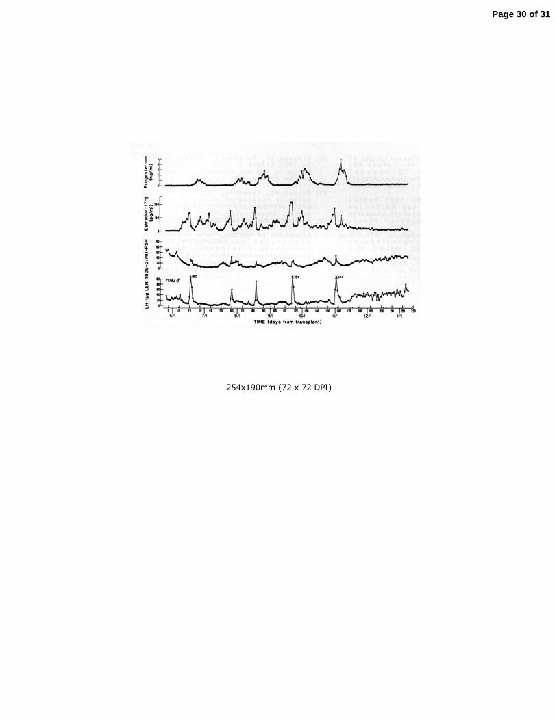
estradiol (Gonzalez-Martinez, et al. 2008). 495
496
One important comparative aspect of perinatal programming of the GnRH surge 497
system in spontaneous ovulators that has emerged since the time of Harris is the 498
recognition that, in the monkey and probably in man, the neural mechanism 499
underlying the pre-ovulatory LH surge is not decommissioned by the testis during 500
early development. This was first demonstrated by Knobil’s laboratory by the 501
finding that unequivocal estrogen-induced LH surges were readily elicited in adult 502
male monkeys castrated postpubertally and implanted with estradiol containing 503
Page 11 of 31

12
Silastic capsules that produced follicular phase levels of the steroid in the agonadal 504
situation (Karsch, et al. 1973). A more sensational demonstration followed when 505
Spies’ laboratory reported that ovarian cycles and ovulation unfolded in an adult 506
male castrate bearing a sc ovarian graft (Figure 7) (Norman and Spies 1986). 507
508
Modulation of GnRH pulsatility 509
510
At the time Harris wrote his monograph in 1955, he discussed the impact of food, 511
temperature, light, social factors and emotional states (in women) on ovarian cycles 512
and fertility, and argued that these modulators of gonadal function are registered by 513
the central nervous system, which then relays the respective exteroceptive or 514
interoceptive cue to the gonad by controlling gonadotropin secretion from the 515
pituitary. Although considerable advances have been made in our understanding of 516
the neural and neuroendocrine mechanisms involved in such modulation, a 517
complete review of these fall beyond the scope of this chapter. Instead, two 518
examples that are of particular conceptual significance are discussed. 519
520
The first concerns the relationship between somatic and sexual development, 521
and specifically the question of how information on growth is relayed to the 522
reproductive axis. In 1963 Kennedy and Mitra (Kennedy and Mitra 1963) proposed 523
that, in the rat, body weight was an initiating factor in the onset of puberty, and 524
inspired by this idea, Frisch and Revelle examined the relationship between body 525
weight and the timing of menarche in girls. The observations of the latter workers 526
led them to propose in 1970 (Frisch and Revelle 1970) “that attainment of body 527
weight in the critical range causes a change in metabolic rate, which, in turn, is 528
responsible for an increase in hypothalamic drive to the pituitary gonadotroph.” An 529
extension of this idea would be to propose that a “metabometer” in the 530
hypothalamus tracks information on metabolism and sends a cue to the GnRH pulse 531
generator when a pubertal metabolic state develops. While this hypothesis has not 532
been systematically examined, interest in the idea of Frisch was re-awakened in 533
1994 by the cloning of the obese gene encoding leptin (Zhang, et al. 1994): an 534
adipocyte hormone that plays a major role in regulating appetite and food intake. 535
Although it is now generally recognized that leptin is not the trigger timing the 536
pubertal re-awakening of the GnRH pulse generator, this hormone is essential, in a 537
permissive sense, for robust GnRH pulsatility in general, and therefore for both 538
pubertal and adult gonadal function. The latter view is best supported by the 539
finding that in young children with leptin deficiency initiation of treatment with 540
recombinant leptin does not induce puberty immediately but rather only after 541
appropriate somatic development has been attained following prolonged exposure 542
to the hormone (Farooqi and O'Rahilly 2014). Although KNDy neurons in the 543
arcuate nucleus of the hypothalamus were initially thought to be the target of 544
leptin’s hypothalamic action to modulate the pituitary-gonadal axis, more recent 545
studies using transgenic mice suggest that the neurobiology underlying the action of 546
leptin to modulate GnRH pulsatility may be considerably more complex (Donato, et 547
al. 2011). 548
549
Page 12 of 31

13
The second modulator of GnRH pulsatility to be discussed is photoperiod; a 550
major factor determining annual changes in gonadal function in mammalian species 551
that breed seasonally. Of all the areas of reproductive neuroendocrinology that 552
interested Harris, the study of season in the post-Harris era has arguably uncovered 553
the most novel of insights into neuroendocrine control systems governing the 554
hypothalamic-pituitary-gonadal axis. The story begins with the recognition in the 555
mid-1960s of the importance of the pineal gland in regulating seasonal changes in 556
gonadal function in the Syrian hamster. Melatonin secreted by the pineal is the 557
major signal relaying information on duration of daylight to the hypothalamus, and 558
a major site of action of melatonin in the hypothalamus is the pars tuberalis, a 559
portion of the anterior pituitary that forms a funnel like structure around the 560
pituitary stalk and median eminence. The initial suggestion that the pars tuberalis 561
was instrumental in mediating seasonal changes in anterior pituitary function came 562
from a study by Lincoln and Clark employing an experimental preparation in which 563
the pituitary of sheep was disconnected from neurovascular control by the 564
hypothalamus (Lincoln and Clarke 1994). Such hypothalamo-pituitary disconnected 565
animals continued to exhibit a seasonal rhythm in prolactin secretion. Importantly, 566
gonadotropin levels on the other hand were routinely low due to the blockade of the 567
GnRH signal by the surgical procedure. At about the same time, Karsch’s laboratory 568
studying the ewe was systematically exploring an earlier idea of others that thyroid 569
hormone was required for seasonal changes in hypothalamic-pituitary function: 570
their work demonstrated that thyroid hormone was permissive for the transition to 571
anovulation during the long days of spring, but not needed for return of 572
reproductive function at the start of the fall breeding season (Karsch, et al. 1995). 573
Insight into the neurobiology underlying the interaction between thyroid hormone 574
and the seasonal melatonin signal came initially from the finding in Japanese quail 575
(a long-day breeder), that expression in the MBH of a thyroid hormone metabolizing 576
enzyme (type 2 diodinase) that increases the local concentration of 577
triiodothyronine , the active cellular metabolite of thyroid hormone, was 578
upregulated by exposure to long days (Yoshimura, et al. 2003). Interestingly, long 579
days also result in an increase in hypothalamic thyroid hormone activity in short 580
day breeders such as sheep. In both cases, the link between melatonin action in the 581
pars tuberalis and deiodinase activity in the MBH appears to be provided by 582
increased synthesis of thyroid stimulating hormone in the pars tuberalis triggered 583
by long days (Figure 8). How low day induced increases in thyroid hormone activity 584
in the MBH either terminate or reactivate pulsatile GnRH release in long and short 585
day breeders, respectively, remains to be established, although KNDy neurons and 586
RFRP expressing neurons have been implicated. As pointed out by Hazelrigg and his 587
colleagues (Hanon, et al. 2008), information in this intriguing pathway moves in the 588
opposite direction to that in Harris’ model (Figure 1), albeit without the need of the 589
hypophysial portal circulation, and therefore is unlikely to have been envisioned by 590
Harris. An excellent review of this fascinating control system has recently been 591
published by Hazlerigg and Simonneauz (2015). 592
593
Bench to bedside 594
595
Page 13 of 31

14
The isolation and characterization of GnRH, together with the demonstration that 596
sustained stimulation of gonadotropin secretion may be achieved with intermittent 597
stimulation of the pituitary whereas continuous exposure of the gland to the peptide 598
leads to desensitization and thus suppression of gonadotropin secretion, provides 599
the conceptual framework for the current therapeutic uses of GnRH receptor 600
analogs. As comprehensively reviewed by Millar and Newton (Millar and Newton 601
2013), GnRH agonists and antagonist are currently used widely to treat gonadal 602
steroid dependent cancers, and disorders of reproductive development including 603
GnRH dependent precocious puberty. These agents are also used extensively in 604
fertility clinics where controlled cycles of follicular development and oocyte 605
maturation are required for egg retrieval and in-vitro fertilization. Millar and 606
Newton also speculate that in the future kisspeptin and neurokinin B analogs may 607
offer additional or improved therapeutic interventions to modulate LH and FSH 608
release. In this regard, a recent report describes successful oocyte maturation 609
following an LH surge induced by a single sc injection of kisspeptin administered at 610
the end of the follicular phase of a controlled cycle of ovarian stimulation (Jayasena, 611
et al. 2014). It remains to be determined, however, whether kisspeptin induced LH 612
release has any advantages over GnRH induced LH release for oocyte maturation in 613
such a clinical setting. 614
615
The future 616
617
Harris’ model of the control of gonadal function has, over the last 60 years, served as 618
a solid framework for those studying mechanisms that govern how the brain 619
regulates the pituitary–gonadal axis: it has stood the test of time and revisions to the 620
paradigm, while quite remarkable, have been primarily those at the level of cellular 621
and molecular detail, rather than those with major conceptual implications. 622
Perhaps an exception to this generalization is the further recognition that during 623
evolution different mammalian species have co-opted different neuroendocrine 624
control systems, or perhaps more precisely utilized a set of neuroendocrine controls 625
to variable degrees, to achieve regulation of the gonad. The implication of the latter 626
view to the future of the field in general is that diversity in the selection of animal 627
models is beneficial, while the interrogation of problems that at the time appear 628
immediately translatable to human health and well being to the exclusion of others 629
is not. With regard to the issue of diversity in selecting animal models, the recent 630
introduction of a technology known as CRISPR-Cas9, which is potentially capable of 631
being used to edit the genome of any mammalian species (Hsu, et al. 2014), is likely 632
to profoundly enhance the value of animal models hitherto considered to be 633
genetically intractable. The power of contemporary molecular human 634
genetics/genomics has already resulted in signal surprises and inevitably will lead 635
to further major advances in our understanding of neural signaling pathways 636
involved in regulating hypothalamic GnRH release. The application of global 637
analyses of hypothalamic gene expression, and thereby the identification of gene 638
networks, associated with neuroendocrine mechanisms governing GnRH release has 639
been introduced by Ojeda’s laboratory (Lomniczi, et al. 2013), and it is likely that 640
Page 14 of 31

15
this systems biology approach will see wider use as high throughput sequencing 641
techniques become less expensive. 642
643
Major questions remain to be addressed and, in the opinion of the writer, the two 644
most important concern the nature of the neurobiological mechanisms that underlie 645
the generation of the pulsatile mode of GnRH release and the mystery of human 646
puberty. The two are related since human puberty is triggered by a re-awakening of 647
GnRH pulse generation. In this regard, optogenetics (Deisseroth 2011), which 648
allows the investigator to selectively activate specific signaling pathways in the 649
brain, would appear to be particularly suitable for interrogating the KNDy neuron 650
model of GnRH pulse generation. Examination of this model would also be greatly 651
aided by a complete “wiring” diagram of the arcuate nucleus, and such a goal is on 652
the horizon as technologies underlying connectomics (Eberle, et al. 2015) become 653
more readily available. Finally, as with many other fields, it will also be of interest 654
to witness over the next decade or so to what extent epigenetic mechanisms 655
contribute to transmission of inheritable traits in hypothalamic control systems 656
governing GnRH release. 657
658
Acknowledgement. The author is most grateful to Dr. Robert L Goodman, 659
Department of Physiology and Pharmacology, West Virginia University, for kindly 660
reading the manuscript and for his insightful comments and suggestions. Work 661
conducted in the author’s laboratory was supported by the National Institutes of 662
Health (Grants HD08610, HD13254 and HD16851). 663
664
665
666
667
668
669
670
671
672
673
674
675
676
677
678
679
680
681
682
683
684
685
686
Page 15 of 31

16
Legends 687
688
Figure 1. Model proposed by Harris in his 1955 monograph to illustrate the 689
relationship between the external environment and the reproductive organs 690
(reprinted from Harris, 1955). 691
692
Figure 2. Demonstration of the need of intermittent GnRH stimulation of the 693
pituitary for sustained secretion of LH (closed data points) and FSH (open data 694
points). A monkey rendered hypogonadotropic by a hypothalamic lesion was 695
treated initially with an intermittent iv infusion of GnRH (1 υg/min for 6 min every 696
h). On day 0 the pulsatile regimen was terminated and replaced with a continuous 697
GnRH infusion (1 υg/min). On day 20 the pulsatile mode of GnRH stimulation was 698
re-instituted (reprinted from Belchetz et al, 1978). 699
700
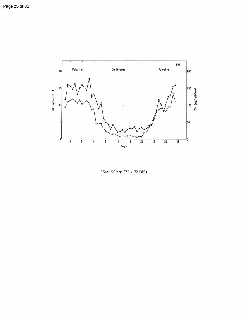
Figure 3. Relationship between pulsatile secretion of GnRH (open data points) into 701
hypophysial portal blood and corresponding episodes of pituitary LH secretion 702
(closed data points) into the systemic circulation in an ovariectomized ewe. 703
Modified from Clarke and Cummins, 1982 – permission pending (Copyright 1982, 704
The Endocrine Society). 705
706
Figure 4. A,B. Typical morphology of mouse GnRH neurons revealed by 707
immunohistochemistry. C. Two GnRH neurons in sheep. D. A GnRH neuron from an 708
adult mouse has been filled in situ with the dye, biocytin, in order to facilitate 709
visualization of the entire length of the dendrites, which in the case of the lower 710
process is seen to extend more than 500 υm. The two higher magnification insets 711
on the right reveal a high density of dendritic spines. Reprinted with permission 712
from Herbison (2015). 713
714
Figure 5. The KNDy neuron model of the GnRH pulse generator proposed by 715
Lehman, Coolen and Goodman. KNDy neurons reside within the arcuate nucleus 716
(dotted purple circle) and express kisspeptin (Ks, green), neurokinin B (NKB, 717
purple) and dynorphin (dyn, red). The model proposes that pulse generation by the 718
network of KNDy neurons in the arcuate nucleus is achieved by a poorly understood 719
reciprocating interplay of stimulatory NKB and inhibitory dyn inputs and an 720
unidentified interneurone (gray). The output of the pulse generator is relayed to 721
the GnRH neuronal network (blue) by a brief kisspeptin signal that evokes a 722
discharge of GnRH into the hypophysial-portal circulation (shown in the lower 723
portion of the figure). Note that the phenotype of each terminal indicates 724
biologically relevant peptide and not selective transport of that peptide to the 725
terminal. Similarly, the triple colored KNDy neurons indicate co-expression of the 3 726
peptides and not location within the cell body. RKs, Ks receptor; RNKB, NKB receptor, 727
RDYN, dyn receptor. Reprinted with permission from Goodman and Inskeep (2015). 728
729
Figure 6. Chronic intermittent neurochemical stimulation of juvenile male monkeys 730
with N-methyl DL aspartate (NMDA) readily induces a precocious pubertal pattern 731
Page 16 of 31

17
of pulsatile GnRH release as reflected by the emergence of corresponding discharges 732
of LH (open data points) and testicular testosterone (closed data points) secretion. 733
Testicular and motile epididymal sperm were typically observed after 16-26 weeks 734
of NMDA stimulation. Means+SE (N=4) are shown. Arrows indicate time of iv 735
injections of NMDA. Reproduced from Plant et al (1989) “Copyright (1989) National 736
Academy of Sciences, U.S.A.” 737
738
Figure 7. Ovarian tissue transplanted sc to the abdomen of an adult male rhesus 739
monkey (#7082) castrated post-pubertally and receiving anti rejection therapy with 740
cyclosporine A exhibits regular cycles of folliculogenesis and ovulation followed by a 741
normal luteal phase. Numbers on the peaks in LH indicate maximum concentration 742
of the gonadotropin on that day. Modified from Norman and Spies (1986) with 743
permission (Copyright 1986, The Endocrine Society). 744
745
Figure 8. A model for the seasonal control of pulsatile GnRH release. The duration 746
of the photoperiod is relayed by melatonin to melatonin receptors (MT1) in the 747
thyrotrophs of the pars tuberalis (PT), and further relayed by thyrotrophin (TSH) to 748
the mediobasal hypothalamus, where the arcuate nucleus KNDy neurons are located. 749
TSH upregulates the expression of the genes encoding deiodinase 2 and 3 (DIO2 and 750
DIO3), in specialized ependymal cells (tanycytes) lining the base of the third 751
ventricle (3v). The diodinase enzymes convert thyroid hormone (T4) into the active 752
metabolite, tri-iodothyronine (T3), and the increase in thyroid hormone activity 753
dictates the level of GnRH pulsatility, which in turn governs the gonadotrophin 754
output from the gonadotrophs in the pars distalis (PD). TH is considered to reach 755
the MBH via the 3v and/or from brain capillaries (Cp). Reprinted with permission 756
from Hazelrigg and Simonneaux (2015). 757
758
759
760
761
762
763
764
765
766
767
768
769
770
771
772
773
774
775
776
777
Page 17 of 31

18
778
779
780
Declaration of interest: I declare that there is no conflict of interest that could be 781
perceived as prejudicing the impartiality of my interpretation of the research 782
reported in this review. 783
784
Funding: Original research is not included in this review! 785
786
787
788
789
790
791
792
793
794
795
796
797
798
799
800
801
802
803
804
805
806
807
808
809
810
811
812
813
814
815
816
817
818
819
820
821
822
823
Page 18 of 31
19
824
825
826
References 827
828
Amoss M, Burgus R, Blackwell R, Vale W, Fellows R & Guillemin R 1971 Purification, 829
amino acid composition and N-terminus of the hypothalamic luteinizing hormone 830
releasing factor (LRF) of ovine origin. Biochem Biophys Res Commun 44 205-210. 831
Barry J, Dubois MP & Poulain P 1973 LRF producing cells of the mammalian 832
hypothalamus. A fluorescent antibody study. Z Zellforsch Mikrosk Anat 146 351-366. 833
Belchetz PE, Plant TM, Nakai Y, Keogh EJ & Knobil E 1978 Hypophysial responses to 834
continuous and intermittent delivery of hypopthalamic gonadotropin-releasing 835
hormone. Science 202 631-633. 836
Cheng G, Coolen LM, Padmanabhan V, Goodman RL & Lehman MN 2010 The 837
kisspeptin/neurokinin B/dynorphin (KNDy) cell population of the arcuate nucleus: 838
sex differences and effects of prenatal testosterone in sheep. Endocrinology 151 839
301-311. 840
Clark SJ, Ellis N, Styne DM, Gluckman PD, Kaplan SL & Grumbach MM 1984 Hormone 841
ontogeny in the ovine fetus. XVII. Demonstration of pulsatile luteinizing hormone 842
secretion by the fetal pituitary gland. Endocrinology 115 1774-1779. 843
Clarke IJ & Cummins JT 1982 The temporal relationship between gonadotropin 844
releasing hormone (GnRH) and luteinizing hormone (LH) secretion in 845
ovariectomized ewes. Endocrinology 111 1737-1739. 846
de Roux N, Genin E, Carel J-C, Matsuda F, Chaussain J-L & Milgrom E 2003 847
Hypogonadotropic hypogonadism due to loss of function of the KiSS1-derived 848
peptide receptor GPR54. Proc Natl Acad Sci U S A 100 10972-10976. 849
Deisseroth K 2011 Optogenetics. Nat Methods 8 26-29. 850
Dierschke DJ, Bhattacharya AN, Atkinson LE & Knobil E 1970 Circhoral oscillations 851
of plasma LH levels in the ovariectomized rhesus monkey. Endocrinology 87 850-852
853. 853
Donato J, Jr., Cravo RM, Frazao R, Gautron L, Scott MM, Lachey J, Castro IA, Margatho 854
LO, Lee S, Lee C, et al. 2011 Leptin's effect on puberty in mice is relayed by the 855
ventral premammillary nucleus and does not require signaling in Kiss1 neurons. J 856
Clin Invest 121 355-368. 857
Dubois SL, Acosta-Martinez M, DeJoseph MR, Wolfe A, Radovick S, Boehm U, Urban 858
JH & Levine JE 2015 Positive, But Not Negative Feedback Actions of Estradiol in 859
Adult Female Mice Require Estrogen Receptor alpha in Kisspeptin Neurons. 860
Endocrinology 156 1111-1120. 861
Eberle AL, Selchow O, Thaler M, Zeidler D & Kirmse R 2015 Mission (im)possible - 862
mapping the brain becomes a reality. Microscopy (Oxf) 64 45-55. 863
Everett JW & Sawyer CH 1950 A 24-hour periodicity in the "LH-release apparatus" 864
of female rats, disclosed by barbiturate sedation. Endocrinology 47 198-218. 865
Everett JW, Sawyer CH & Markee JE 1949 A neurogenic timing factor in control of 866
the ovulatory discharge of luteinizing hormone in the cyclic rat. Endocrinology 44 867
234-250. 868
Page 19 of 31

20
Farooqi IS & O'Rahilly S 2014 20 years of leptin: human disorders of leptin action. J 869
Endocrinol 223 T63-70. 870
Forest MG, Sizonenko PC, Cathiard AM & Betrand J 1974 Hypophysogonadal 871
function in humans during the first year of life. I. Evidence for testicular activity in 872
early infancy. J Clin Invest 53 819-828. 873
Frisch RE & Revelle R 1970 Height and weight at menarche and a hypothesis of 874
critical body weights and adolescent events. Science 169 397-399. 875
Goding JR, Catt KJ, Brown JM, Kaltenbach CC, Cumming IA & Mole BJ 1969 876
Radioimmunoassay for ovine luteinizing hormone. Secretion of luteinizing hormone 877
during estrus and following estrogen administration in sheep. Endocrinology 85 878
133-142. 879
Gonzalez-Martinez D, De Mees C, Douhard Q, Szpirer C & Bakker J 2008 Absence of 880
gonadotropin-releasing hormone 1 and Kiss1 activation in alpha-fetoprotein 881
knockout mice: prenatal estrogens defeminize the potential to show preovulatory 882
luteinizing hormone surges. Endocrinology 149 2333-2340. 883
Goodman RL 1978 The site of the positive feedback action of estradiol in the rat. 884
Endocrinology 102 151-159. 885
Goodman RL, Inskeep, E.K. 2015 Control of the Ovarian Cycle of the Sheep. In Knobil 886
and Neill's Physiology of Reproduction 4th Edition, pp 1259-1305. Eds TM Plant and 887
AJ Zeleznik, San Diego, CA, USA: Elsevier Inc. 888
Goodman RL, Lehman MN, Smith JT, Coolen LM, de Oliveira CV, Jafarzadehshirazi 889
MR, Pereira A, Iqbal J, Caraty A, Ciofi P, et al. 2007 Kisspeptin neurons in the arcuate 890
nucleus of the ewe express both dynorphin A and neurokinin B. Endocrinology 148 891
5752-5760. 892
Halasz B & Gorski RA 1967 Gonadotrophic hormone secretion in female rats after 893
partial or total interruption of neural afferents to the medial basal hypothalamus. 894
Endocrinology 80 608-622. 895
Hanon EA, Lincoln GA, Fustin JM, Dardente H, Masson-Pevet M, Morgan PJ & 896
Hazlerigg DG 2008 Ancestral TSH mechanism signals summer in a photoperiodic 897
mammal. Curr Biol 18 1147-1152. 898
Harris GW 1955 Neural control of the Pituitary Gland. London: Edward Arnold. 899
Harris GW 1969 Ovulation. Am J Obstet Gynecol 105 659-669. 900
Hazlerigg D, Simonneaux, V. 2015 Seasonal Reproduction in Mammals. In Knobil and 901
Neill's Physiology of Reproduction 4th Edition, pp 1575-1604. Eds TM Plant and AJ 902
Zeleznik, San Diego, CA, USA: Elsevier Inc. 903
Herbison AE 2015 Physiology of the Adult Gonadotropin-Releasing Hormone 904
Neuronal Network. In Knobil and Neill's Physiology of Reproduction 4th Edition, pp 905
399-467. Eds TM Plant and AJ Zeleznik, San Diego, CA, USA: Elsevier Inc. 906
Herde MK, Iremonger KJ, Constantin S & Herbison AE 2013 GnRH neurons elaborate 907
a long-range projection with shared axonal and dendritic functions. J Neurosci 33 908
12689-12697. 909
Hsu PD, Lander ES & Zhang F 2014 Development and applications of CRISPR-Cas9 910
for genome engineering. Cell 157 1262-1278. 911
Jayasena CN, Abbara A, Comninos AN, Nijher GM, Christopoulos G, Narayanaswamy 912
S, Izzi-Engbeaya C, Sridharan M, Mason AJ, Warwick J, et al. 2014 Kisspeptin-54 913
Page 20 of 31
21
triggers egg maturation in women undergoing in vitro fertilization. J Clin Invest 124 914
3667-3677. 915
Karsch FJ 1980 Twenty-fifth Annual Bowditch Lecture. Seasonal reproduction: a 916
sage of reversible fertility. Physiologist 23 29-38. 917
Karsch FJ, Dahl GE, Hachigian TM & Thrun LA 1995 Involvement of thyroid 918
hormones in seasonal reproduction. J Reprod Fertil Suppl 49 409-422. 919
Karsch FJ, Dierschke DJ & Knobil E 1973 Sexual differentiation of pituitary function: 920
apparent difference bewteen primates and rodents. Science 179 484-486. 921
Kennedy GC & Mitra J 1963 Body weight and food intake as initiating factors for 922
puberty in the rat. J Physiol 166 408-418. 923
Knobil E 1980 The neuroendocrine control of the menstrual cycle. Recent Prog Horm 924
Res 36 53-88. 925
Knobil E 1992 Remembrance: the discovery of the hypothalamic gonadotropin-926
releasing hormone pulse generator and of its physiological significance. 927
Endocrinology 131 1005-1006. 928
Knobil E, Plant TM, Wildt L, Belchetz PE & Marshall G 1980 Control of the rhesus 929
monkey menstrual cycle: permissive role of hypothalamic gonadotropin-releasing 930
hormone. Science 207 1371-1373. 931
Krey LC, Butler WR & Knobil E 1975 Surgical disconnection of the medial basal 932
hypothalamus and pituitary function in the rhesus monkey. I. Gonadotropin 933
secretion. Endocrinology 96 1073-1087. 934
Lincoln GA & Clarke IJ 1994 Photoperiodically-induced cycles in the secretion of 935
prolactin in hypothalamo-pituitary disconnected rams: evidence for translation of 936
the melatonin signal in the pituitary gland. J Neuroendocrinol 6 251-260. 937
Lomniczi A, Wright H, Castellano JM, Sonmez K & Ojeda SR 2013 A system biology 938
approach to identify regulatory pathways underlying the neuroendocrine control of 939
female puberty in rats and nonhuman primates. Horm Behav 64 175-186. 940
Majumdar SS, Mikuma N, Ishwad PC, Winters SJ, Attardi BJ, Perera AD & Plant TM 941
1995 Replacement with recombinant human inhibin immediately after 942
orchidectomy in the hypophysiotropically clamped male rhesus monkey (Macaca 943
mulatta) maintains follicle-stimulating hormone (FSH) secretion and FSH beta 944
messenger ribonucleic acid levels at precastration values. Endocrinology 136 1969-945
1977. 946
Matsuo H, Baba Y, Nair RM, Arimura A & Schally AV 1971 Structure of the porcine 947
LH- and FSH-releasing hormone. I. The proposed amino acid sequence. Biochem 948
Biophys Res Commun 43 1334-1339. 949
McArdle CA, Roberson, M.S. 2015 Gonadotropes and Gonadotropin-Releasing 950
Hormone Signaling. In Knobil and Neill's Physiology of Reproduction 4th Edition, pp 951
335-397. Eds TM Plant and AJ Zeleznik, San Diego, CA, USA: Elsevier Inc. 952
Millar RP & Newton CL 2013 Current and future applications of GnRH, kisspeptin 953
and neurokinin B analogues. Nat Rev Endocrinol 9 451-466. 954
Norman RL & Spies HG 1986 Cyclic ovarian function in a male macaque: additional 955
evidence for a lack of sexual differentiation in the physiological mechanisms that 956
regulate the cyclic release of gonadotropins in primates. Endocrinology 118 2608-957
2610. 958
Page 21 of 31
22
Oakley AE, Clifton DK & Steiner RA 2009 Kisspeptin signaling in the brain. Endocr 959
Rev 30 713-743. 960
Plant TM 1980 The effects of neonatal orchidectomy on the developmental pattern 961
of gonadotropin secretion in the male rhesus monkey (Macaca mulatta). 962
Endocrinology 106 1451-1454. 963
Plant TM 2012 A comparison of the neuroendocrine mechanisms underlying the 964
initiation of the preovulatory LH surge in the human, Old World monkey and rodent. 965
Front Neuroendocrinol 33 160-168. 966
Plant TM, Gay VL, Marshall GR & Arslan M 1989 Puberty in monkeys is triggered by 967
chemical stimulation of the hypothalamus. Proc Natl Acad Sci U S A 86 2506-2510. 968
Plant TM & Marshall GR 2001 The functional significance of FSH in spermatogenesis 969
and the control of its secretion in male primates. Endocr Rev 22 764-786. 970
Plant TM, Terasawa, E., Witchel, S.F. 2015 Puberty in non-human primates and man. 971
In Knobil and Neill's Physiology of Reproduction 4th Edition, pp 1487-1536. Eds TM 972
Plant and AJ Zeleznik, San Diego, CA, USA: Elsevier Inc. 973
Pohl CR & Knobil E 1982 The role of the central nervous system in the control of 974
ovarian function in higher primates. Annu Rev Physiol 44 583-593. 975
Rance NE, Krajewski SJ, Smith MA, Cholanian M & Dacks PA 2010 Neurokinin B and 976
the hypothalamic regulation of reproduction. Brain Res 1364 116-128. 977
Sarkar DK, Chiappa SA, Fink G & Sherwood NM 1976 Gonadotropin-releasing 978
hormone surge in pro-oestrous rats. Nature 264 461-463. 979
Schwanzel-Fukuda M, Bick D & Pfaff DW 1989 Luteinizing hormone-releasing 980
hormone (LHRH)-expressing cells do not migrate normally in an inherited 981
hypogonadal (Kallmann) syndrome. Brain Res Mol Brain Res 6 311-326. 982
Seeburg PH & Adelman JP 1984 Characterization of cDNA for precursor of human 983
luteinizing hormone releasing hormone. Nature 311 666-668. 984
Seminara SB, Messager S, Chatzidaki EE, Thresher RR, Acierno JS, Jr., Shagoury JK, 985
Bo-Abbas Y, Kuohung W, Schwinof KM, Hendrick AG, et al. 2003 The GPR54 gene as 986
a regulator of puberty. N Engl J Med 349 1614-1627. 987
Shivers BD, Harlan RE, Morrell JI & Pfaff DW 1983 Absence of oestradiol 988
concentration in cell nuclei of LHRH-immunoreactive neurones. Nature 304 345-989
347. 990
Sklar CA, Mueller PL, Gluckman PD, Kaplan SL, Rudolph AM & Grumbach MM 1981 991
Hormone ontogeny in the ovine fetus. VII. Circulating luteinizing hormone and 992
follicle-stimulating hormone in mid- and late gestation. Endocrinology 108 874-880. 993
Szentagothai J FB, Mess B, Halasz B 1968 Hypothalamic Control of the Anterior 994
Pituitary, 3rd Edition. Budapest: Akademiai Kiado. 995
Terasawa E, Guerrier KA & Plant TM 2013 Kisspeptin and puberty in mammals. Adv 996
Exp Med Biol 784 253-273. 997
Topaloglu AK, Reimann F, Guclu M, Yalin AS, Kotan LD, Porter KM, Serin A, Mungan 998
NO, Cook JR, Ozbek MN, et al. 2009 TAC3 and TACR3 mutations in familial 999
hypogonadotropic hypogonadism reveal a key role for Neurokinin B in the central 1000
control of reproduction. Nat Genet 41 354-358. 1001
Tsutsui K, Saigoh E, Ukena K, Teranishi H, Fujisawa Y, Kikuchi M, Ishii S & Sharp PJ 1002
2000 A novel avian hypothalamic peptide inhibiting gonadotropin release. Biochem 1003
Biophys Res Commun 275 661-667. 1004
Page 22 of 31

23
Tsutsui K, Ubuka T, Bentley GE & Kriegsfeld LJ 2012 Gonadotropin-inhibitory 1005
hormone (GnIH): discovery, progress and prospect. Gen Comp Endocrinol 177 305-1006
314. 1007
Vale W, Rivier C, Hsueh A, Campen C, Meunier H, Bicsak T, Vaughan J, Corrigan A, 1008
Bardin W, Sawchenko P, et al. 1988 Chemical and biological characterization of the 1009
inhibin family of protein hormones. Recent Prog Horm Res 44 1-34. 1010
Wakabayashi Y, Nakada T, Murata K, Ohkura S, Mogi K, Navarro VM, Clifton DK, Mori 1011
Y, Tsukamura H, Maeda K, et al. 2010 Neurokinin B and dynorphin A in kisspeptin 1012
neurons of the arcuate nucleus participate in generation of periodic oscillation of 1013
neural activity driving pulsatile gonadotropin-releasing hormone secretion in the 1014
goat. J Neurosci 30 3124-3132. 1015
Waldhauser F, Weissenbacher G, Frisch H & Pollak A 1981 Pulsatile secretion of 1016
gonadotropins in early infancy. Eur J Pediatr 137 71-74. 1017
Wierman ME, Kiseljak-Vassiliades K & Tobet S 2011 Gonadotropin-releasing 1018
hormone (GnRH) neuron migration: initiation, maintenance and cessation as critical 1019
steps to ensure normal reproductive function. Front Neuroendocrinol 32 43-52. 1020
Wilson RC, Kesner JS, Kaufman JM, Uemura T, Akema T & Knobil E 1984 Central 1021
electrophysiologic correlates of pulsatile luteinizing hormone secretion in the 1022
rhesus monkey. Neuroendocrinology 39 256-260. 1023
Wray S, Grant P & Gainer H 1989 Evidence that cells expressing luteinizing 1024
hormone-releasing hormone mRNA in the mouse are derived from progenitor cells 1025
in the olfactory placode. Proc Natl Acad Sci U S A 86 8132-8136. 1026
Yeo SH & Herbison AE 2014 Estrogen-negative feedback and estrous cyclicity are 1027
critically dependent upon estrogen receptor-alpha expression in the arcuate nucleus 1028
of adult female mice. Endocrinology 155 2986-2995. 1029
Yoshimura T, Yasuo S, Watanabe M, Iigo M, Yamamura T, Hirunagi K & Ebihara S 1030
2003 Light-induced hormone conversion of T4 to T3 regulates photoperiodic 1031
response of gonads in birds. Nature 426 178-181. 1032
Zeleznik AJ, Plant, T.M. 2015 Control of the Menstrual Cycle. In Knobil and Neill's 1033
Physiology of Reproduction 4th Edition, pp 1307-1361. Eds TM Plant and AJ Zeleznik, 1034
San Diego, CA, USA: Elsevier Inc. 1035
Zhang Y, Proenca R, Maffei M, Barone M, Leopold L & Friedman JM 1994 Positional 1036
cloning of the mouse obese gene and its human homologue. Nature 372 425-432. 1037
1038
1039
Page 23 of 31

152x190mm (300 x 300 DPI)
Page 24 of 31
254x190mm (72 x 72 DPI)
Page 25 of 31
254x190mm (72 x 72 DPI)
Page 26 of 31

99x151mm (300 x 300 DPI)
Page 27 of 31
101x98mm (300 x 300 DPI)
Page 28 of 31

254x190mm (72 x 72 DPI)
Page 29 of 31
254x190mm (72 x 72 DPI)
Page 30 of 31

190x275mm (72 x 72 DPI)
Page 31 of 31