07 radiology in surgery tutorial hajhamad m msu
137
Radiology in surgery diagnosis and therapy Dr. Mohammed Hajhamad MB.ChB. (Egypt) M.S (Malaysia) Department of Surgery International Medical School Management and Science University 1
-
Upload
mohammed-m-h-hajhamad -
Category
Health & Medicine
-
view
404 -
download
0
Transcript of 07 radiology in surgery tutorial hajhamad m msu

Radiology in surgerydiagnosis and therapy
Dr. Mohammed HajhamadMB.ChB. (Egypt) M.S (Malaysia)
Department of SurgeryInternational Medical School
Management and Science University
1

May 1, 2023
Objectives
1. Become familiar with the basic techniques and principles of
radiological investigation
2. Understand the principles of selection of the most
appropriate radiological technique for a given clinical
problem.
3. Identify the key roles of radiology in the diagnosis and
management of surgical disorders.
4. Understand the principles and indications for radiotherapy in
surgical diseases.
2

May 1, 2023
Remember …
No radiological technique replaces clinical skills.
Do not base clinical decision making on imaging findings alone.
“ Treat the patient and not the X-ray”
3

May 1, 2023
How are radiological techniques used in surgery?
To aid in the diagnosis of a surgical disorderAs an interventional technique to treat a surgical disorder or one of its complicationsTo guide a surgical procedure.
4

May 1, 2023
Today’s topics
X-raysUltrasoundComputed tomography (CT)Magnetic resonance imaging (MRI)Nuclear medicine.Interventional radiologyRadiotherapy
5

May 1, 2023
History
WILHELM CONRAD RONTGENFather of radiologyInvented X-ray in 1895
6

History
SIR GODFREY HOUNSFIELDFather of CTInvented CT in 1975

May 1, 2023
Radiographs, the basics
The five radiographic densities are in order of increasing brightness:
I. Air2. Fat3. Fluid4. Bone5. Metal.
8

May 1, 2023
Radiographs, the basics
Too darkOver exposure
9

May 1, 2023
Radiographs, the basics
Too brightUnder exposure
10

May 1, 2023
Radiographs, the basics
Sharpness Experiment
11

May 1, 2023
CT scan and MRILamp and the desk are spinning rapidly around your finger.
(experiment)
This situation is analogous to a
In CT scan (Computed Tomography), the x-ray tube (lamp) and the
detector (wall) spin rapidly around the patient.
Information is transferred to a computer and multiple images are
reconstructed.
CT images give the impression of looking at cross-sectional slices of
the patient.
MRI (Magnetic Resonance Imaging) generates cross-sectional images
using a large magnetic field.
12

May 1, 2023
CT and MRI
13

May 1, 2023
Ultrasonography, the basics
Piez0-electric effectPiezo (in Greek) means: squeeze.
Piezoelectric Effect is the ability of certain materials to generate an electric charge in response to applied mechanical stress.
14

May 1, 2023
Ultrasonography, the basics
An ultrasound wave is generated when an electric field is applied to an array of piezoelectric crystals located on the transducer surface. Electrical stimulation causes mechanical distortion of the crystals resulting in vibration and production of sound waves.
15

May 1, 2023
Ultrasonography, the basics
Ultrasound: sound waves at higher frequencies than can be detected by human being (>20,000 Hz)In medicine, we use very high frequency between 2-20 MHz.
16

May 1, 2023
Ultrasonography, the basics
17

May 1, 2023
Ultrasonography, the basics
Acoustic impedance (Z) is a physical property of tissue. It describes how much resistance an ultrasound beam encounters as it passes through a tissue. Acoustic impedance depends on: the density of the tissue (d, in kg/m3) the speed of the sound wave (c, in m/s)
18

May 1, 2023
Ultrasonography, the basics
How difficult its for a sound wave to penetrate material?
19

May 1, 2023
Ultrasonography, the basics
20

May 1, 2023
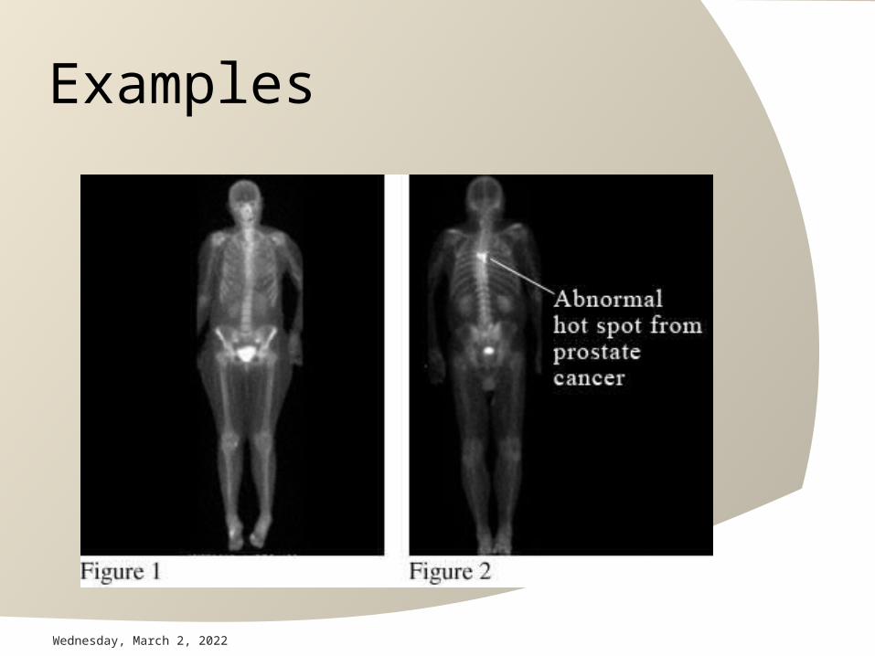
Nuclear imaging
Radio-isotops (radiopharmaceuticals) are given intravenously or orally. Then, external detectors (gamma cameras) capture and form images from the radiation emitted by the radiopharmaceuticals. There are several techniques of diagnostic nuclear medicine.
21

May 1, 2023
Examples Whole body bone scan – used to identify metastatic cancer involving the bone.Thyroid uptake scan – used to visualize the thyroid gland when disease of the thyroid is suspected.Renal scan – used to indicate the perfusion, function and structure of the kidneys. It is also used to indicate the presence of obstruction or renovascular hypertension.Parathyroid scan – done primarily to detect tumors in the parathyroid gland.Liver/spleen scan – allows for visualization of the liver and spleen. It is indicated for patients with cancer to rule out metastatic tumor in the liver.
22
May 1, 2023
Examples technetium-99m rubidium-82 thallium-201 fluoro-deoxy glucose (FDG)

May 1, 2023
Examples

May 1, 2023
Examples

May 1, 2023
Examples

May 1, 2023
Examples
May 1, 2023
Examples

May 1, 2023
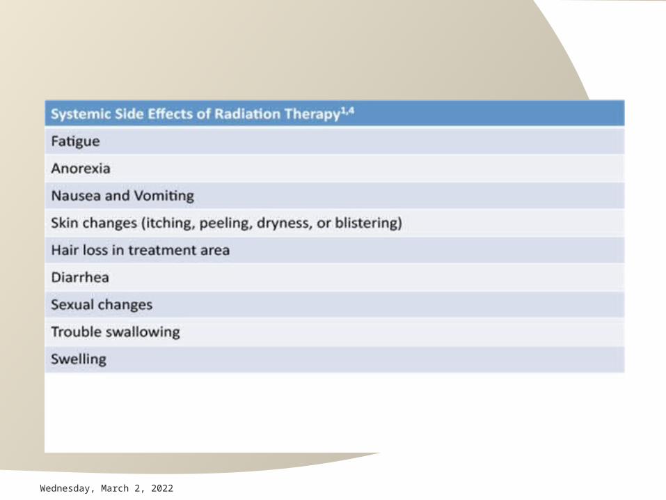
Theraputic radiologyRadiotherapy A clinical modality dealing with the
use of ionizing radiation in the treatment of patients
Cancer treatment

May 1, 2023
How Radiotherapy Works ? DNA
Translocations Deletions

May 1, 2023
Types of Radiotherapy
•External Beam Radiotherapy: radiation source is outside the patient
•Brachytherapy: radiation source is inside the patient: Intra-cavitary (cervix cancer) Intra-lumenal (esophagus, bronchus) Interstitial (Head Neck, Breast)

May 1, 2023
External Beam Radiotherapy

May 1, 2023
Interstitial

May 1, 2023

May 1, 2023
May 1, 2023

May 1, 2023
Interventional radiology

May 1, 2023
What is interventional radiology?
Interventional radiology is a subspecialty which provides minimally invasive diagnosis and/or treatment using imaging (ultrasound, CT, or fluoroscopy) to target the intervention and show the results of the intervention.

May 1, 2023
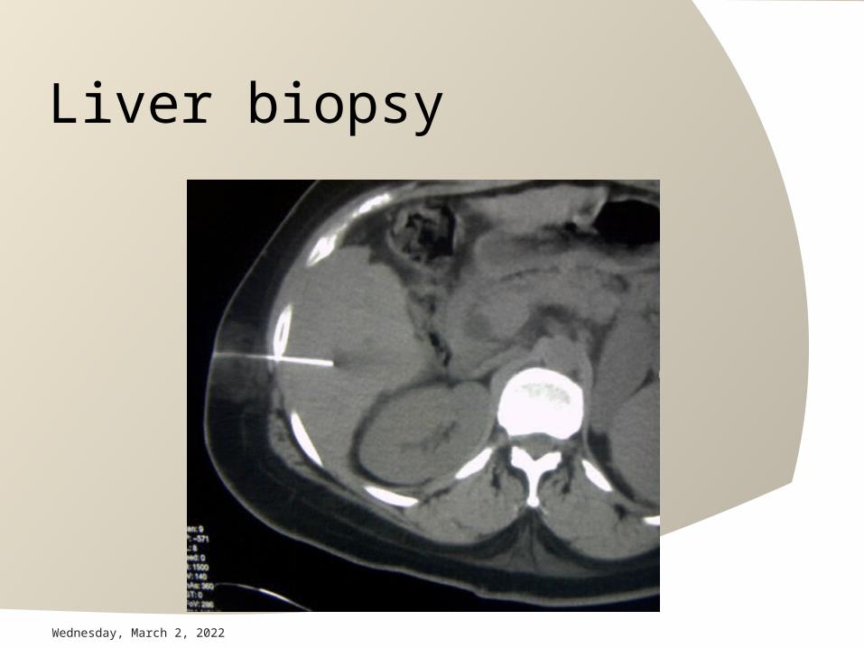
1. Percutaneous biopsy
US, CT or fluoroscopyRandom sampling or sampling of a massLung, mediastinum, pleura, chest wall, nodesLiver, adrenal gland, pancreas kidneys, lymph nodes
May 1, 2023
Liver biopsy

May 1, 2023
Lung biopsy

May 1, 2023
2. Percutaneous abscess drainage
US, CT or fluoroscopyAspiration or drainage tube placementUsually for infectionPleura, lungHepatic (intra/sub), pericolic gutters, perisplenic, peri/intrapancreatic, pouch of Douglas, psoas, abdominal wall

May 1, 2023
3. Arteriography
Injection of contrast media directly into arteries and vis via fluoroscopyUsually immediately precedes and intervention is angioplasty, stenting, embolization, thrombolysisAorta, pelvis, lower and upper extremities, kidneys, gut, lungs

May 1, 2023
Aortic angiography

May 1, 2023
Pelvic embolization post trauma

May 1, 2023
13. Foreign body retrieval
Most frequently guidewires or cathetersUsually in the right heart or pulmonary arteryRetrieval under fluoroscopic guidance using snares needed given infection, arrhythmia risk

May 1, 2023
Cholangiogram (L), internal external drainage from the L (R)

May 1, 2023
Jom ... practice

49
RADIOLOGY DIAGRAM
X-ray image
Pathologyimage

Zenker's Diverticulum

Esophageal Cancer

Esophageal Cancer

53
Benign Esophageal Stricture

54
Achalasia


NORMALESOPHAGUS
DIAPHRAGM
HIATAL HERNIA
DIAPHRAGM
*Note distended distal esophagus with herniation of gastric fundus into chest through esophageal hiatus.
This allows for reflux of gastric contents into esophagus.
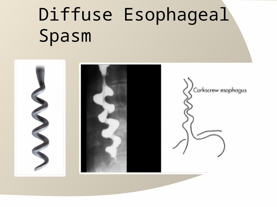
Diffuse Esophageal Spasm

58
Gastric Ulcer
Barium collects in ulcer crater
Endoscopic view of ulcer

Malignant Ulcer Benign Ulcer

Gastric Carcinoma

61
Duodenal Ulcer
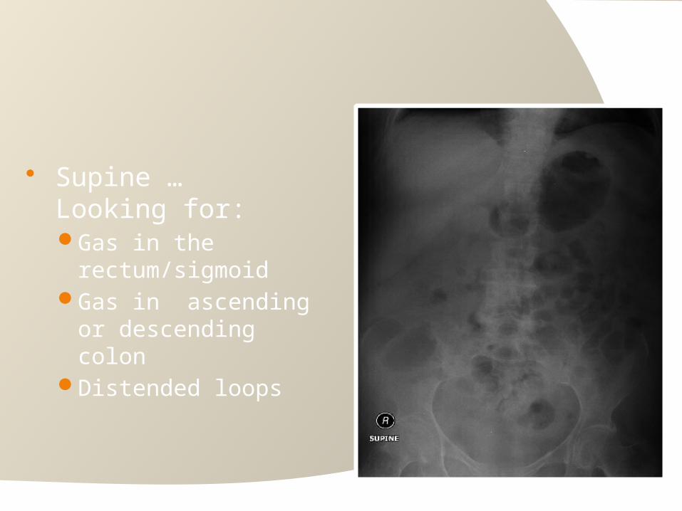
Supine … Looking for:Gas in the
rectum/sigmoidGas in ascending or
descending colonDistended loops

Erect .. looking for:free air “under
diaphrams”air-fluid levels

Normal Gas Pattern
○ Stomach Always (except
supine)○ Small Bowel
Two or three loops of non-distended bowel
Normal diameter = 2.5 cm = 1 US quarter
○ Large BowelIn rectum or sigmoid –
almost always

stomach
colon
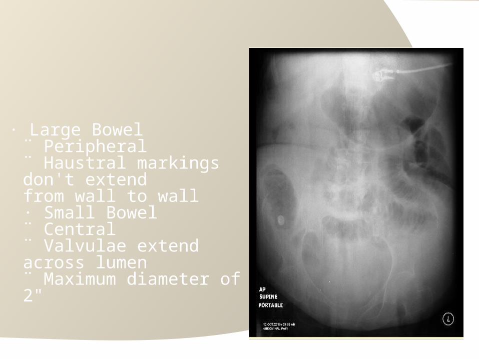
· Large Bowel¨ Peripheral¨ Haustral markings don't extendfrom wall to wall· Small Bowel¨ Central¨ Valvulae extend across lumen¨ Maximum diameter of 2"

HEPATIC FLEXURE
SPLENIC FLEXURE
TRANSVERSE COLON
CECUM
ASCENDING
COLON
DESENDIN
G COLO
NTERMINAL ILEUM
Normal Colon

68
An erect CXR (not AXR) is the best projection to diagnose a pneumoperitoneum (gas in the peritoneal cavity).

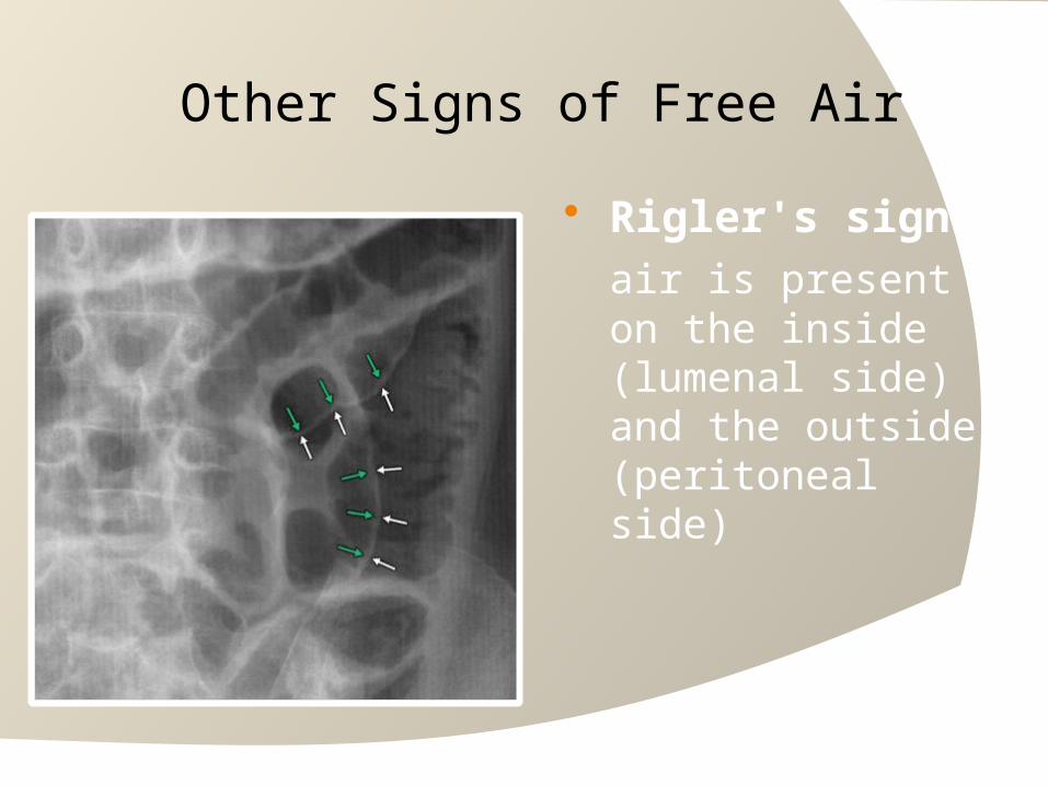
Other Signs of Free Air Rigler's sign
air is present on the inside (lumenal side) and the outside (peritoneal side)

ERECT
Ng tube
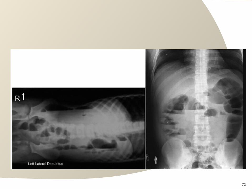
Small Bowel Obstruction
72

Small Bowel Obstruction

Small Bowel Obstruction
DilatedBowel
Non DilatedBowel
OBSTRUCTION*

75
Hernia

76
ColonObstruction

77
Colon Cancer

78

Sigmoid Volvulus
“coffee bean sign”

80
“beak sign”
Barium fills to point of obstruction and
twist of sigmoid colon
Colon Volvulus


Intussusception Meniscus sign

Chrohn’s Disease
normal

84
Chrohn’s Disease
String Signcobblestone mucosa

85
Ulcerative Colitis
Lead Pipe Colon ahaustral appearance


Diverticulosis

88
Diverticulitis

89
Appendicolith

90
Ileostomy

91
Gall Stones

Porcelain Gallbladder

Nephrocalcinosis
•Hyperparathyroidism •Medullary sponge kidney

ERCP - Normal

ERCP

96
Bile Duct Stricture

97
MRCP

Small Bowel ObstructionPROXIMAL DILATED
BOWEL
DISTALNORMALBOWEL

99
Peritoneal Free air

Retroperitoneal Air

Pneumoretroperitoneum

102
Polyp of Transverse Colon

103
Gastric Tumor
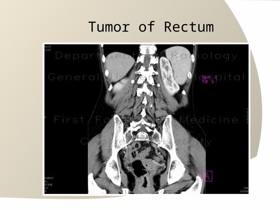
Tumor of Rectum

105

106
Tumor of Rectum
107
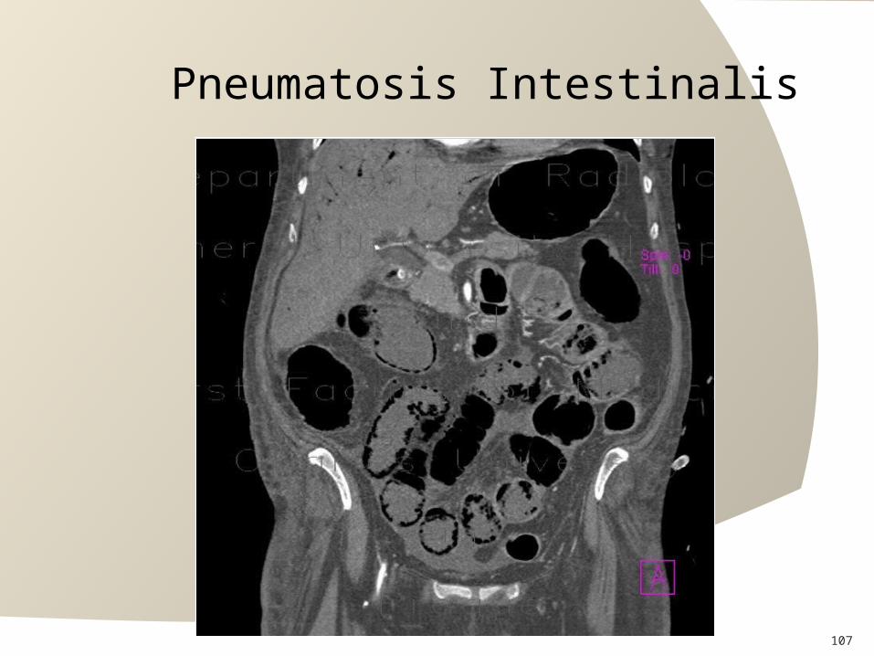
Pneumatosis Intestinalis
108
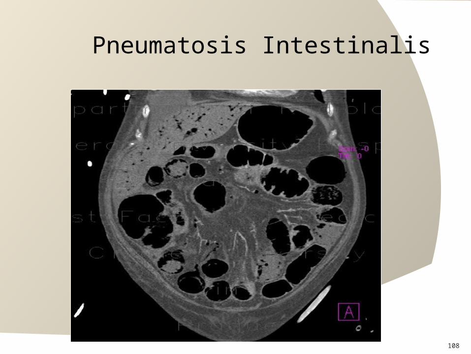
Pneumatosis Intestinalis

109

110
Pneumobilia

111
GS ilues

112
Amebic Liver abscess
113
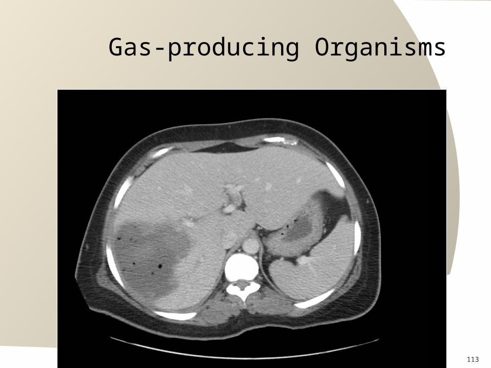
Gas-producing Organisms

114
Pyogenic Abscess

115
Liver Metastases

116
Hepatocellular Carcinoma

117
Hernia

118
Inguinal Hernia
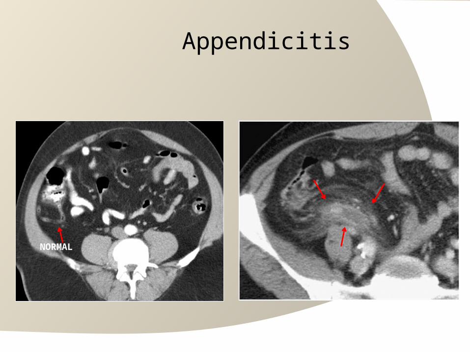
Appendicitis
NORMAL

120
DrainageAbscess

121
AAA

Pancreatitis

Pancreatitis

Pancreatitis

Carotid Body Tumor
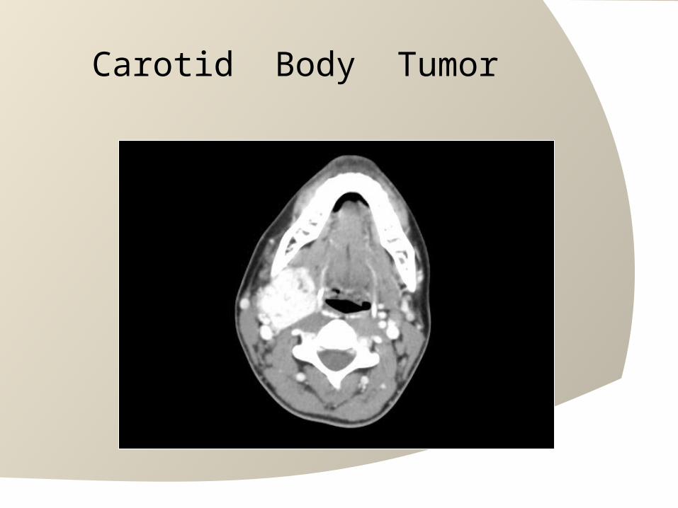
Carotid Body Tumor

127
T1

T2

129
Gallbladder Stones + Wall Thickening

130
Gallbladder Stones + Wall Thickening

Gallbladder Stones + Wall Thickening
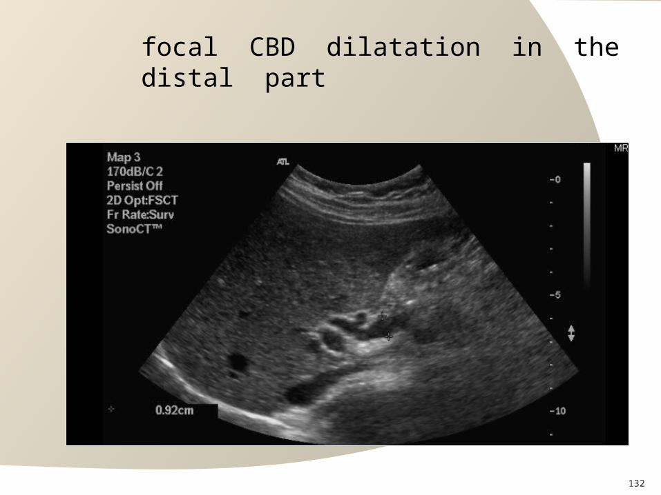
132
focal CBD dilatation in the distal part

133
Anechoic Liver Lesions

CBD is dilated measuring 1 cm with tiny stone

multiple gallstones with mild thickening

136
Malignant Breast Lesion

Pathological Axillary Lymph Node