04a Homeostasis & Diagnostic Tests€¦ · Homeostasis and Diagnostic Tests, HASPI Medical Biology...
10
Homeostasis and Diagnostic Tests, HASPI Medical Biology Lab 04a 159 Name(s): Period: Date: HASPI Medical Biology Lab 04a Background/Introduction Homeostasis & Feedback Mechanisms Imagine drinking three bottles of water every day for a week, and not being able to excrete any of the water from your body? What do you think might happen? Bloating, pain, illness, nothing? In actuality, it would cause death! Balancing substances, even a substance as simple as water, is crucial to keeping the body healthy. This balancing act of substances is known as homeostasis. Examples of substances and/or conditions that need to be balanced include temperature, energy, pressure, calcium, pH, sugar, fats, iron, water, oxygen, carbon dioxide, blood, and even sleep. How does the body maintain homeostasis? It uses feedback mechanisms or loops. Feedback mechanisms respond when imbalance occurs by attempting to correct the balance, and returning the body to homeostasis. http://cnx.org/content/m45989/latest/106_Pregnancy-Positive_Feedback.jpg Positive Feedback – Rare and most commonly occurs in illness or special circumstances, such as labor. In positive feedback the imbalance continues to increase, hence the use of the word “positive” to describe the feedback. An example of positive feedback is contractions during labor. 1. During labor the pressure of the baby’s head on the cervix initiates the brain to start producing a hormone called oxytocin. 2. Oxytocin stimulates the uterus to contract and push the baby, placing more pressure on the cervix. 3. The pressure on the cervix initiates the brain to produce more oxytocin, and the cycle continues until the pressure is relieved when the baby is delivered. Negative Feedback – The most common type of feedback in the body. In negative feedback the imbalance is corrected back to its normal value. An example of negative feedback is regulating temperature in the body, called thermoregulation. The normal core human body temperature is 36-38° C. 1. When the external temperature is warmer than the body temperature, the body warms slightly. a. An organ within the brain, called the hypothalamus, reacts by releasing hormones that increase sweating and dilate blood vessels allowing heat to be released. b. This results in a body temperature decrease. 2. When the external temperature is colder than the body temperature, the body cools slightly. a. The hypothalamus reacts by releasing hormones that cause shivering and constrict blood vessels allowing heat to be conserved. b. This results in a body temperature increase. http://bio1152.nicerweb.com/Locked/media/ch40/40_21HypothalamThermostat.jpg
-
Upload
nguyenkhue -
Category
Documents
-
view
266 -
download
2
Transcript of 04a Homeostasis & Diagnostic Tests€¦ · Homeostasis and Diagnostic Tests, HASPI Medical Biology...
Homeostasis and Diagnostic Tests, HASPI Medical Biology Lab 04a 159
Name(s): Period: Date:
HASPI Medical Biology Lab 04a Background/Introduction Homeostasis & Feedback Mechanisms Imagine drinking three bottles of water every day for a week, and not being able to excrete any of the water from your body? What do you think might happen? Bloating, pain, illness, nothing? In actuality, it would cause death! Balancing substances, even a substance as simple as water, is crucial to keeping the body healthy. This balancing act of substances is known as homeostasis. Examples of substances and/or conditions that need to be balanced include temperature, energy, pressure, calcium, pH, sugar, fats, iron, water, oxygen, carbon dioxide, blood, and even sleep. How does the body maintain homeostasis? It uses feedback mechanisms or loops. Feedback mechanisms respond when imbalance occurs by attempting to correct the balance, and returning the body to homeostasis.
http://cnx.org/content/m45989/latest/106_Pregnancy-Positive_Feedback.jpg
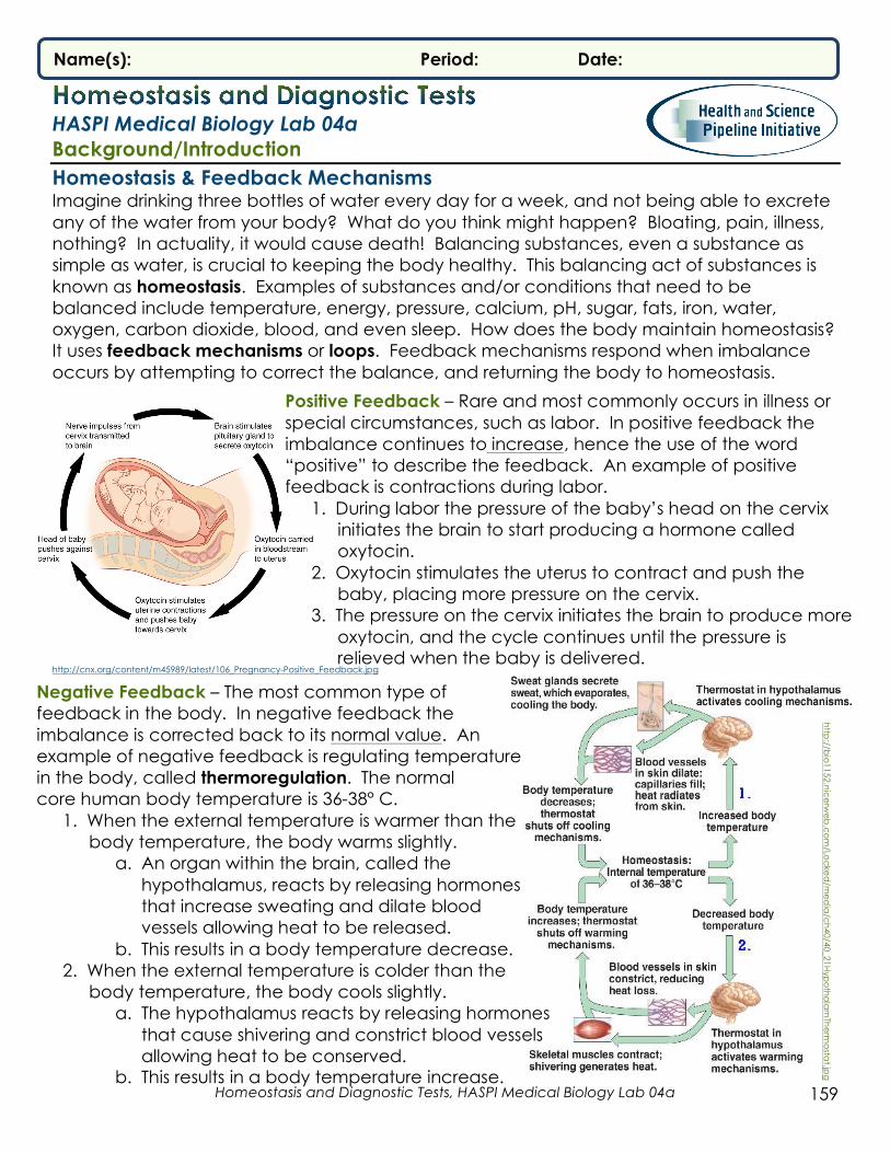
Positive Feedback – Rare and most commonly occurs in illness or special circumstances, such as labor. In positive feedback the imbalance continues to increase, hence the use of the word “positive” to describe the feedback. An example of positive feedback is contractions during labor.
1. During labor the pressure of the baby’s head on the cervix initiates the brain to start producing a hormone called oxytocin.
2. Oxytocin stimulates the uterus to contract and push the baby, placing more pressure on the cervix.
3. The pressure on the cervix initiates the brain to produce more oxytocin, and the cycle continues until the pressure is relieved when the baby is delivered.
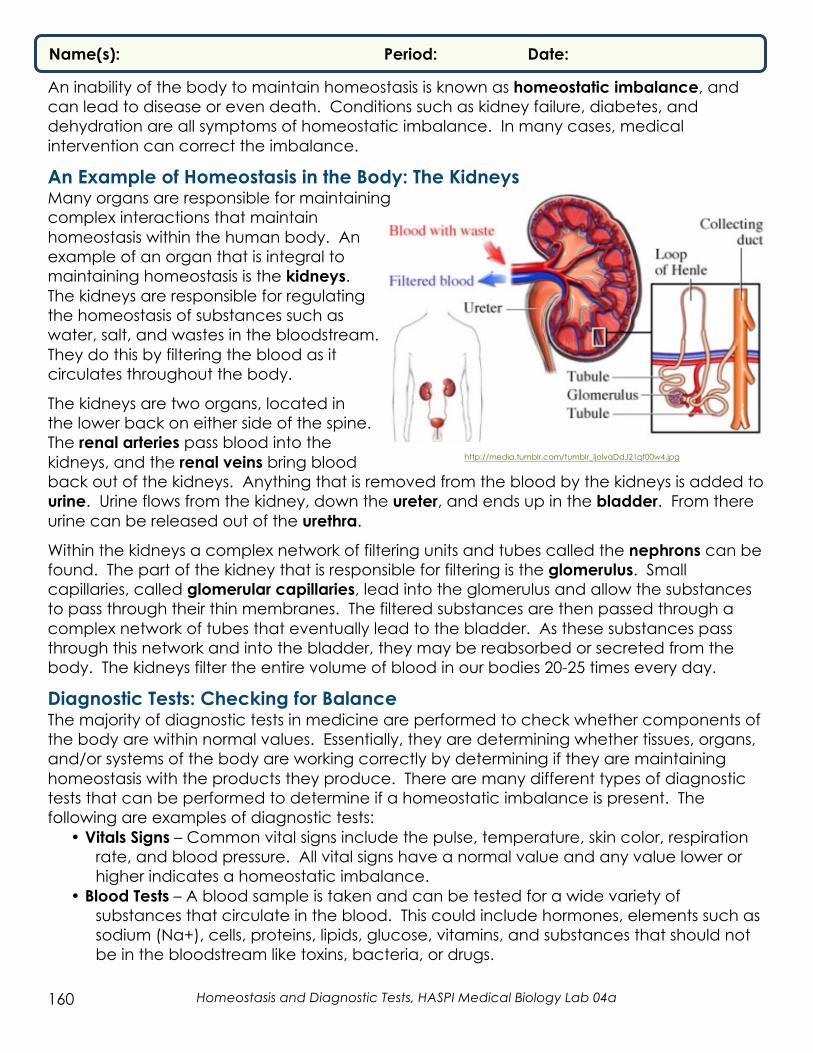
Negative Feedback – The most common type of feedback in the body. In negative feedback the imbalance is corrected back to its normal value. An example of negative feedback is regulating temperature in the body, called thermoregulation. The normal core human body temperature is 36-38° C.
1. When the external temperature is warmer than the body temperature, the body warms slightly.
a. An organ within the brain, called the hypothalamus, reacts by releasing hormones that increase sweating and dilate blood vessels allowing heat to be released.
b. This results in a body temperature decrease. 2. When the external temperature is colder than the
body temperature, the body cools slightly. a. The hypothalamus reacts by releasing hormones
that cause shivering and constrict blood vessels allowing heat to be conserved.
b. This results in a body temperature increase.
http
://bio
1152.nic
erw
eb
.co
m/Lo
cke
d/m
ed
ia/c
h40/40_21H
ypo
tha
lam
The
rmo
stat.jp
g
Homeostasis and Diagnostic Tests, HASPI Medical Biology Lab 04a 160
Name(s): Period: Date:
An inability of the body to maintain homeostasis is known as homeostatic imbalance, and can lead to disease or even death. Conditions such as kidney failure, diabetes, and dehydration are all symptoms of homeostatic imbalance. In many cases, medical intervention can correct the imbalance.
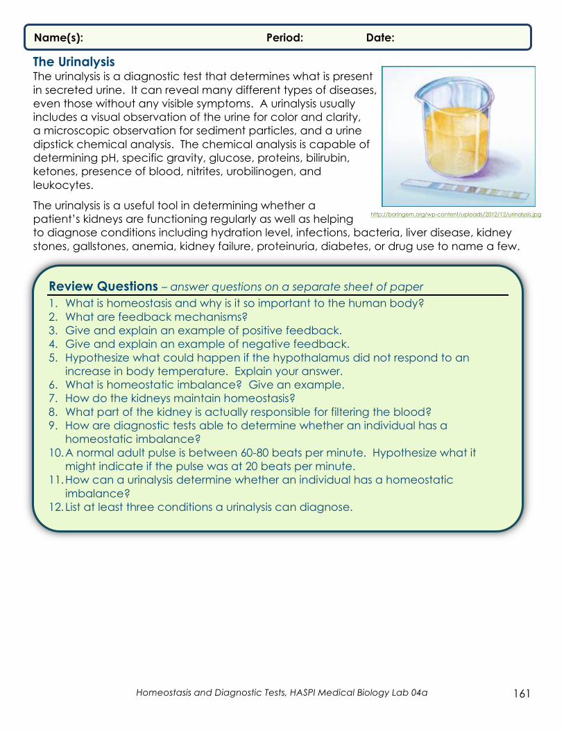
An Example of Homeostasis in the Body: The Kidneys Many organs are responsible for maintaining complex interactions that maintain homeostasis within the human body. An example of an organ that is integral to maintaining homeostasis is the kidneys. The kidneys are responsible for regulating the homeostasis of substances such as water, salt, and wastes in the bloodstream. They do this by filtering the blood as it circulates throughout the body.
The kidneys are two organs, located in the lower back on either side of the spine. The renal arteries pass blood into the kidneys, and the renal veins bring blood back out of the kidneys. Anything that is removed from the blood by the kidneys is added to urine. Urine flows from the kidney, down the ureter, and ends up in the bladder. From there urine can be released out of the urethra.
Within the kidneys a complex network of filtering units and tubes called the nephrons can be found. The part of the kidney that is responsible for filtering is the glomerulus. Small capillaries, called glomerular capillaries, lead into the glomerulus and allow the substances to pass through their thin membranes. The filtered substances are then passed through a complex network of tubes that eventually lead to the bladder. As these substances pass through this network and into the bladder, they may be reabsorbed or secreted from the body. The kidneys filter the entire volume of blood in our bodies 20-25 times every day.
Diagnostic Tests: Checking for Balance The majority of diagnostic tests in medicine are performed to check whether components of the body are within normal values. Essentially, they are determining whether tissues, organs, and/or systems of the body are working correctly by determining if they are maintaining homeostasis with the products they produce. There are many different types of diagnostic tests that can be performed to determine if a homeostatic imbalance is present. The following are examples of diagnostic tests:
• Vitals Signs – Common vital signs include the pulse, temperature, skin color, respiration rate, and blood pressure. All vital signs have a normal value and any value lower or higher indicates a homeostatic imbalance.
• Blood Tests – A blood sample is taken and can be tested for a wide variety of substances that circulate in the blood. This could include hormones, elements such as sodium (Na+), cells, proteins, lipids, glucose, vitamins, and substances that should not be in the bloodstream like toxins, bacteria, or drugs.
http://media.tumblr.com/tumblr_ljolvaDdJ21qf00w4.jpg
Homeostasis and Diagnostic Tests, HASPI Medical Biology Lab 04a 161
Name(s): Period: Date:
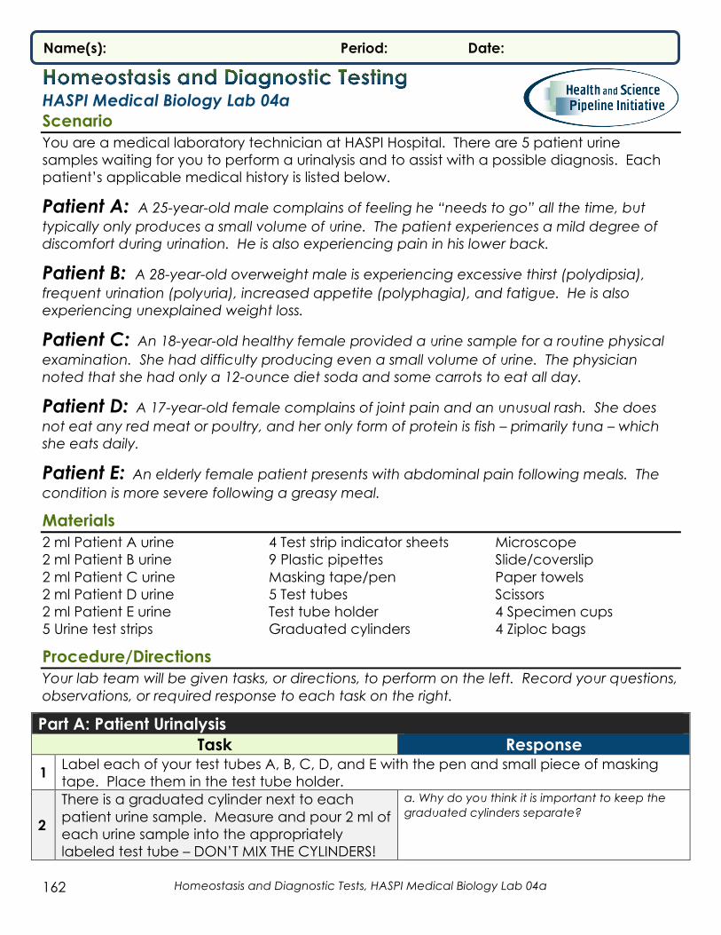
The Urinalysis The urinalysis is a diagnostic test that determines what is present in secreted urine. It can reveal many different types of diseases, even those without any visible symptoms. A urinalysis usually includes a visual observation of the urine for color and clarity, a microscopic observation for sediment particles, and a urine dipstick chemical analysis. The chemical analysis is capable of determining pH, specific gravity, glucose, proteins, bilirubin, ketones, presence of blood, nitrites, urobilinogen, and leukocytes.
The urinalysis is a useful tool in determining whether a patient’s kidneys are functioning regularly as well as helping to diagnose conditions including hydration level, infections, bacteria, liver disease, kidney stones, gallstones, anemia, kidney failure, proteinuria, diabetes, or drug use to name a few.
Review Questions – answer questions on a separate sheet of paper 1. What is homeostasis and why is it so important to the human body? 2. What are feedback mechanisms? 3. Give and explain an example of positive feedback. 4. Give and explain an example of negative feedback. 5. Hypothesize what could happen if the hypothalamus did not respond to an
increase in body temperature. Explain your answer. 6. What is homeostatic imbalance? Give an example. 7. How do the kidneys maintain homeostasis? 8. What part of the kidney is actually responsible for filtering the blood? 9. How are diagnostic tests able to determine whether an individual has a
homeostatic imbalance? 10.A normal adult pulse is between 60-80 beats per minute. Hypothesize what it
might indicate if the pulse was at 20 beats per minute. 11.How can a urinalysis determine whether an individual has a homeostatic
imbalance? 12.List at least three conditions a urinalysis can diagnose.
http://boringem.org/wp-content/uploads/2012/12/urinalysis.jpg
Homeostasis and Diagnostic Tests, HASPI Medical Biology Lab 04a 162
Name(s): Period: Date:
HASPI Medical Biology Lab 04a Scenario You are a medical laboratory technician at HASPI Hospital. There are 5 patient urine samples waiting for you to perform a urinalysis and to assist with a possible diagnosis. Each patient’s applicable medical history is listed below.
Patient A: A 25-year-old male complains of feeling he “needs to go” all the time, but typically only produces a small volume of urine. The patient experiences a mild degree of discomfort during urination. He is also experiencing pain in his lower back.
Patient B: A 28-year-old overweight male is experiencing excessive thirst (polydipsia), frequent urination (polyuria), increased appetite (polyphagia), and fatigue. He is also experiencing unexplained weight loss.
Patient C: An 18-year-old healthy female provided a urine sample for a routine physical examination. She had difficulty producing even a small volume of urine. The physician noted that she had only a 12-ounce diet soda and some carrots to eat all day.
Patient D: A 17-year-old female complains of joint pain and an unusual rash. She does not eat any red meat or poultry, and her only form of protein is fish – primarily tuna – which she eats daily.
Patient E: An elderly female patient presents with abdominal pain following meals. The condition is more severe following a greasy meal.
Materials 2 ml Patient A urine 2 ml Patient B urine 2 ml Patient C urine 2 ml Patient D urine 2 ml Patient E urine 5 Urine test strips
4 Test strip indicator sheets 9 Plastic pipettes Masking tape/pen 5 Test tubes Test tube holder Graduated cylinders
Microscope Slide/coverslip Paper towels Scissors 4 Specimen cups 4 Ziploc bags
Procedure/Directions Your lab team will be given tasks, or directions, to perform on the left. Record your questions, observations, or required response to each task on the right.
Part A: Patient Urinalysis Task Response
1 Label each of your test tubes A, B, C, D, and E with the pen and small piece of masking tape. Place them in the test tube holder.
2
There is a graduated cylinder next to each patient urine sample. Measure and pour 2 ml of each urine sample into the appropriately labeled test tube – DON’T MIX THE CYLINDERS!
a. Why do you think it is important to keep the graduated cylinders separate?

Homeostasis and Diagnostic Tests, HASPI Medical Biology Lab 04a 163
Name(s): Period: Date:
3
Using the scissors, cut the 5 urine test strips lengthwise. You should have 10 small strips. Five strips will be used for Part A and 4 for Part B. Return any extra strips to your instructor. The same results can be read on a full or half strip.
4
Observe each patient urine sample for color and clarity. Refer to the Background Section for reference. Record your observations in Data Table 1.
a. Do you think food or drink choices could drastically change the color of your urine? Explain your answer.
5
Place a drop of “Patient A urine” onto the slide and place the cover slip over it. View the urine under the microscope to identify any sediment particles within the urine. If you see ANY sediment or crystals in the urine record a + in Data Table 1. If there are no visible particles record a – in Data Table 1.
6 Clean off the slide and cover slip. Repeat step 5 for Patient Urine B-E.
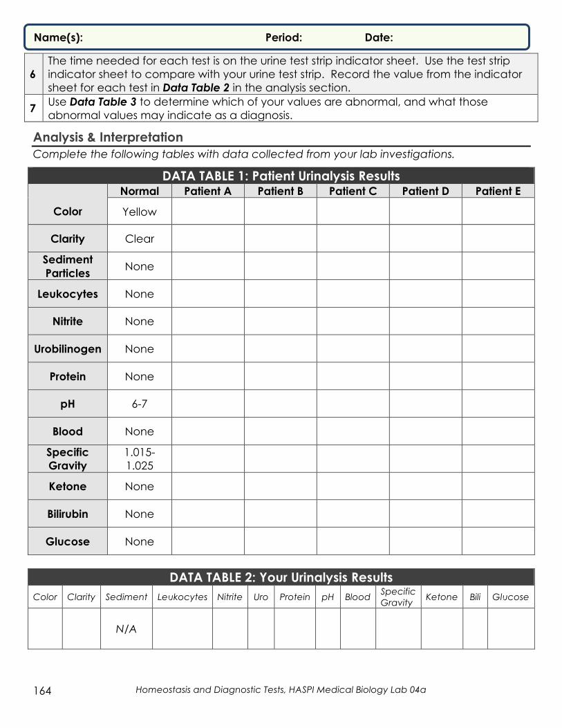
7
Place one of the urine test strips on a paper towel. Use the plastic dropper for the urine sample for Patient A to place urine on each of the 10 boxes on one strip. Each box requires 30-120 seconds; the time needed for each test is on the urine test strip indicator sheet. Use the test strip indicator sheet to compare with your urine test strip. Record the value from the indicator sheet for each test in Data Table 1 in the analysis section.
a. Do you think a single test parameter (color, pH, etc.) that is imbalanced would make it easier to diagnose disease than multiple imbalances? Why?
8 Repeat step 7 for the remaining four urine samples.
9 Use Data Table 3 to determine which of the patient values are abnormal, and what those abnormal values may indicate as a diagnosis. Note: Physicians make diagnoses, but the work of lab technicians provides important information that the doctor often needs to do so.
Part B: How’s My Homeostasis? Task
1 Get a Ziploc bag. Place a pipette, specimen cup, urine test strip (1/2 of test strip cut in Part A), and a test strip indicator sheet into the Ziploc bag.
2 Take the bag with all of the supplies home to perform your own urinalysis using the steps below. ALL of the following steps are to be performed at home. DO NOT bring any of your test supplies or urine samples back to the classroom! Discard all supplies after use.
3 Carefully fill approximately ½ of your specimen cup with your own urine by placing the cup midstream during urination.
4 Place your urine test strip flat on a paper towel on a flat surface.
5 Using the pipette, place drops of urine on each of the 10 test boxes of the urine strip until they are saturated. You do not have a microscope, so you will not be recording any sediment for your urine.
Homeostasis and Diagnostic Tests, HASPI Medical Biology Lab 04a 164
Name(s): Period: Date:
6 The time needed for each test is on the urine test strip indicator sheet. Use the test strip indicator sheet to compare with your urine test strip. Record the value from the indicator sheet for each test in Data Table 2 in the analysis section.
7 Use Data Table 3 to determine which of your values are abnormal, and what those abnormal values may indicate as a diagnosis.
Analysis & Interpretation Complete the following tables with data collected from your lab investigations.
DATA TABLE 1: Patient Urinalysis Results
Color
Normal Patient A Patient B Patient C Patient D Patient E
Yellow
Clarity
Clear
Sediment Particles
None
Leukocytes
None
Nitrite
None
Urobilinogen
None
Protein
None
pH
6-7
Blood
None
Specific Gravity
1.015-1.025
Ketone
None
Bilirubin
None
Glucose
None
DATA TABLE 2: Your Urinalysis Results
Color
Clarity
Sediment
Leukocytes
Nitrite
Uro
Protein
pH
Blood Specific Gravity
Ketone
Bili
Glucose
N/A

Homeostasis and Diagnostic Tests, HASPI Medical Biology Lab 04a 165
Name(s): Period: Date:
DATA TABLE 3: Abnormal Vs. Normal Urine
Visual Observations
Appearance Possible Condition(s) Yellow and Clear Normal
Dark Yellow May indicate concentrated urine from dehydration Brownish or Green May indicate bile pigments
Reddish-Amber May indicate urobilinogen, which is produced in the intestine by bacteria in bile; evidence of liver disease, Addison’s, or other conditions
Cloudy May indicate phosphates, white blood cells (WBCs), bacteria, epithelial cells or fat
Milky May indicate presence of WBCs, bacteria, or fat
Microscopic Observations
Appearance Possible Conditions
Crystals Can be normally occurring; persistent crystals may indicate kidney stones or infection
Large Particles (casts)
May indicate chronic kidney failure, proteinuria, or be present after strenuous exercise
Chemical Analysis
Property Possible Conditions Leukocytes Indicates inflammation of the urinary tract usually caused by infection
Nitrite Normal urine has very little to no nitrite. Presence may indicate bacterial contamination/urinary tract infection (UTI)
Urobilinogen Urobilinogen is produced in the intestine by bacteria in bile; may indicate liver disease, Addison’s, or other conditions
Protein Normal urine has no protein. Presence may indicate kidney trauma, ingestion of heavy metals, bacterial toxins, nephritis, or hypertension; may also arise under conditions of physical stress or strenuous exercise
pH
Normal urine pH is between 6-7; below 6 may indicate gout, fever, or kidney stones; above 7 may indicate urinary tract infection, vegetarian diet, bowel obstruction, vomiting, or drug use
Red Blood Cells (erythrocytes)
May indicate irritation of the urinary tract organs by calculi or physical trauma; may also indicate hemolysis of red blood cells due to such conditions as anemia, transfusion reactions, burns, or renal disease
Specific Gravity
Normal urine specific gravity is between 1.015 and 1.025 Less than 1.015: may indicate excess fluid intake, diabetes insipidis or chronic renal failure; More than 1.025: may indicate limited fluid intake, dehydration, fever, kidney inflammation
Ketones
Normal urine has no ketones. Presence may indicate starvation or abnormal metabolic processes such as excessive metabolism of fats or proteins in the body; possibly indicates diabetic ketosis
Bilirubin Normal urine has no bilirubin. Presence may indicate liver pathology such as hepatitis or cirrhosis
Glucose Normal urine has no glucose. Presence may result from excessive carbohydrate intake or diabetes mellitus
DATA TABLE 4: Diagnosis and Treatment On a separate sheet of paper create a data table that summarizes the results, diagnosis, and possible treatments for patients A-E and your own results. Include the following in your table:
• Abnormal results (if there are none, write normal) • Possible diagnosis (if any) • Possible treatment options (you will need to research your diagnosis for this answer)

Homeostasis and Diagnostic Tests, HASPI Medical Biology Lab 04a 166
Name(s): Period: Date:
Analysis Questions – answer questions on a separate sheet of paper PART A
1. Why is it important to develop a case history of the physical symptoms of each patient to be used along with the physical tests performed on the patient’s urine specimen?
2. You receive two urine samples – one from the patient in the morning, and one from the afternoon. Would you expect the urinalyses to have the same results? Why or why not?
3. The presence of blood in the urine can indicate a serious kidney problem. Why are kidney problems so serious?
4. Suppose a urine sample revealed abnormal results, such as protein in the urine. If there is a result differing from the norm, should a physician always make an immediate diagnosis of a disorder? Why or why not?
5. How would the specific gravity of urine be different after a vigorous workout without the consumption of significant quantities of water?
6. The presence of ketones is often high in the urine of people with diabetes, and people who suffer from anorexia. What characteristics do these two groups have in common?
7. Plan and conduct an investigation to provide evidence that feedback mechanisms maintain homeostasis.
PART B 8. Were any of your results outside of the normal values? If yes, hypothesize what could
be causing the abnormal values. 9. Explain how what you ate and drank in the last 24 hours could impact your pH and
specific gravity values. 10.Provide and explain an example as evidence that feedback mechanisms maintain
homeostasis in the human body. Use examples of positive AND negative feedback (the examples cannot be contractions or temperature regulation from the Background information). This may require textbook or internet research. CITE YOUR SOURCES!

Homeostasis and Diagnostic Tests, HASPI Medical Biology Lab 04a 167
Name(s): Period: Date:
Connections & Applications Your instructor may assign or allow you to choose any of the following activities. As per NGSS/CCSS, these extensions allow students to explore outside activities recommended by the standards.
1. RESEARCH A BODY SYSTEM: Choose one of the following body systems - digestive, cardiovascular, endocrine, or respiratory. Investigate and choose three diseases of the system you chose. Cite at least two sources. Answer the following for EACH disease: a. Explain specifically HOW the disease interferes with feedback mechanisms b. Symptoms of the disease c. Diagnostic tests that can be used to diagnose the disease d. At least two treatment options and how they can correct the imbalance
2. INTERPRETING TEST RESULTS: A distressed relative has brought her diagnostic blood test
results to you for help interpreting what they could mean. She can’t talk to her doctor until he returns from vacation, and she is very stressed about the results. Analyze the results and determine the following: a. What substances are outside of normal reference intervals and by how much? b. Research and determine what problems these abnormal values might indicate and
any symptoms that could be associated c. Research and determine the treatment options for this problem to help alleviate your
relative’s concern
http://www.healthtestingcenters.com/themes/kktest/images/Results/Basic-1o.gif

Homeostasis and Diagnostic Tests, HASPI Medical Biology Lab 04a 168
Name(s): Period: Date:
3. DEVELOP A MODEL KIDNEY: Develop a model that simulates the function of the kidneys. The model can be a physical model, a computer model, or an illustration. Provide picture evidence that your model can: a. Filter out small substances like water and salt b. Keep large substances like blood cells from being filtered out c. Maintain homeostasis of water (this means not all of the water can be filtered at once;
water can be added to your model as it functions). Provide an explanation of how your model maintains homeostasis of water along with the picture evidence.
Resources & References • Freudenrich, C. 2001. How Your Kidneys Work. Discovery Fit & Health.
http://health.howstuffworks.com/human-body/systems/kidney-urinary/kidney.htm.
• Martin, T. and Baustian, M. 2003. Interpreting Medical Tests: What’s in Urine? Cornell Institute for Biology Teachers, pp. 1-9.
• Neo/SCI. Urinalysis and Disease Identification Investigation. www.neosci.com.