Etching ECE/ChE 4752: Microelectronics Processing Laboratory Gary S. May February 19, 2004.

!"#$%&'%()*%+*,,%-&,./*%01%02%/*/3452*%3&.26%&4752*,,*1%%

!"#$%&'(")*++*%,*'-&'./%/,*#01#%-2)#3&)/%4'(")*++*% % %5"0)%-2)#3&) %%% %67'2#72'*%%%%%%%%%%%%%%%%%%%%%%%4'(")0/./%%8$+&'&,+"/7 % %,$&7&/9)7$*/0/%%%%%%%%%% %:&2;+*<.*.;= %%%>+")7/?%,'&3/7/%%!@ %%%%%%%% % %.&:01#"3&)?%-&+:0)(%%%%%% %/0)(+*<.*.;=%%%%% %%%A++%%
%%%%%%% % %&-%)*B%,'&7*0)/?%+0,0:/%%C&+(0 %%%%%%%%% % %%/&'3)(?%.&:01#"3&)%%% %%%/0)(+*<.*.;=%%%% %%A++%*2D"'9&7*/%
% % %%&-%)*B%,'&7*0)/%%507& %%%%%%%%%%% % %%*)*'(9%,'&:2#3&)%%%%%%%%%% %%:&2;+*<.*.;=%%%% %%A++%*2D"'9&7*/%%E"#2&+*%%%% %%%%%%%%%%%%%%%%%%%%/7&'"(*%F%$&2/*#+*")0)(%%%%%%%%%/0)(+*<.*.;=%%%% %%A++%*2D"'9&7*/%%G2#+*2/%%%%%% % %HGA%."0)7*)")#*%F%%%%%%%%%%%%%%%%%%:&2;+*<.*.;=%%%% %%A++%*2D"'9&7*/%
%%%%%%%% % %@GA%7'")/#'0,3&)%
C*)*'"+%'2+*I%4'(")*++*/%"'*%(*)*'"7*:%-'&.%7$*%/".*%79,*%&-%&'(")*++*%
%.07&#$&):'0"% 507&#$&):'0"%J.")9K'0;;&)L%
#$+&'&,+"/7/% 8$+&'&,+"/7/%J.")9L%
*):&,+"/.0#%'*3#2+2.%J!@L% *):&,+"/.0#%'*3#2+2.%J!@L%JML%
('&B7$% :0N0/0&)%
)2#+*2/% G2#+*2/%JML%

Cytoplasmic !!!!Nuclear!!!!Mitochondrial!!!Plasma membrane !Secreted !Resident ER and Golgi !Endosomes, Lysosomes!
NO SIGNAL SEQUENCE!
NUCLEAR LOCALIZATION SEQUENCE!
MITOCHONDRIAL SIGNAL SEQUENCE!
ER SIGNAL SEQUENCE!

The endoplasmic reticulum (ER) -- Largest membrane-bound organelle. -- Supply proteins to other organelles in the endomembrane system. -- ~1/3 of total cellular proteins.
The secretory pathway
ER Golgi
cis medial trans ribosome
8M%
PM
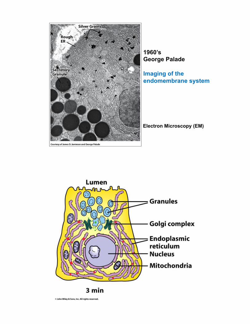
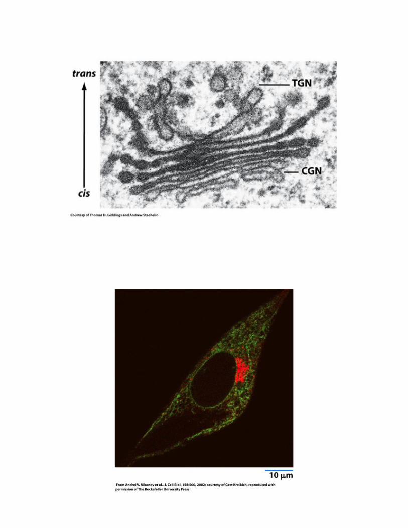
Electron Microscopy (EM)
1960’s George Palade Imaging of the endomembrane system


Trafficking of GFP-VSVG
Temperature sensitive vesicular stomatitis virus G protein (VSVG) mutant begins to traffic at permissive temperature.
Temperature shift from 40 oC to 32 oC
Endomembrane system

The endoplasmic reticulum (ER)
Figure 12-8 Molecular Biology of the Cell (© Garland Science 2008)
-- Distinct functions -- One continuous organelle -- Ratio varies across cell types

The rough ER – site for protein synthesis
Pancreatic acinar cell (Similar in other professional secretory cells)
(Lumen)
The smooth ER
Leydig cell from testis
-- Lipid synthesis -- Adapted to specific biological functions -- Detoxification (e.g. cytochrome p450 in liver )
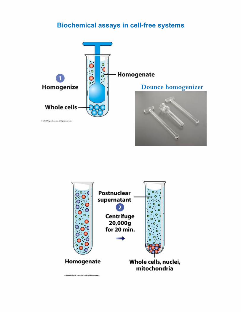
Biochemical assays in cell-free systems
Dounce homogenizer

Figure 12-37b Molecular Biology of the Cell (© Garland Science 2008)

Rough ER
Smooth ER
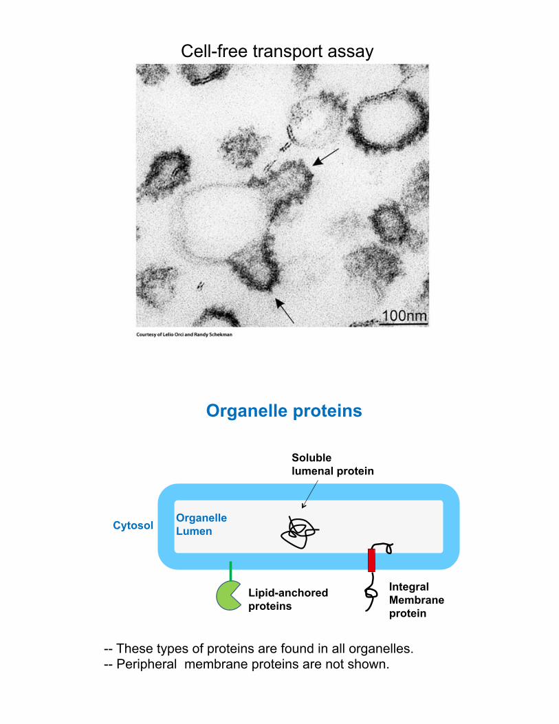
Cell-free transport assay
Integral Membrane protein
Soluble lumenal protein
Organelle proteins
Cytosol Organelle Lumen
-- These types of proteins are found in all organelles. -- Peripheral membrane proteins are not shown.
Lipid-anchored proteins

Integral membrane protein
Soluble lumenal protein
How are proteins targeted to organelles?
Cytosol Organelle Lumen
Ribosome
Signals must exist to direct proteins:!!1. Signal sequences for translocation!
2. Sorting or retention signals!

Signal !sequence!
Translocation across the ER membrane !
trans-membrane!(TM) segment!
Soluble!protein!
Membrane!protein!
Cytoplasmic !!!!Nuclear!!!!Mitochondrial!!!Plasma membrane !Secreted !Resident ER and Golgi !Endosomes, Lysosomes!
NO SIGNAL SEQUENCE!
NUCLEAR LOCALIZATION SEQUENCE!
MITOCHONDRIAL SIGNAL SEQUENCE!
ER SIGNAL SEQUENCE!

Table 12-3 Molecular Biology of the Cell (© Garland Science 2008)
Signal sequence directs protein targeting
Each organelle has its own signal sequences for targeting
Figure 12-35a Molecular Biology of the Cell (© Garland Science 2008)
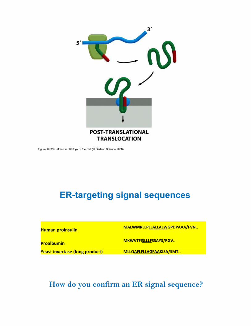
Figure 12-35b Molecular Biology of the Cell (© Garland Science 2008)
8./52%94&021.,02% :;<=:><<?<<;<<;<=@?A?;;;BCDEFF%%%
?4&5,3./02% :G=DHCIJ<<<CJJ;KJB>@DFF%%
K*51(%02-*4(51*%L,&27%94&6.+(M% :<<N;C<C<<;@C;;GIJ;BJ:HFF%%
ER-targeting signal sequences
How do you confirm an ER signal sequence?

Signal !sequence!
Translocation across the ER membrane !
trans-membrane!(TM) segment!
Soluble!protein!
Membrane!protein!
Recognition of signal sequence on the ER
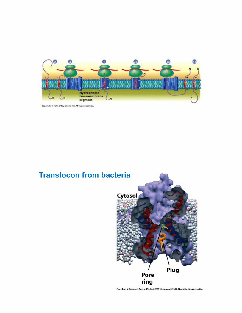
Translocon from bacteria
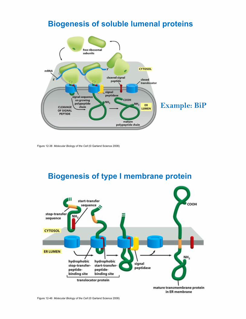
Figure 12-38 Molecular Biology of the Cell (© Garland Science 2008)
Biogenesis of soluble lumenal proteins
Example: BiP
Figure 12-46 Molecular Biology of the Cell (© Garland Science 2008)
Biogenesis of type I membrane protein

N!
N!
C!
C!
Type I!membrane!protein !
cytosol!
Membrane!
lumen!
N!C!
N!
C!
Other topologies!
_!
Topologies of ER proteins
Soluble !lumenal !protein!
Juxtamembrane residues may influence protein orientation

N!
N!
N!
C!
C!
C!
Membrane proteins have different topologies in ER membrane!
cleaved!signal!sequence!
Type I! Type II!
N!
C!Type III!
cytosol!
ER!
lumen!
-cleavable signal seq.!(7-15 apolar residues)!-transmembrane anchor!stops translocation!
-signal anchor !seq. (18-25 apolar)!-not cleaved!
-reverse signal!anchor!
+! +!
_!_!
+!+!
_! _!
+! +!
+! +!
?!
The topology of a protein is maintained:

819O#1%
O$*%."0)%,'&7*0)%&-%7$0/%-".0+9%0/%P0??%B$0#$%7"D*/%,"'7%0)%.")9%"/,*#7/%&-%!@%P2"+079%#&)7'&+%JQ8L=%R7%;0):/%7&%%N"'0&2/%)"/#*)7%"):%)*B+9%/9)7$*/0S*:%,'&7*0)/%"):%"//0/7/%7$*0'%-&+:0)(=%R)%"::03&)?%07%0/%0)N&+N*:%0)%7$*%,'&#*//*/%%&-%!@<"//�"7*:%:*('":"3&)%"):%7$*%2)-&+:*:%,'&7*0)%'*/,&)/*=%O$*%-2)#3&)/%&-%7$*%/*#&):%T/,UV%-".0+9%%.*.;*'?%(+2#&/*<'*(2+"7*:%,'&7*0)%JC@>LMUV?%"'*%'*+"3N*+9%2)*W,+&'*:=%%819Q#1%X0N*%!@%,'&7*0)/%&-%7$*%T/,YV%-".0+9%J!@:ZM[\L%"'*%D)&B)=%O$*9%#&)7"0)%"%+2.0)"++9%*W,&/*:%]<:&."0)%"):%#")%%/3.2+"7*%^0>%AO>"/*%"#3N079%0)%N07'&=%%819R#%
O$*%&)+9%D)&B)%T/,_V%-".0+9%.*.;*'%0/%@>?RQ=%H*/,07*%;*0)(%";2):")7%0)%7$*%!@?%07%0/%)&7%*//*)3"+%-&'%#*++%%N0";0+079%"):%/**./%7&%+0.07%07/%0)7*'"#3&)/%7&%"%/."++%/*7%&-%/2;/7'"7*/=%%S5,2*T02%"):%+5,4*U+.,02%O$*/*%7B&%+*#3)%#$",*'&)*/%0)7*'"#7%B07$%"):%"//0/7%7$*%-&+:0)(%&-%,'&7*0)/%7$"7%#"''9%.&)&(+2#&/9+"7*:%G<+0)D*:%%(+9#")/=%%
ER chaperones and folding enzymes
Disulfide bonds are common among ER proteins
!"#$%&'(()&*+,-)%((.*#/%"0*%(1!2.3%
Disulfide bonds stabilize proteins and promote folding

Figure 12-50 Molecular Biology of the Cell (© Garland Science 2008)
N-glycosylation site: N-X-S/T
N-glycosylation in the ER

N-glycosylation stabilizes proteins and promotes folding
Properly folded ER proteins can be transported to other organelles in the endomembrane system

Incorrectly folded proteins are retained in the ER
Figure 12-54 Molecular Biology of the Cell (© Garland Science 2008)
Incorrectly folded proteins are eventually degraded in the cytosol

Ron and Walter
ER stress response/unfolded protein response
Vesicular transport
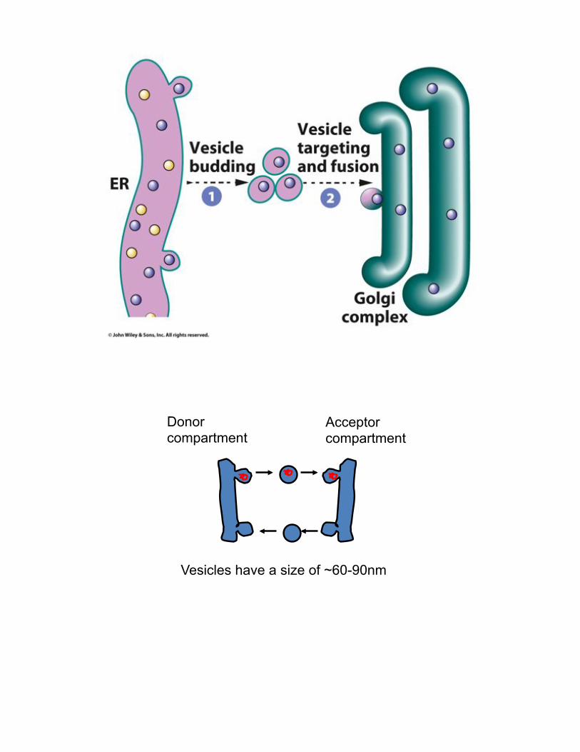
Donor compartment
Acceptor compartment
Vesicles have a size of ~60-90nm
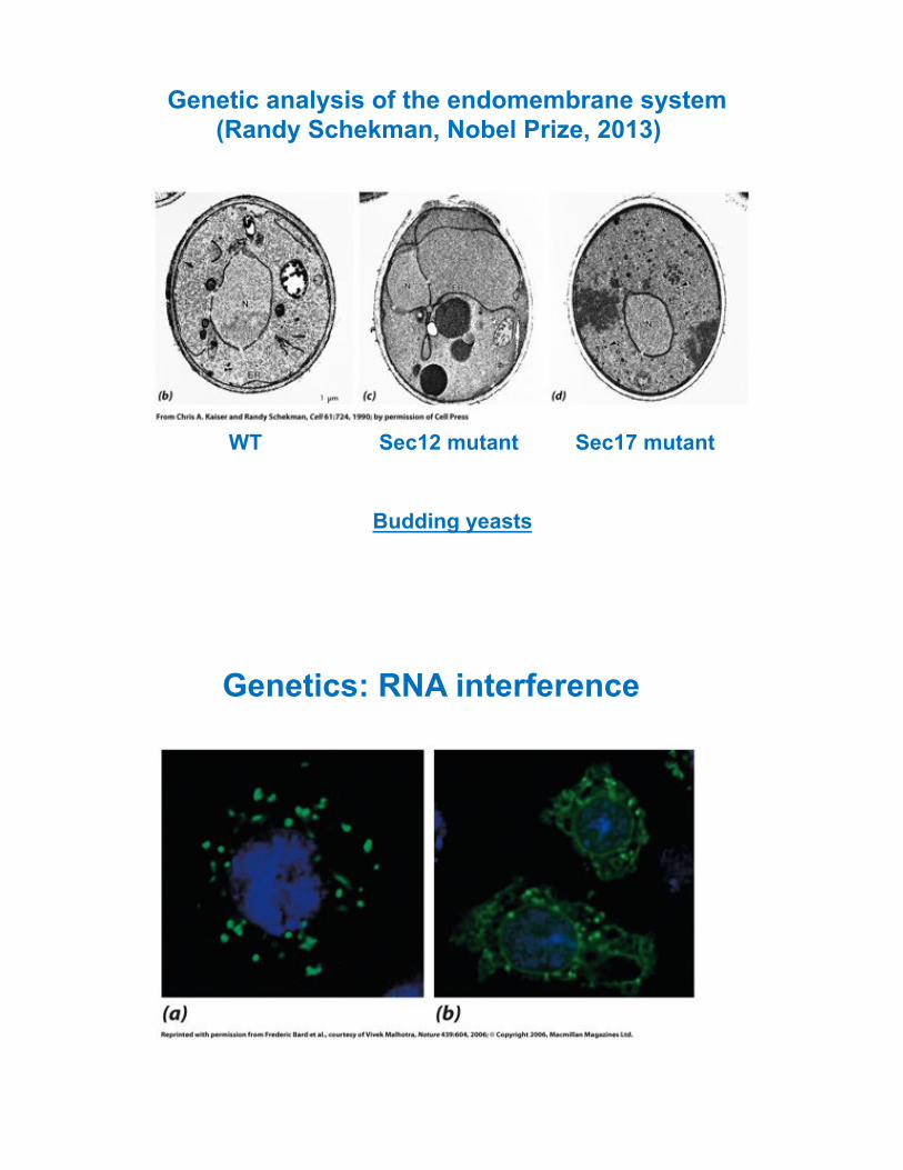
Genetic analysis of the endomembrane system (Randy Schekman, Nobel Prize, 2013)
Budding yeasts
WT Sec12 mutant Sec17 mutant
Genetics: RNA interference
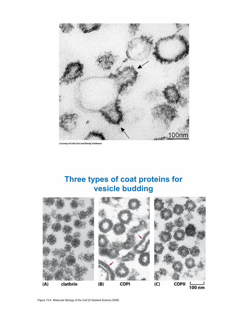
Figure 13-4 Molecular Biology of the Cell (© Garland Science 2008)
Three types of coat proteins for vesicle budding

Figure 13-20 Molecular Biology of the Cell (© Garland Science 2008)
Proteins destined for export are captured by COPII coats
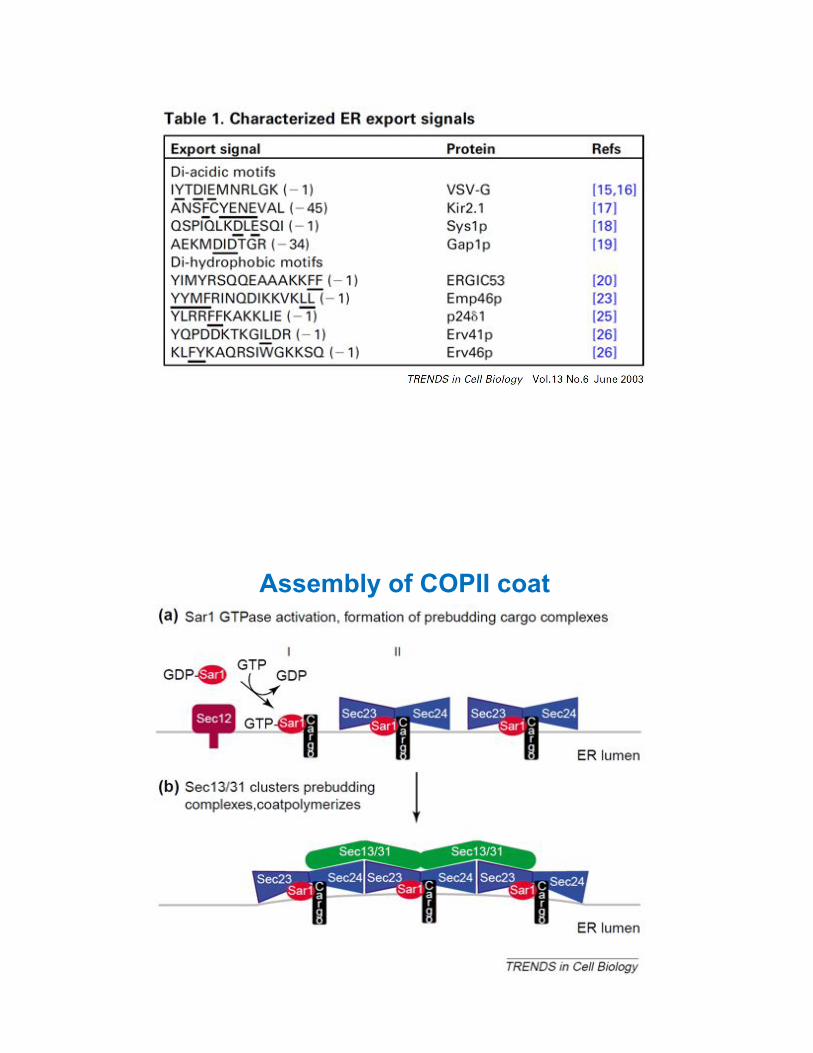
Assembly of COPII coat

Figure 13-13d Molecular Biology of the Cell (© Garland Science 2008)
Dissociation

Lipid transfer protein
Transport vesicles

The Golgi
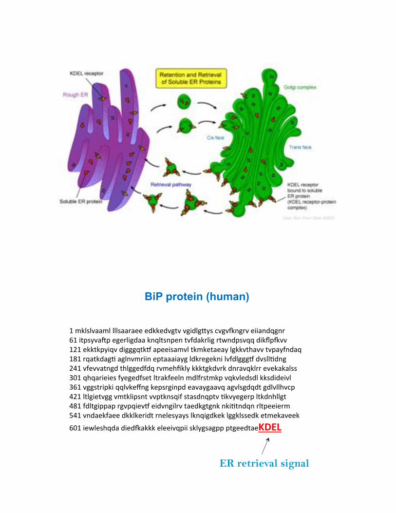
M%.D+/+N"".+%+++/""'"**%*:DD*:N(7N%N(0:+(`9/%#N(Na)('N%*00"):P()'%%bM%07,/9N"c,%*(*'+0(:""%D)P+7/),*)%7N-:"D'+0(%'7B):,/NPP%:0Dd,aNN%%MeM%*DD7D,90PN%:0(((P7Df%",**0/".N+%7D.D*7"*"9%+(DDN7$"NN%7N,"9-):"P%%MgM%'P"7D:"(3%"(+)N.'00)%*,7"""0"9(%+:D'*(*D)0%+N-:+(((f%:N/++3:)(%%eYM%N-*NN"7)(:%7$+((*:-:P%'N.*$1D+9%DDD7(D:N'D%:)'"NPD+''%*N*D"D"+//%%hVM%P$P"'0*0*/%-9*(*:-/*7%+7'"D-**+)%.:+-'/7.D,%NPDN+*:/:+%DD/:0:*0N+%%hbM%N((/7'0,D0%PP+ND*i)(%D*,/'(0),:%*"N"9(""NP%"(N+/(:P:7%(:+N++$N#,%%YeM%+7+(0*7N((%N.7D+0,/)7%NN,7D)/P0-%/7"/:)P,7N%3DN9*(*',%+7D:)$++(7%%YgM%-:+7(0,,",%'(N,P0*Nf%*0:N)(0+'N%7"*:D(7()D%)D037):P)%'+7,**0*'.%%\YM%N):"*D-"**%:DD+D*'0:7%')*+*/9"9/%+D)P0(:D*D%+((D+//*:D%*7.*D"N**D%%
bVM%0*B+*/$P:"%:0*:a"DDD%*+**0NP,00%/D+9(/"(,,%,7(**:7"*GAV<%%
BiP protein (human)
ER retrieval signal
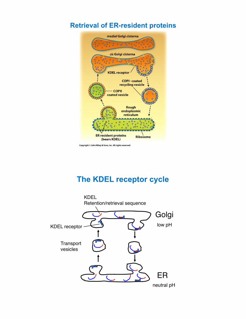
Retrieval of ER-resident proteins
Golgi!
ER!
Transport!vesicles!
KDEL!Retention/retrieval sequence!
KDEL receptor! low pH!
neutral pH!
The KDEL receptor cycle

Figure 13-4 Molecular Biology of the Cell (© Garland Science 2008)
Three types of coat proteins for vesicle budding

COPI coated vesicles mediate Golgi-to-ER and intra-Golgi transport

W1/0./%(*(4&T06*%%%%%%%%%%%%%%%:522&10651*%II%%%%%%%%%%%%%%%%%%%E.+,*&106*%609)&19)5(51*%%%
Modification of oligosaccharides in the Golgi

Figure 13-32 Molecular Biology of the Cell (© Garland Science 2008)
O-linked glycosylation occurs in the Golgi
or serine
Transport through the Golgi

Exocytic pathway
Donor compartment
Acceptor compartment
Vesicles have a size of ~60-90nm

Figure 13-14 Molecular Biology of the Cell (© Garland Science 2008)
Vesicle docking and fusion
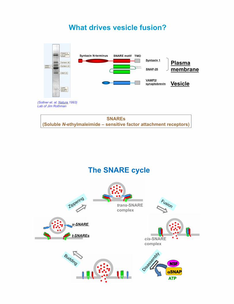
What drives vesicle fusion?
SNAREs (Soluble N-ethylmaleimide – sensitive factor attachment receptors)
(Sollner et. al. Nature 1993) Lab of Jim Rothman
Plasma membrane
Vesicle
The SNARE cycle
trans-SNARE complex
cis-SNARE complex
ATP
NSF
!SNAP
NSF
!SNAP
NSF
ATP !SNAP
NSF
t-SNAREs
v-SNARE
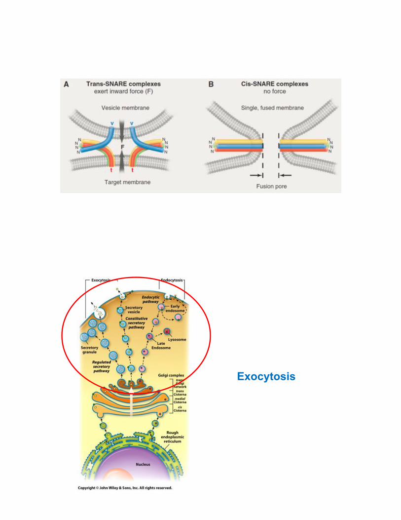
Exocytosis

Figure 13-4 Molecular Biology of the Cell (© Garland Science 2008)
Three types of coat proteins for vesicle budding
COPI coated vesicles mediate Golgi-to-ER and intra-Golgi transport

Two types of exocytosis
Stimuli
Golgi
Extracellular Space
Neurotransmitter release in neurons
Insulin secretion from pancreas
Constitutive exocytosis
Regulated exocytosis
All cell types
Neurotransmitter release in synaptic transmission
[Ca2+] Wormbook
Presynaptic Terminal
Postsynaptic Terminal

H*(52.1%(&T02%
The endocytic pathway - Endocytosis
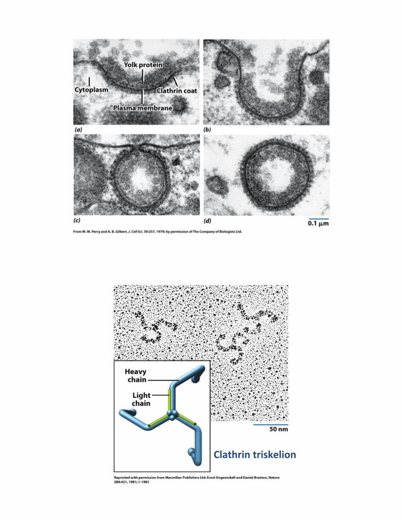
S,5()402%(401X*,0&2%
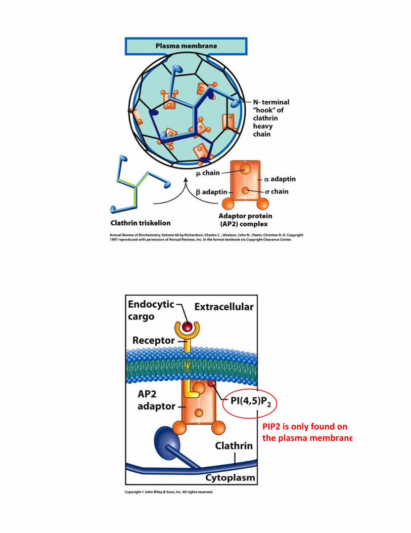
?I?Y%01%&2,Z%'&.26%&2%%()*%9,51/5%/*/3452*%

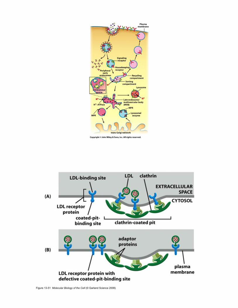
Figure 13-51 Molecular Biology of the Cell (© Garland Science 2008)

Figure 13-53 Molecular Biology of the Cell (© Garland Science 2008)
Figure 13-57 Molecular Biology of the Cell (© Garland Science 2008)

The endocytic pathway - Lysosome

Figure 13-36 Molecular Biology of the Cell (© Garland Science 2008)

Figure 13-43 Molecular Biology of the Cell (© Garland Science 2008)

Transport through the Golgi