0 )1'2 -3# '3 -%&) '&4 )5- .,'2 -3# '3 -%&) %0...
5
http://www.jstor.org !"#$%&’( *"+,’&-./. %0 1’2-3#’3-%& ’&4 5-.,’2-3#’3-%& %0 3," 6-((78-3,4$’9’( :"0("; -& <=(>.-’ <#3,%$?.@A B-&+"&3 C’.3"((#++-D 1’$%(4 E-&.F"$D G$H-&I J#=0"$/’&&D K$-+ :L J’&4"( M%#$+"A M+-"&+"D !"9 M"$-".D B%(L NOPD !%L QRSOD ?*’$L SPD NRPT@D ==L NPUV7NPUW E#2(-.,"4 2>A </"$-+’& <..%+-’3-%& 0%$ 3," <4H’&+"/"&3 %0 M+-"&+" M3’2(" X:YA http://www.jstor.org/stable/1728293 <++".."4A TWZTPZSTTW STAUT Your use of the JSTOR archive indicates your acceptance of JSTOR's Terms and Conditions of Use, available at http://www.jstor.org/page/info/about/policies/terms.jsp. JSTOR's Terms and Conditions of Use provides, in part, that unless you have obtained prior permission, you may not download an entire issue of a journal or multiple copies of articles, and you may use content in the JSTOR archive only for your personal, non-commercial use. Please contact the publisher regarding any further use of this work. Publisher contact information may be obtained at http://www.jstor.org/action/showPublisher?publisherCode=aaas. Each copy of any part of a JSTOR transmission must contain the same copyright notice that appears on the screen or printed page of such transmission. JSTOR is a not-for-profit organization founded in 1995 to build trusted digital archives for scholarship. We work with the scholarly community to preserve their work and the materials they rely upon, and to build a common research platform that promotes the discovery and use of these resources. For more information about JSTOR, please contact [email protected].
Transcript of 0 )1'2 -3# '3 -%&) '&4 )5- .,'2 -3# '3 -%&) %0...
http://www.jstor.org
!"#$%&'()*"+,'&-./.)%0)1'2-3#'3-%&)'&4)5-.,'2-3#'3-%&)%0)3,")6-((78-3,4$'9'():"0(";)-&<=(>.-'<#3,%$?.@A)B-&+"&3)C'.3"((#++-D)1'$%(4)E-&.F"$D)G$H-&I)J#=0"$/'&&D)K$-+):L)J'&4"(M%#$+"A)M+-"&+"D)!"9)M"$-".D)B%(L)NOPD)!%L)QRSOD)?*'$L)SPD)NRPT@D)==L)NPUV7NPUWE#2(-.,"4)2>A)</"$-+'&)<..%+-'3-%&)0%$)3,")<4H'&+"/"&3)%0)M+-"&+"M3'2(")X:YA)http://www.jstor.org/stable/1728293<++".."4A)TWZTPZSTTW)STAUT
Your use of the JSTOR archive indicates your acceptance of JSTOR's Terms and Conditions of Use, available athttp://www.jstor.org/page/info/about/policies/terms.jsp. JSTOR's Terms and Conditions of Use provides, in part, that unlessyou have obtained prior permission, you may not download an entire issue of a journal or multiple copies of articles, and youmay use content in the JSTOR archive only for your personal, non-commercial use.
Please contact the publisher regarding any further use of this work. Publisher contact information may be obtained athttp://www.jstor.org/action/showPublisher?publisherCode=aaas.
Each copy of any part of a JSTOR transmission must contain the same copyright notice that appears on the screen or printedpage of such transmission.
JSTOR is a not-for-profit organization founded in 1995 to build trusted digital archives for scholarship. We work with thescholarly community to preserve their work and the materials they rely upon, and to build a common research platform thatpromotes the discovery and use of these resources. For more information about JSTOR, please contact [email protected].
2
Habituated (8th stimulus)
4 J/
2
Habituated (8th stimulus)
4 J/
Nerve unblocked (20th stimulus)
1 sec
Nerve unblocked (20th stimulus)
1 sec
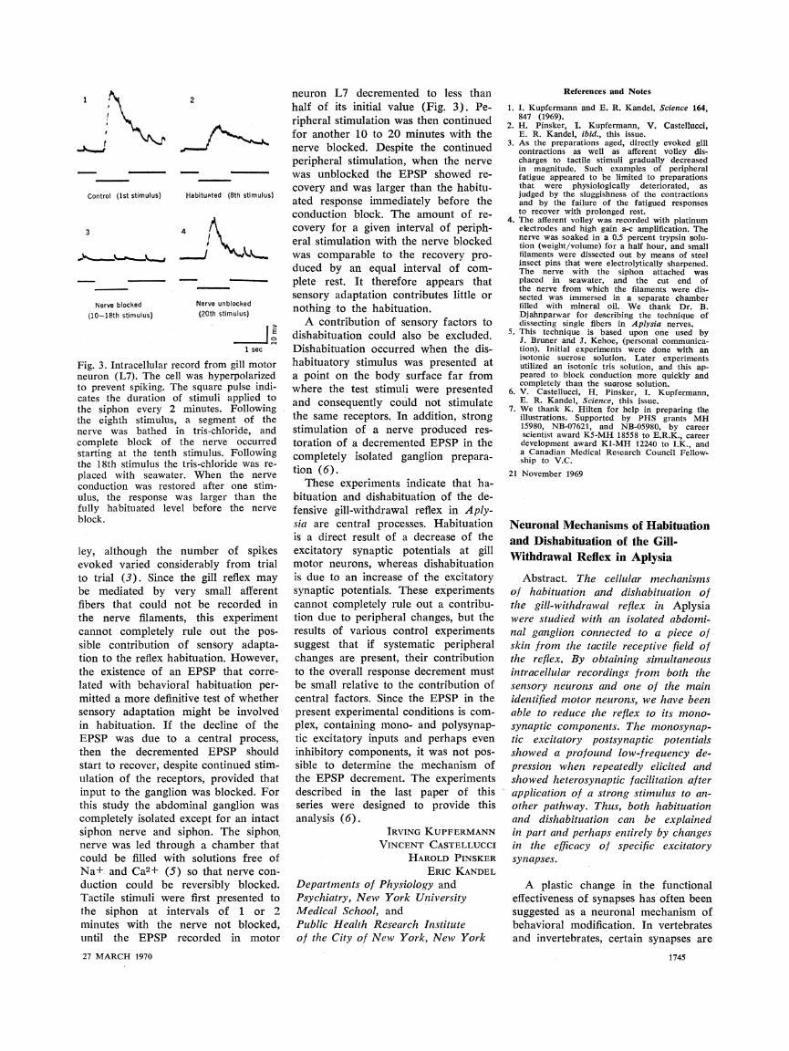
Fig. 3. Intracellular record from gill motor neuron (L7). The cell was hyperpolarized to prevent spiking. The square pulse indi- cates the duration of stimuli applied to the siphon every 2 minutes. Following the eighth stimulus, a segment of the nerve was bathed in tris-chloride, and complete block of the nerve occurred starting at the tenth stimulus. Following the 18th stimulus the tris-chloride was re- placed with seawater. When the nerve conduction was restored after one stim- ulus, the response was larger than the fully habituated level before the nerve block.
ley, although the number of spikes evoked varied considerably from trial to trial (3). Since the gill reflex may be mediated by very small afferent fibers that could not be recorded in the nerve filaments, this experiment cannot completely rule out the pos- sible contribution of sensory adapta- tion to the reflex habituation. However, the existence of an EPSP that corre- lated with behavioral habituation per- mitted a more definitive test of whether sensory adaptation might be involved in habituation. If the decline of the EPSP was due to a central process, then the decremented EPSP should start to recover, despite continued stim- ulation of the receptors, provided that input to the ganglion was blocked. For this study the abdominal ganglion was completely isolated except for an intact siphon nerve and siphon. The siphon, nerve was led through a chamber that could be filled with solutions free of Na+ and Ca2+ (5) so that nerve con- duction could be reversibly blocked. Tactile stimuli were first presented to the siphon at intervals of 1 or 2 minutes with the nerve not blocked, until the EPSP recorded in motor 27 MARCH 1970
neuron L7 decremented to less than half of its initial value (Fig. 3). Pe- ripheral stimulation was then continued for another 10 to 20 minutes with the nerve blocked. Despite the continued peripheral stimulation, when the nerve was unblocked the EPSP showed re- covery and was larger than the habitu- ated response immediately before the conduction block. The amount of re- covery for a given interval of periph- eral stimulation with the nerve blocked was comparable to the recovery pro- duced by an equal interval of com- plete rest. It therefore appears that sensory adaptation contributes little or nothing to the habituation.
A contribution of sensory factors to dishabituation could also be excluded. Dishabituation occurred when the dis- habituatory stimulus was presented at a point on the body surface far from where the test stimuli were presented and consequently could not stimulate the same receptors. In addition, strong stimulation of a nerve produced res- toration of a decremented EPSP in the completely isolated ganglion prepara- tion (6).
These experiments indicate that ha- bituation and dishabituation of the de- fensive gill-withdrawal reflex in Aply- sia are central processes. Habituation is a direct result of a decrease of the excitatory synaptic potentials at gill motor neurons, whereas dishabituation is due to an increase of the excitatory synaptic potentials. These experiments cannot completely rule out a contribu- tion due to peripheral changes, but the results of various control experiments suggest that if systematic peripheral changes are present, their contribution to the overall response decrement must be small relative to the contribution of central factors. Since the EPSP in the present experimental conditions is com- plex, containing mono- and polysynap- tic excitatory inputs and perhaps even inhibitory components, it was not pos- sible to determine the mechanism of the EPSP decrement. The experiments described in the last paper of this series were designed to provide this analysis (6).
IRVING KUPFERMANN VINCENT CASTELLUCCI
HAROLD PINSKER ERIC KANDEL
Departments of Physiology and
neuron L7 decremented to less than half of its initial value (Fig. 3). Pe- ripheral stimulation was then continued for another 10 to 20 minutes with the nerve blocked. Despite the continued peripheral stimulation, when the nerve was unblocked the EPSP showed re- covery and was larger than the habitu- ated response immediately before the conduction block. The amount of re- covery for a given interval of periph- eral stimulation with the nerve blocked was comparable to the recovery pro- duced by an equal interval of com- plete rest. It therefore appears that sensory adaptation contributes little or nothing to the habituation.
A contribution of sensory factors to dishabituation could also be excluded. Dishabituation occurred when the dis- habituatory stimulus was presented at a point on the body surface far from where the test stimuli were presented and consequently could not stimulate the same receptors. In addition, strong stimulation of a nerve produced res- toration of a decremented EPSP in the completely isolated ganglion prepara- tion (6).
These experiments indicate that ha- bituation and dishabituation of the de- fensive gill-withdrawal reflex in Aply- sia are central processes. Habituation is a direct result of a decrease of the excitatory synaptic potentials at gill motor neurons, whereas dishabituation is due to an increase of the excitatory synaptic potentials. These experiments cannot completely rule out a contribu- tion due to peripheral changes, but the results of various control experiments suggest that if systematic peripheral changes are present, their contribution to the overall response decrement must be small relative to the contribution of central factors. Since the EPSP in the present experimental conditions is com- plex, containing mono- and polysynap- tic excitatory inputs and perhaps even inhibitory components, it was not pos- sible to determine the mechanism of the EPSP decrement. The experiments described in the last paper of this series were designed to provide this analysis (6).
IRVING KUPFERMANN VINCENT CASTELLUCCI
HAROLD PINSKER ERIC KANDEL
Departments of Physiology and Psychiatry, New York University Medical School, and Public Health Research Institute of the City of New York, New York
Psychiatry, New York University Medical School, and Public Health Research Institute of the City of New York, New York
References and Notes
1. I. Kupfermann and E. R. Kandel, Science 164, 847 (1969).
2. H. Pinsker, I. Kupfermann, V. Castellucci, E. R. Kandel, ibid., this issue.
3. As the preparations aged, directly evoked gill contractions as well as afferent volley dis- charges to tactile stimuli gradually decreased in magnitude. Such examples of peripheral fatigue appeared to be limited to preparations that were physiologically deteriorated, as judged by the sluggishness of the contractions and by the failure of the fatigued responses to recover with prolonged rest.
4. The afferent volley was recorded with platinum electrodes and high gain a-c amplification. The nerve was soaked in a 0.5 percent trypsin solu- tion (weight/volume) for a half hour, and small filaments were dissected out by means of steel insect pins that were electrolytically sharpened. The nerve with the siphon attached was placed in seawater, and the cut end of the nerve from which the filaments were dis- sected was immersed in a separate chamber filled with mineral oil. We thank Dr. B. Djahnparwar for describing the technique of dissecting single fibers in Aplysia nerves.
5. This technique is based upon one used by J. Bruner and J. Kehoe, (personal communica- tion). Initial experiments were done with an isotonic sucrose solution. Later experiments utilized an isotonic tris solution, and this ap- peared to block conduction more quickly and completely than the suorose solution.
6. V. Castellucci, H. Pinsker, I. Kupfermann, E. R. Kandel, Science, this issue.
7. We thank K. Hilten for help in preparing the illustrations. Supported by PHS grants MH 15980, NB-07621, and NB-05980, by career scientist award K5-MH 18558 to E.R.K., career development award Kl-MH 12240 to I.K., and a Canadian Medical Research Council Fellow- ship to V.C.
21 November 1969
Neuronal Mechanisms of Habituation and Dishabituation of the Gill- Withdrawal Reflex in Aplysia
Abstract. The cellular mechanisms of habituation and dishabituation of the gill-withdrawal reflex in Aplysia were studied with an isolated abdomi- nal ganglion connected to a piece of skin from the tactile receptive field of the reflex. By obtaining simultaneous intracellular recordings from both the sensory neurons and one of the main identified motor neurons, we have been able to reduce the reflex to its mono- synaptic components. The monosynap- tic excitatory postsynaptic potentials showed a profound low-frequency de- pression when repeatedly elicited and showed heterosynaptic facilitation after application of a strong stimulus to an- other pathway. Thus, both habituation and dishabituation can be explained in part and perhaps entirely by changes in the efficacy of specific excitatory synapses.
A plastic change in the functional
References and Notes
1. I. Kupfermann and E. R. Kandel, Science 164, 847 (1969).
2. H. Pinsker, I. Kupfermann, V. Castellucci, E. R. Kandel, ibid., this issue.
3. As the preparations aged, directly evoked gill contractions as well as afferent volley dis- charges to tactile stimuli gradually decreased in magnitude. Such examples of peripheral fatigue appeared to be limited to preparations that were physiologically deteriorated, as judged by the sluggishness of the contractions and by the failure of the fatigued responses to recover with prolonged rest.
4. The afferent volley was recorded with platinum electrodes and high gain a-c amplification. The nerve was soaked in a 0.5 percent trypsin solu- tion (weight/volume) for a half hour, and small filaments were dissected out by means of steel insect pins that were electrolytically sharpened. The nerve with the siphon attached was placed in seawater, and the cut end of the nerve from which the filaments were dis- sected was immersed in a separate chamber filled with mineral oil. We thank Dr. B. Djahnparwar for describing the technique of dissecting single fibers in Aplysia nerves.
5. This technique is based upon one used by J. Bruner and J. Kehoe, (personal communica- tion). Initial experiments were done with an isotonic sucrose solution. Later experiments utilized an isotonic tris solution, and this ap- peared to block conduction more quickly and completely than the suorose solution.
6. V. Castellucci, H. Pinsker, I. Kupfermann, E. R. Kandel, Science, this issue.
7. We thank K. Hilten for help in preparing the illustrations. Supported by PHS grants MH 15980, NB-07621, and NB-05980, by career scientist award K5-MH 18558 to E.R.K., career development award Kl-MH 12240 to I.K., and a Canadian Medical Research Council Fellow- ship to V.C.
21 November 1969
Neuronal Mechanisms of Habituation and Dishabituation of the Gill- Withdrawal Reflex in Aplysia
Abstract. The cellular mechanisms of habituation and dishabituation of the gill-withdrawal reflex in Aplysia were studied with an isolated abdomi- nal ganglion connected to a piece of skin from the tactile receptive field of the reflex. By obtaining simultaneous intracellular recordings from both the sensory neurons and one of the main identified motor neurons, we have been able to reduce the reflex to its mono- synaptic components. The monosynap- tic excitatory postsynaptic potentials showed a profound low-frequency de- pression when repeatedly elicited and showed heterosynaptic facilitation after application of a strong stimulus to an- other pathway. Thus, both habituation and dishabituation can be explained in part and perhaps entirely by changes in the efficacy of specific excitatory synapses.
A plastic change in the functional effectiveness of synapses has often been suggested as a neuronal mechanism of behavioral modification. In vertebrates and invertebrates, certain synapses are
1745
effectiveness of synapses has often been suggested as a neuronal mechanism of behavioral modification. In vertebrates and invertebrates, certain synapses are
1745
1. 1.
Control (lst stimulus) Control (lst stimulus)
Nerve blocked (10-18th stimulus)
Nerve blocked (10-18th stimulus)
capable of undergoing functional modi- fication (1-3). However, the relevance of synaptic plasticity to a specific in- stance of behavioral modification has never been demonstrated. We have de- scribed behavioral parameters of ha- bituation and dishabituation of the gill- withdrawal reflex in the intact Aplysia (4), and we have examined their cellu- lar correlates in a semi-intact prepara- tion (5). We now describe experiments in the isolated ganglion in which we have simplified the neural circuit of the reflex in order to investigate the cellu- lar mechanisms. Our data indicate that both habituation and dishabituation of the gill-withdrawal reflex in Aplysia in- volve changes in the effectiveness of a specific set of central excitatory syn- apses between the sensory neuron and the motor neuron. These plastic changes result from homosynaptic de- pression and heterosynaptic facilitation, respectively.
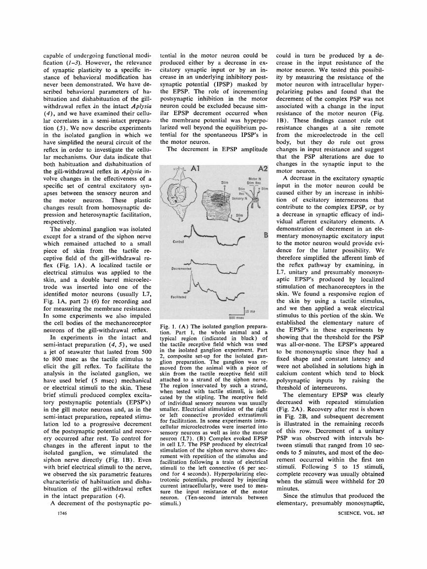
The abdominal ganglion was isolated except for a strand of the siphon nerve which remained attached to a small piece of skin from the tactile re- ceptive field of the gill-withdrawal re- flex (Fig. 1A). A localized tactile or electrical stimulus was applied to the skin, and a double barrel microelec- trode was inserted into one of the identified motor neurons (usually L7, Fig. 1A, part 2) (6) for recording and for measuring the membrane resistance. In some experiments we also impaled the cell bodies of the mechanoreceptor neurons of the gill-withdrawal reflex.
In experiments in the intact and semi-intact preparation (4, 5), we used a jet of seawater that lasted from 500 to 800 msec as the tactile stimulus to elicit the gill reflex. To facilitate the analysis in the isolated ganglion, we have used brief (5 msec) mechanical or electrical stimuli to the skin. These brief stimuli produced complex excita- tory postsynaptic potentials (EPSP's) in the gill motor neurons and, as in the semi-intact preparation, repeated stimu- lation led to a progressive decrement of the postsynaptic potential and recov- ery occurred after rest. To control for changes in the afferent input to the isolated ganglion, we stimulated the siphon nerve directly (Fig. lB). Even with brief electrical stimuli to the nerve, we observed the six parametric features characteristic of habituation and disha- bituation of the gill-withdrawal reflex in the intact preparation (4).
A decrement of the postsynaptic po- 1746
tential in the motor neuron could be produced either by a decrease in ex- citatory synaptic input or by an in- crease in an underlying inhibitory post- synaptic potential (IPSP) masked by the EPSP. The role of incrementing postsynaptic inhibition in the motor neuron could be excluded because sim- ilar EPSP decrement occurred when the membrane potential was hyperpo- larized well beyond the equilibrium po- tential for the spontaneous IPSP's in the motor neuron.
The decrement in EPSP amplitude
. Al A2 ,^--*^L^t~~~ 1^^ Motor N
/ /^y'-}, istim Stim Rec ' Stim Stim
B Control
Decremented \ f
Facilitated
10 mv 500 msec
Fig. 1. (A) The isolated ganglion prepara- tion. Part 1, the whole animal and a typical region (indicated in black) of the tactile receptive field which was used in the isolated ganglion experiment. Part 2, composite set-up for the isolated gan- glion preparation. The ganglion was re- moved from the animal with a piece of skin from the tactile receptive field still attached to a strand of the siphon nerve. The region innervated by such a strand, when tested with tactile stimuli, is indi- cated by the stipling. The receptive field of individual sensory neurons was usually smaller. Electrical stimulation of the right or left connective provided extrastimuli for facilitation. In some experiments intra- cellular microelectrodes were inserted into sensory neurons as well as into the motor neuron (L7). (B) Complex evoked EPSP in cell L7. The PSP produced by electrical stimulation of the siphon nerve shows dec- rement with repetition of the stimulus and facilitation following a train of electrical stimuli to the left connective (6 per sec- ond for 4 seconds). Hyperpolarizing elec- trotonic potentials, produced by injecting current intracellularly, were used to mea- sure the input resistance of the motor neuron. (Ten-second intervals between stimuli.)
could in turn be produced by a de- crease in the input resistance of the motor neuron. We tested this possibil- ity by measuring the resistance of the motor neuron with intracellular hyper- polarizing pulses and found that the decrement of the complex PSP was not associated with a change in the input resistance of the motor neuron (Fig. 1B). These findings cannot rule out resistance changes at a site remote from the microelectrode in the cell body, but they do rule out gross changes in input resistance and suggest that the PSP alterations are due to changes in the synaptic input to the motor neuron.
A decrease in the excitatory synaptic input in the motor neuron could be caused either by an increase in inhibi- tion of excitatory interneurons that contribute to the complex EPSP, or by a decrease in synaptic efficacy of indi- vidual afferent excitatory elements. A demonstration of decrement in an ele- mentary monosynaptic excitatory input to the motor neuron would provide evi- dence for the latter possibility. We therefore simplified the afferent limb of the reflex pathway by examining, in L7, unitary and presumably monosyn- aptic EPSP's produced by localized stimulation of mechanoreceptors in the skin. We found a responsive region of the skin by using a tactile stimulus, and we then applied a weak electrical stimulus to this portion of the skin. We established the elementary nature of the EPSP's in these experiments by showing that the threshold for the PSP was all-or-none. The EPSP's appeared to be monosynaptic since they had a fixed shape and constant latency and were not abolished in solutions high in calcium content which tend to block polysynaptic inputs by raising the threshold of interneurons.
The elementary EPSP was clearly decreased with repeated stimulation (Fig. 2A). Recovery after rest is shown in Fig. 2B, and subsequent decrement is illustrated in the remaining records of this row. Decrement of a unitary PSP was observed with intervals be- tween stimuli that ranged from 10 sec- onds to 5 minutes, and most of the dec- rement occurred within the first ten stimuli. Following 5 to 15 stimuli, complete recovery was usually obtained when the stimuli were withheld for 20 minutes.
Since the stimulus that produced the elementary, presumably monosynaptic,
SCIENCE, VOL. 167
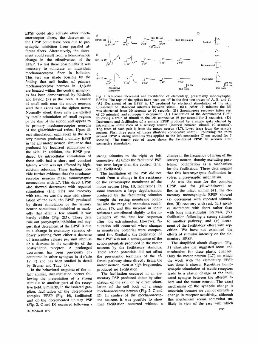
EPSP could also activate other mech- anoreceptor fibers, the decrement in the EPSP could have been due to pre- synaptic inhibition from parallel af- ferent fibers. Alternatively, the decre- ment could result from a homosynaptic change in the effectiveness of the EPSP. To test these possibilities it was necessary to stimulate an individual mechanoreceptor fiber in isolation. This test was made possible by the finding that cell bodies of primary mechanoreceptor neurons in Aplysia are located within the central ganglion, as has been demonstrated by Nicholls and Baylor (7) in the leech. A cluster of small cells near the motor neurons send their axons out the siphon nerve. Normally silent, these cells are excited by tactile stimulation of small regions of the skin of the siphon and appear to be primary mechanoreceptor neurons of the gill-withdrawal reflex. Upon di- rect stimulation, each spike in the sen- sory neuron produced a unitary EPSP in the gill motor neuron, similar to that produced by localized stimulation of the skin. In addition, the EPSP pro- duced by intracellular stimulation of these cells had a short and constant latency which was not affected by high- calcium solutions. These findings pro- vide further evidence that the mechano- receptor neurons make monosynaptic connections with L7. This direct EPSP also showed decrement with repeated stimulation (Fig. 2D) and recovery with rest. As was the case with stimu- lation of the skin, the EPSP produced by direct stimulation of the sensory neuron sometimes diminished so mark- edly that after a few stimuli it was barely visible (Fig. 2D). These data rule out presynaptic inhibition and sug- gest that decrement of the EPSP is due to a change in excitatory synaptic ef- ficacy resulting from either a decrease of transmitter release per unit impulse or a decrease in the sensitivity of the postsynaptic receptor. A prolonged decrement has been previously en- countered in other synapses in Aplysia (2, 3) and has been studied in detail by Bruner and Tauc (3).
In the behavioral response of the in- tact animal, dishabituation occurs fol- lowing the presentation of a strong stimulus to another part of the recep- tive field. Similarly, in the isolated gan- glion, facilitation of the decremented complex EPSP (Fig. 1iB, facilitated) and of the decremented unitary PSP (Fig. 2, C and D) occurred following a
27 MARCH 1970
A ISI=30 secJ 1
00o1^
D
0 30"
II= 10 sec
0' 0 .
"1'10"
ISI=10 sec _ --J , 010o' 0140
Connective stimulation
IS= 110 sec f, ~ _^--^ ^f --- Rest 20 minutes
7'30"' 19'20" 100 msec
1'20" 1' 30" 2'r50 1130"1 2'50"
0150" 1140" 3'30" 15 mv
100 msec
Control
Decremented
Facilitated 10 mv
i20 mv 100 msec
Fig. 2. Response decrement and facilitation of elementary, presumably monosynaptic, EPSP's. The tops of the spikes have been cut off in the first two traces of A, B, and C. (A) Decrement of an EPSP in L7 produced by electrical stimulation of the skin (30-second or 10-second intervals between stimuli, ISI). After 19 minutes the ISI was shortened from 30 seconds to 10 seconds. (B) Spontaneous recovery (after rest of 20 minutes) and subsequent decrement. (C) Facilitation of the decremented EPSP following a train of stimuli to the left connective (9 per second for 2 seconds). (D) Decrement and facilitation of a unitary EPSP produced by a single spike elicited by intracellular stimulation of a sensory neuron (interval between stimuli, 10 seconds). Top trace of each pair is from the motor neuron (L7), lower trace from the sensory neuron. First three pairs of traces illustrate consecutive stimuli. Following the third evoked EPSP a strong stimulus was applied to the left connective (7 per second for 5 seconds). The fourth pair of traces shows the facilitated EPSP 30 seconds after connective stimulation.
strong stimulus to the right or left connective. At times the facilitated PSP was even larger than the control (Fig. 2D, facilitated).
The facilitation of the PSP did not result from a change in the resistance of the extrasynaptic membrane of the motor neuron (Fig. 1B, facilitated). In some instances a large depolarization produced by the facilitating stimulus brought the resting membrane poten- tial into the range of anomalous rectifi- cation (8), and this increase in input resistance contributed slightly to the in- crements of the first few responses after the extrastimulus. However, fa- cilitation still occurred when changes in membrane potential were compen- sated for. Similarly, the facilitation of the EPSP was not a consequence of the action potentials produced in the motor neurons by the facilitatory stimulus. These action potentials did not affect the presynaptic terminals of the af- ferent pathway since directly firing the motor neuron, even at high frequencies, produced no facilitation.
The facilitation occurred in an ele- mentary PSP produced either by stim- ulation of the skin or by direct stimu- lation of the cell body of a single mechanoreceptor neuron (Fig. 2, C and D). In studies of the mechanorecep- tor neurons it was possible to show that facilitation occurred without a
change in the frequency of firing of the sensory neuron, thereby excluding post- tetanic potentiation as a mechanism for the facilitation. These data suggest that this heterosynaptic facilitation in- volves a presynaptic mechanism.
As was the case for the complex EPSP and for gill-withdrawal re- flex in the intact animal (4), the ele- mentary monosynaptic EPSP showed (i) decrement with repeated stimula- tion, (ii) recovery with rest, (iii) great- er decrement with short rather than with long interstimulus intervals, (iv) facilitation following a strong stimulus to another pathway, and (v) decre- ment of the facilitatory effect with rep- etition. We have not examined the effects of stimulus intensity on the ele- mentary EPSP.
The simplified circuit diagram (Fig. 3) illustrates the suggested locus and mechanism for these plastic changes. Only the motor neuron (L7) on which the work with the elementary EPSP was done is shown. Repetitive homo- synaptic stimulation of tactile receptors leads to a plastic change at the indi- cated synapse between the afferent fi- bers and the motor neuron. The exact mechanism of the synaptic change is uncertain because we cannot exclude a change in receptor sensitivity, although this mechanism seems somewhat un- likely in view of the ease with which
1747
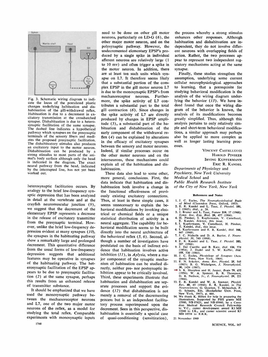
Fig. 3. Schematic wiring diagram to indi- cate the locus of the postulated plastic changes underlying habituation and dis- habituation of the gill-withdrawal reflex. Habituation is due to a decrement in ex- citatory transmission at the crosshatched synapse. Dishabituation is due to a hetero- synaptic facilitation of the same synapse. The dashed line indicates a hypothetical pathway which synapses on the presynaptic terminals of the sensory fibers and medi- ates the proposed presynaptic facilitation. The dishabituatory stimulus also produces an excitatory input to the motor neuron. Dishabituation can be produced by a strong stimulus to most parts of the ani- mal's body surface although only the head is indicated in the diagram. The exact neural pathway from the head, indicated by the interrupted line, has not yet been worked out.
heterosynaptic facilitation occurs. By analogy to the brief low-frequency syn- aptic depression that has been analyzed in detail at the vertebrate and at the crayfish neuromuscular junction (9), we suggest that the decrement of the elementary EPSP represents a decrease in the release of excitatory transmitter from the presynaptic terminal. How- ever, unlike the brief low-frequency de- pression evident at many synapses (10), the synapses in the habituating pathway show a remarkably large and prolonged decrement. This quantitative difference from the usual forms of low-frequency depression suggests that additional features may be operative in synapses of the habituating pathway. The het- erosynaptic facilitation of the EPSP ap- pears to be due to presynaptic facilita- tion (2) at the same synapse, perhaps this results from an enhanced release of transmitter substance.
It should be emphasized that we have used the monosynaptic pathway be- tween the mechanoreceptor neurons and L7, one of the two major motor neurons of the reflex, as a model for studying the total reflex. Comparable experiments with monosynaptic inputs
1748
need to be done on other gill motor neurons, particularly on LD-G (6), the other major motor neuron, and on the polysynaptic pathway. However, the undecremented elementary EPSP's pro- duced by a single spike in individual afferent neurons are relatively large (1 to 10 mv) and often trigger a spike in the motor neuron. In addition, there are at least ten such units which syn- apse on L7. It therefore seems likely that a substantial portion of the com- plex EPSP in the gill motor neuron L7 is due to the monosynaptic EPSP's from mechanoreceptor neurons. Further- more, the spike activity of L7 con- tributes a substantial part to the total gill contraction (6). Since changes in the spike activity of L7 are directly produced by changes in EPSP ampli- tude (5), a substantial part of the ha- bituation and dishabituation of the early component of the withdrawal re- flex (4) can be explained by alterations in the efficacy of excitatory synapses between the sensory and motor neurons. Indeed, if similar processes occur on the other motor neurons and on the interneurons, these mechanisms could explain all of the habituation and dis- habituation.
These data also lead to some other, more general, conclusions. First, the data indicate that habituation and dis- habituation both involve a change in the functional effectiveness of previ- ously existing excitatory connections. Thus, at least in these simple cases, it seems unnecessary to explain the be- havioral modifications by invoking elec- trical or chemical fields or a unique statistical distribution of activity in a neural aggregate. The capability for be- havioral modification seems to be built directly into the neural architecture of the behavioral reflex (5, 6). Second, al- though a number of investigators have postulated on the basis of indirect evi- dence that habituation involves active inhibition (11), in Aplysia, where a ma- jor component of the synaptic mecha- nism of habituation can be studied di- rectly, neither pre- nor postsynaptic in- hibition appear to be critically involved. Third, these experiments illustrate that habituation and dishabituation are sep- arate processes and support the evi- dence (12) that dishabituation is not merely a removal of the decrementing process but is an independent facilita- tory process superimposed upon the decrement. Seen in this perspective, dis- habituation is essentially a special case of quasi-conditioning (sensitization),
the process whereby a strong stimulus enhances other responses. Although habituation and dishabituation are in- dependent, they do not involve differ- ent neurons with overlapping fields of action. Rather, the two processes ap- pear to represent two independent reg- ulatory mechanisms acting at the same synapse.
Finally, these studies strengthen the assumption, underlying some current cellular neurophysiological approaches to learning, that a prerequisite for studying behavioral modification is the analysis of the wiring diagram under- lying the behavior (13). We have in- deed found that once the wiring dia- gram of the behavior is known, the analysis of its modifications becomes greatly simplified. Thus, although this analysis pertains to only relatively sim- ple and short-term behavioral modifica- tions, a similar approach may perhaps also be applied to more complex as well as longer lasting learning proc- esses.
VINCENT CASTELLUCCI HAROLD PINSKER
IRVING KUPFERMANN ERIC R. KANDEL
Departments of Physiology and Psychiatry, New York University Medical School and Public Health Research Institute of the City of New York, New York
References and Notes
1. J. C. Eccles, The Neurophysiological Basis of Mind (Clarendon Press, Oxford, 1953).
2. E. R. Kandel and L. Tauc, J. Physiol. 181, 1 (1965); ibid., p. 28.
3. J. Bruner and L. Tauc, Nature 210, 37 (1966); Symp. Soc. Exp. Biol. 20, 457 (1966).
4. H. Pinsker, I. Kupfermann, V. Castellucci, E. Kandel, Science, this issue.
5. I. Kupfermann, V. Castellucci, H. Pinsker, E. Kandel, ibid., this issue.
6. I. Kupfermann and E. R. Kandel, ibid. 164, 847 (1969).
7. J. C. Nicholls and D. A. Baylor, J. Neuro- physiol. 31, 740 (1968).
8. E. R. Kandel and L. Tauc, J. Physiol. 183, 287 (1966).
9. J. Del Castillo and B. Katz, ibid. 124, 574 (1954); J. Dudel and S. W. Kuffler, ibid. 155, 530 (1961).
10. J. C. Eccles, Physiology of Synapses (Aca- demic Press, New York, 1964).
11. E. N. Sokolov, Annu. Rev. Physiol. 25, 545 (1963); B. G. Wickelgren, J. Neurophysiol. 30, 1424 (1967).
12. S. K. Sharpless and H. Jasper, Brain 79, 655 (1956); W. A. Spencer, R. R. Thompson, D. R. Neilson, Jr., J. Neurophysiol. 29, 221 (1966).
13. E. R. Kandel and W. A. Spencer, Physiol. Rev. 48, 65 (1968); E. R. Kandel, in The Neurosciences, G. Quarton, T. Melnechuk, P. 0. Schmitt, Eds. (Rockefeller Univ. Press, New York, 1967), p. 666.
14. We thank K. Hilten for help in preparing the illustrations. Supported by PHS grants MH 15980, NB 07621, and NB 05980, by a Cana- dian Medical Research Council Fellowship to V.C., career development award K1 MH 12240 to I.K., and career scientist award K5 MH 18558 to E.R.K.
21 November 1969
SCIENCE, VOL. 167


![[] Arnaldo Castellucci - Endodontics (vol. 2).pdf](https://static.fdocuments.us/doc/165x107/55cf9b86550346d033a665ed/wwwfisierulmeuro-arnaldo-castellucci-endodontics-vol-2pdf.jpg)