
, Yanxia Chen , Yan Zhao , David Christopher Lung ...
45
Accepted Manuscript © The Author(s) 2021. Published by Oxford University Press for the Infectious Diseases Society of America. This is an Open Access article distributed under the terms of the Creative Commons Attribution- NonCommercial-NoDerivs licence (http://creativecommons.org/licenses/by-nc-nd/4.0/), which permits non-commercial reproduction and distribution of the work, in any medium, provided the original work is not altered or transformed in any way, and that the work is properly cited. For commercial re-use, please contact [email protected] Intravenous injection of COVID-19 mRNA vaccine can induce acute myopericarditis in mouse model Can Li 1,a , Yanxia Chen 1,a , Yan Zhao 1,a , David Christopher Lung 2 , Zhanhong Ye 1 , Wenchen Song 1 , Fei-Fei Liu 1 , Jian-Piao Cai 1 , Wan-Man Wong 1 , Cyril Chik-Yan Yip 1 , Jasper Fuk-Woo Chan 1,3,4 , Kelvin Kai-Wang To 1,3 , Siddharth Sridhar 1,3 , Ivan Fan-Ngai Hung 3,5 , Hin Chu 1 , Kin-Hang Kok 1 , Dong-Yan Jin 6 , Anna Jinxia Zhang 1,b , Kwok-Yung Yuen 1,3,4,b a Joint first authors b Joint last authors 1 State Key Laboratory of Emerging Infectious Diseases, Carol Yu Centre for Infection, Department of Microbiology, Li Ka Shing Faculty of Medicine, The University of Hong Kong, Pokfulam, Hong Kong Special Administrative Region, China. 2 Department of Pathology, Queen Elizabeth Hospital and Hong Kong Children’s Hospital, Hong Kong, Hong Kong Special Administrative Region, China. 3 Department of Clinical Microbiology and Infection Control, The University of Hong Kong- Shenzhen Hospital, Shenzhen, Guangdong, China. 4 Hainan Medical University-The University of Hong Kong Joint Laboratory of Tropical Infectious Diseases, The University of Hong Kong, Pokfulam, Hong Kong Special Administrative Region, China. 5 Department of Medicine, Li Ka Shing Faculty of Medicine, The University of Hong Kong, Pokfulam, Hong Kong Special Administrative Region, China. Downloaded from https://academic.oup.com/cid/advance-article/doi/10.1093/cid/ciab707/6353927 by guest on 22 August 2021
Transcript of , Yanxia Chen , Yan Zhao , David Christopher Lung ...

Accep
ted M
anus
cript
© The Author(s) 2021. Published by Oxford University Press for the Infectious Diseases Society of America. This is an Open Access article distributed under the terms of the Creative Commons Attribution-NonCommercial-NoDerivs licence (http://creativecommons.org/licenses/by-nc-nd/4.0/), which permits non-commercial reproduction and distribution of the work, in any medium, provided the original work is not altered or transformed in any way, and that the work is properly cited. For commercial re-use, please contact [email protected]
Intravenous injection of COVID-19 mRNA vaccine can induce acute myopericarditis in
mouse model
Can Li1,a
, Yanxia Chen1,a
, Yan Zhao1,a
, David Christopher Lung2, Zhanhong Ye
1, Wenchen
Song1, Fei-Fei Liu
1, Jian-Piao Cai
1, Wan-Man Wong
1, Cyril Chik-Yan Yip
1, Jasper Fuk-Woo
Chan1,3,4
, Kelvin Kai-Wang To1,3
, Siddharth Sridhar1,3
, Ivan Fan-Ngai Hung3,5
, Hin Chu1,
Kin-Hang Kok1, Dong-Yan Jin
6, Anna Jinxia Zhang
1,b, Kwok-Yung Yuen
1,3,4,b
a Joint first authors
b Joint last authors
1State Key Laboratory of Emerging Infectious Diseases, Carol Yu Centre for Infection,
Department of Microbiology, Li Ka Shing Faculty of Medicine, The University of Hong
Kong, Pokfulam, Hong Kong Special Administrative Region, China.
2Department of Pathology, Queen Elizabeth Hospital and Hong Kong Children’s Hospital,
Hong Kong, Hong Kong Special Administrative Region, China.
3Department of Clinical Microbiology and Infection Control, The University of Hong Kong-
Shenzhen Hospital, Shenzhen, Guangdong, China.
4Hainan Medical University-The University of Hong Kong Joint Laboratory of Tropical
Infectious Diseases, The University of Hong Kong, Pokfulam, Hong Kong Special
Administrative Region, China.
5Department of Medicine, Li Ka Shing Faculty of Medicine, The University of Hong Kong,
Pokfulam, Hong Kong Special Administrative Region, China.
Dow
nloaded from https://academ
ic.oup.com/cid/advance-article/doi/10.1093/cid/ciab707/6353927 by guest on 22 August 2021

Accep
ted M
anus
cript
2
6School of Biomedical Sciences, Li Ka Shing Faculty of Medicine, The University of Hong
Kong, Pokfulam, Hong Kong Special Administrative Region, China.
Correspondence: K.-Y. Yuen, State Key Laboratory of Emerging Infectious Diseases, Carol
Yu Centre for Infection, Department of Microbiology, Li Ka Shing Faculty of Medicine, The
University of Hong Kong, Pokfulam, Hong Kong Special Administrative Region, China; and
Department of Clinical Microbiology and Infection Control, The University of Hong Kong-
Shenzhen Hospital, Shenzhen, China ([email protected]).
Summary: Intravenous injection of COVID-19 mRNA vaccine may induce gross and
histopathological changes of acute myopericarditis in Balb/c mice.
Dow
nloaded from https://academ
ic.oup.com/cid/advance-article/doi/10.1093/cid/ciab707/6353927 by guest on 22 August 2021

Accep
ted M
anus
cript
3
ABSTRACT
Background. Post-vaccination myopericarditis is reported after immunization with COVID-
19 mRNA-vaccines. The effect of accidental intravenous injection of this vaccine on the heart
is unknown.
Methods. We compared the clinical manifestations, histopathological changes, tissue mRNA
expression and serum levels of cytokine/chemokine in Balb/c mice at different time points
after intravenous(IV) or intramuscular(IM) vaccine injection with normal saline(NS) control.
Results. Though significant weight loss and higher serum cytokine/chemokine levels were
found in IM group at 1 to 2 days post-injection(dpi), only IV group developed
histopathological changes of myopericarditis as evidenced by cardiomyocyte degeneration,
apoptosis and necrosis with adjacent inflammatory cell infiltration and calcific deposits on
visceral pericardium, while evidence of coronary artery or other cardiac pathologies was
absent. SARS-CoV-2 spike antigen expression by immunostaining was occasionally found in
infiltrating immune cells of the heart or injection site, in cardiomyocytes and intracardiac
vascular endothelial cells, but not skeletal myocytes. The histological changes of
myopericarditis after the first IV-priming dose persisted for 2 weeks and were markedly
aggravated by a second IM- or IV-booster dose. Cardiac tissue mRNA expression of IL-1β,
IFN-β, IL-6 and TNF-α increased significantly from 1dpi to 2dpi in IV but not IM group,
compatible with presence of myopericarditis in IV group. Ballooning degeneration of
hepatocytes was consistently found in IV group. All other organs appeared normal.
Dow
nloaded from https://academ
ic.oup.com/cid/advance-article/doi/10.1093/cid/ciab707/6353927 by guest on 22 August 2021

Accep
ted M
anus
cript
4
Conclusions. This study provided in-vivo evidence that inadvertent intravenous injection of
COVID-19 mRNA-vaccines may induce myopericarditis. Brief withdrawal of syringe
plunger to exclude blood aspiration may be one possible way to reduce such risk.
Keywords: mouse model; SARS-CoV-2; mRNA vaccine; intravenous; intramuscular;
COVID-19.
Dow
nloaded from https://academ
ic.oup.com/cid/advance-article/doi/10.1093/cid/ciab707/6353927 by guest on 22 August 2021

Accep
ted M
anus
cript
5
Introduction
Safe and effective whole-population vaccination against severe acute respiratory syndrome
coronavirus 2 (SARS-CoV-2) is the only long term solution to the ongoing Coronavirus
Disease 2019 (COVID-19) pandemic[1], which has caused about 200 million cases of
COVID-19 globally and over 4 million deaths by 17 July 2021[2]. However the one-dose
vaccination rate in US and UK was only 54.9% and 67.8% respectively as of 10 July 2021[3].
Vaccine hesitancy among the general public is a significant problem, and is partially driven
by the apprehension of rare but potentially severe side effects of these rapidly developed
novel vaccines. An example of such a side effect is myopericarditis following mRNA
COVID-19 vaccines, which has a crude incidence of 40.6 cases per million second doses
administered to males aged 12−29 years[4]. The pathogenesis of this unexpected
complication remains elusive.
The World Health Organization (WHO)[5] and Centers for Disease Control and Prevention
(CDC) [6] no longer recommend aspiration of syringe plunger during intramuscular
injections, especially during vaccination when a rapid injection of a small volume may reduce
discomfort[6]. However, a self- reporting study of registered nurses showed that 40%
reported blood aspiration at least once, and 4% reported blood aspiration 13 times or more
during intramuscular injection. The finding suggests that inadvertent intravenous injection of
vaccine is possible[7]. Recently, inadvertent intravenous injection of adenovirus-vector based
COVID-19 vaccine was implicated to trigger platelet-adenovirus aggregates taken up by
spleen which mountedl B cell response of binding antibodies against platelets[8]. In this
study, we investigated the differences in the cardiac pathology induced by intravenous(IV) or
intramuscular(IM) BNT162b2 mRNA COVID-19 vaccine when compared with normal
saline(NS) injection in a Balb/c mouse model.
Dow
nloaded from https://academ
ic.oup.com/cid/advance-article/doi/10.1093/cid/ciab707/6353927 by guest on 22 August 2021

Accep
ted M
anus
cript
6
METHODS
Animal Model
Female Balb/c (substrain OlaHsd) mice at age of 6-8 weeks were obtained from the Centre
for Comparative Medicine Research of The University of Hong Kong and kept in Biosafety
Level (BSL)-2 animal laboratory with 12-hour light-dark cycle and free access to water and
diet. The animals were randomly assigned to three groups for the administration of IV or IM
COVID-19 mRNA vaccine, or normal saline(NS) control(Figure 1A). COVID-19 mRNA
vaccine (BNT162b2 lot number 1B004A, BioNTech, Germany) dose of 0.25ug per gram of
body weight was injected (about 5µg in 50µL per mouse; dose according to an
immunogenicity study) via tail vein or thigh muscle, respectively, with the control group
having the same volume of NS[9]. Another group of male mice was subsequently tested by
the same protocol after preliminary positive results. The mice were monitored by clinical
signs and body weight changes for 14 days. Necropsies were performed at 1, 2, 7 and 14 days
post-injection(dpi). Organs and blood were sampled for histological and real-time
quantitative RT-PCR, or cytokine/chemokine levels respectively. A group of mice received a
second boosting dose 14 days after the first priming dose and examined at 2dpi after
boosting. The procedures for animal experiments in this study were approved by the HKU
Committee on the Use of Live Animals in Teaching and Research.
Histopathology and Immunohistochemical Staining of Tissues
Formalin-fixed and paraffin-embedded mouse heart, lung, liver, spleen, kidney and brain
tissues were cut into 4µm sections and stained with hematoxylin and eosin(H&E) for
histopathological examination. Immunohistochemistry staining for leukocyte biomarkers,
SARS-CoV-2 spike receptor binding domain (S-RBD) and terminal deoxynucleotidyl
transferase dUTP nick end labelling(TUNEL) were performed as we described
Dow
nloaded from https://academ
ic.oup.com/cid/advance-article/doi/10.1093/cid/ciab707/6353927 by guest on 22 August 2021

Accep
ted M
anus
cript
7
previously[10]. Primary antibodies including rabbit anti-SARS-CoV-2 S-RBD used in our
previous study [11], rabbit anti-mouse CD45, CD68, or CD3(Abcam) were used in this
study. DNA fragmentation in cardiomyocytes was labelled using Click-iT® Plus TUNEL
assay kit(Thermo Fisher Scientific) for the detection of apoptosis[10]. The slides were
mounted and examined under light microscope. Representative images were captured with
Olympus BX53 semi-motorised fluorescence microscope. The measurement of cytokine and
chemokines mRNA expression levels in different tissues, serum cytokine/chemokine levels,
serum troponin levels, and the statistical analysis(Supplementary method).
RESULTS
Intravenous SARS-CoV-2 mRNA Vaccine Administration Induced Grossly Visible
Pathology in Heart
Groups of female Balb/c mice at age of 6-8 week were given BNT162b2 COVID-19 mRNA
vaccine either IV or IM, or same volume of NS(Figure 1A). None of the animals showed
clinical signs of lethargy, ruffled furs, hunched back posture, and rapid breathing throughout
the course of observation. Significant decrease in body weight was observed in IM mRNA
vaccine group(mean 3.6%±2.1%) starting from 1dpi; animals recovered their initial weight at
7 dpi(Figure 1B). Autopsy at 1 to 2dpi showed white patches over the visceral pericardium in
37.5%(1dpi, n=8) to 38.5%(2dpi, n=13) of IV vaccine group but none in the IM vaccine or
NS control groups(Figure 1C and 1D;Table 1;p<0.05). No grossly visible changes were
observed in other organs of the animals(Supplementary Figure1).
Dow
nloaded from https://academ
ic.oup.com/cid/advance-article/doi/10.1093/cid/ciab707/6353927 by guest on 22 August 2021

Accep
ted M
anus
cript
8
Histopathological Changes in Mouse Heart After IV mRNA Vaccine Administration
Low power scanning of heart sections showed blue stained thickened visceral pericardium
over the right atrium and ventricle at 1dpi of IV vaccine, which became more prominent at
2dpi(Figure 2A). At higher magnification, calcific deposits were seen in these thickened
pericardial tissues(Figure 2D). Multifocal pericardial and myocardial inflammatory cell
infiltrates and interstitial oedema were also observed(Figure 2C). Frequent foci of
cardiomyocytes had degenerative changes as evident by the loss of the normal pattern of
cross striation and occasionally sarcoplasmic vacuolation, and necrotic changes as
distinguished by the attainment of a homogenous appearance, sarcoplasmic fragmentation, or
pyknosis(Figure 2E). These changes were significantly more frequent in the IV vaccine group
at 2dpi(Table 1), and more often in the right atrium and right ventricles of the affected
animals and especially prominent on their pericardial side. Immunohistochemical staining
with anti-CD45(biomarker for immune cells of lymphoid or myeloid origin) indicated that
these were leukocytes, of which many were macrophages or histiocytes positive for CD68.
CD3-positive T cells were less often seen(Figure 2G).The numbers of leukocytes were more
than 14 per square millimetre in the affected myocardial foci. These findings suggested that
mice given IV mRNA COVID-19 vaccine can develop acute myopericarditis. Similar
histopathological changes and severity were found in male mice(supplementary figure 2B,
2C)
Dow
nloaded from https://academ
ic.oup.com/cid/advance-article/doi/10.1093/cid/ciab707/6353927 by guest on 22 August 2021

Accep
ted M
anus
cript
9
IV mRNA Vaccine Administration Induced Apoptosis of Cardiomyocytes, Tissue
mRNA and protein Expression of SARS-CoV-2 Spike
To determine if apoptosis was induced in cardiomyocytes, TUNEL immunofluorescent
staining was used for detection of DNA fragmentation in apoptotic cells(Figure 3A).
Apoptotic cardiomyocytes were significantly more often found in the IV group than IM
group or NS group. Apoptotic cardiomyocytes distributed both sporadically or as large foci
were found in 75% of IV group at 1dpi(6/8) and 38.5% at 2dpi(5/13) (p<0.05; Table 1, Figure
3B).
To understand whether mRNA vaccine can transfect cardiomyocytes to express SARS-CoV-
2 spike protein, we used immunostaining to detect SARS-CoV-2 S-RBD and showed
occasional positive cardiomyocytes, infiltrating immune cells and vascular endothelial cells
within the myocardium and pericardium in the IV but not IM vaccine or NS control
group(Figures 4A, 4B, 4C).. Using RT-qPCR[12], the amount of COVID-19 mRNA Spike-
RBD gene copies in heart tissues was significantly higher in IV than IM group at
1dpi(supplementary figure 3). Though no statistical significant differences were found, the
mean amount of Spike-RBD mRNA was higher in IV than IM group at all other time points.
IM mRNA Vaccine Administration Only Induced Mild Myocardial Congestion and
Edema
No grossly visible change of the heart in IM group was seen on autopsy or low power
scan(Figure 5A). H&E sections showed some myocardial vascular congestion and mild
interstitial edema at 1dpi(Figure 5B). Degenerative changes of cardiomyocytes were
occasionally found at 2dpi(Figure 5B). No obvious immune cell infiltration, cardiomyocyte
Dow
nloaded from https://academ
ic.oup.com/cid/advance-article/doi/10.1093/cid/ciab707/6353927 by guest on 22 August 2021

Accep
ted M
anus
cript
10
necrosis, or TUNEL-positive apoptotic cells were found at 1 or 2dpi(Figure 5C). The changes
were not sufficient to satisfy the histological criteria of myocarditis. Notably SARS-CoV-2
spike protein expression was only found in the infiltrating immune cells in the thigh muscle
1dpi in IM group. No degeneration, necrosis or SARS-CoV-2 protein expression was evident
in the skeletal myocytes(Figures 5D, 5E).
mRNA Vaccine Administration Induced Inflammatory Cytokine/Chemokine Response
in the Heart, and increased Serum Troponin and Cytokine/Chemokine levels
RT-qPCR assay showed increased mRNA expression of IFN-α/β, IL-6, TNF-α, CXCL10,
CCL3 in heart tissue homogenates of the IV group, among which IFN-β, IL-6, and TNF-α
were significantly higher at 2dpi than 1dpi(Figure 6A). As for the IM group, all tested
inflammatory cytokines/chemokines were transiently upregulated in heart tissues at 1dpi, and
then decreased at 2dpi(Figure 6B). But IL-1 β expression was significantly higher in cardiac
tissues of male than female mice at 2dpi(supplementary figure 2A).
Beads-based multiplex cytokine/chemokine flow cytometry assay showed that the IM group
had significantly higher serum concentrations of cytokines/chemokines at 1dpi which
decreased at 2dpi(Figure 6C). Increased serum cytokine/chemokine concentrations were
found in the IV group at 1dpi, but only CXCL10 and CCL5 were significantly higher than the
NS control group(Figure 6C). Enzyme immunoassay showed that serum troponin levels of IV
group (1328.2+325.8pg/ml) was significantly higher than IM group (237.5+121.2pg/ml) and
NS group(215+115.9pg/ml). (p<0.0001; Table 1)
Dow
nloaded from https://academ
ic.oup.com/cid/advance-article/doi/10.1093/cid/ciab707/6353927 by guest on 22 August 2021

Accep
ted M
anus
cript
11
Histopathological Changes of the Heart at 7dpi and 14dpi after First Dose, and at 2
Days after the Second Dose Given at 14 Days After First Dose
At 7dpi, the heart of mice in IV group showed persistent changes of myopericarditis(Figure
7A), while IM group only showed vascular congestion, myocardial edema and occasional
foci of cardiomyocyte degeneration(Figure 7A). At 14dpi, 4/6 (66.7%) of the mice in IV
group showed grossly visible white patches over the visceral pericardium, and 6/6(100%)
showed changes of myopericarditis, compared with only mild degenerative changes in IM
group(Figure 7B; Table 1).
Two days after the second dose of mRNA vaccine, 4/9(44.7%) and 2/6(33.3%) of the mice in
IM/IV group and IV/IM group developed grossly visible white patches over the visceral
pericardium, respectively. Both groups showed more diffuse and severe changes of
myopericarditis with foci of mild myocardial haemorrhage which affected both right and left
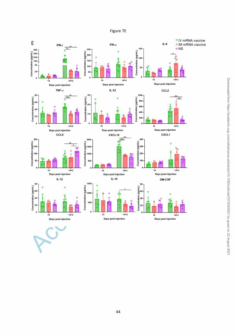
heart(Figure 7D). Moreover, serum cytokine/chemokine levels by beads-based flow
cytometry assay showed significantly increased IFN-, TNF-α and CXCL10 at 2dpi after IV
boosting which suggested that IV vaccine increased inflammatory responses, while CCL2
was increased after both IV and IM boosting(Figure 7E).
IV mRNA Vaccine Administration Induced Histopathological changes in Liver
H&E-stained sections of the liver tissues of the IV group showed diffuse ballooning
degeneration of hepatocytes and focal hepatocyte necrosis at 1 to 2dpi(Figure 8A), while the
liver tissues of IM group showed much milder changes without hepatocyte necrosis at 1 to
2dpi(Supplementary figure 1B). SARS-CoV-2 spike RBD expression by immunostaining
was occasionally seen in hepatocytes of the IV but not IM group(Figure 8B). Except for some
Dow
nloaded from https://academ
ic.oup.com/cid/advance-article/doi/10.1093/cid/ciab707/6353927 by guest on 22 August 2021

Accep
ted M
anus
cript
12
vascular congestion in lungs, H&E stained sections of spleen, brain, and kidney at 1 to 2dpi
were unremarkable(Supplementary figure 1C).
DISCUSSION
In a Balb/c mouse model with both male and female mice, IV but not IM
administration of COVID-19 mRNA vaccine induced a rapid onset of multifocal
myopericarditis with elevated serum troponin, cardiomyocyte degeneration and changes of
both necrosis and apoptosis, adjacent inflammatory infiltrate of mononuclear cells, interstitial
edema, and visceral pericardial calcification within 2dpi. Moreover, the IL-1β, IFN-β, IL-6
and TNF-α expression levels generally increased significantly from 1dpi to 2dpi in the IV but
not IM group. Overall, the findings have satisfied the Dallas and immunohistochemical
criteria of myocarditis[13]. Similar to findings of cardiac magnetic resonance imaging in
human myocarditis, the most prominent site of focal involvement was the pericardial side of
atrial and ventricular walls [14]. Notably, the myopericarditis was subclinical and the changes
persisted but did not progress within 14dpi. But these histopathological changes of
myopericarditis deteriorated and became rather diffuse after the second dose boosting with
either IV or IM administration 14 days after the first dose of priming. While Balb/c mice
have been extensively used for modelling myocarditis due to viral, protozoal and
autoimmune insults[15], myocardial mineralization can occur spontaneously with age in
inbred laboratory rodents. However, we have excluded this possibility by the demonstration
of highly significant differences in the gross pathological and histological changes of
myopericarditis between mice challenged by IV versus IM vaccine or NS control(Table
1)[16].
Dow
nloaded from https://academ
ic.oup.com/cid/advance-article/doi/10.1093/cid/ciab707/6353927 by guest on 22 August 2021

Accep
ted M
anus
cript
13
Acute myocardial injury due to hypersensitivity myocarditis was reported after
smallpox vaccine at a rate of 12.3 to 463 cases per 100,000[17] and very rarely associated
with other vaccines for yellow fever and influenza[17-19]. COVID-19 mRNA vaccines were
associated with myopericarditis at a rate of 12.6 to 24 cases per million following a second
dose[20]. The clinical manifestations of acute chest pain, dyspnea, arrhythmia, raised serum
troponin, electrocardiographic and gadolinium enhanced cardiac MRI changes often started 3
to 5 days after the second dose of vaccination or occasionally after first dose[21, 22]. Two
patients without measurable SARS-CoV-2 spike IgG presented shortly after their first
vaccine dose, suggesting that myocardial damage can happen with just one dose of mRNA
vaccine as demonstrated in our present study[23]. Moreover the second dose of mRNA
vaccine given 14 days after first dose by either IM or IV route has markedly exacerbated the
myopericarditis which is also compatible with the clinical findings in human subjects.
The pathogenesis of an early onset of myopericarditis in IV vaccinated mice is
unclear. Here, we showed that SARS-CoV-2 spike protein was occasionally expressed in
cardiomyocytes 1dpi of IV mRNA vaccine, though such expression was more often in the
infiltrating immune cells in the myocardium and visceral pericardium. We have previously
shown that the replication of SARS-CoV-1 leads to substantial accumulation of heavily
modified transmembrane viral proteins such as unfolded spike at the endoplasmic reticulum
which rapidly exceed its folding capacity leading to stress and the unfolded protein response.
When the damage to the endoplasmic reticulum is severe or persistent, the unfolded protein
response triggers apoptosis[24]. The same in vitro phenomenon was reported with SARS-
CoV-2[25]. More studies on pathogenesis are warranted.
Another possible causative mechanism of the mRNA vaccine induced myopericarditis
could be the overly activation of cytokine production, which was also reported to cause
reversible myopericarditis and cardiomyopathy in patients treated with interferon for chronic
Dow
nloaded from https://academ
ic.oup.com/cid/advance-article/doi/10.1093/cid/ciab707/6353927 by guest on 22 August 2021

Accep
ted M
anus
cript
14
myeloid leukaemia, viral hepatitis and multiple sclerosis[26, 27]. Intravenous injection of
inactivated typhoid vaccine was associated with progressive radiological cardiomegaly within
two weeks[28]. However, a sufficient degree of myocardial damage satisfying the
histopathological criteria of myopericarditis was not observed in our IM vaccine group
despite significantly higher serum proinflammatory cytokine/chemokine levels and body
weight loss. Cardiac damage due to hypersensitivity towards other components of mRNA
vaccine is unlikely as similar degree of damage should happen in both IV and IM groups.
Both Pfizer/BioNTech and Moderna have clearly stated that their vaccines should
only be given via IM route[29, 30]. However current CDC[6] and WHO guidelines[5] no
longer recommend precautionary measures during IM vaccine administration. Brief
aspiration for blood return during intramuscular injection of medication as a preventive
measure against accidental IV injection was previously present in most guidelines[31]. This
practice becomes controversial as scientific evidence of the perceived benefit of this
procedure is lacking for IM injection of vaccine. The CDC Pink Book 2020[6] and WHO
2015 position paper[5] have recommended against aspiration prior to vaccine injection so as
to minimize pain[32]. The veins and arteries within the reach of a syringe needle in the
deltoid region are considered too small to allow a rapid IV injection of vaccine without
blowing out the vessel[6]. However this speculation also lacks supportive scientific evidence.
Another possibility of getting a high blood mRNA vaccine level is the rapid
movement of the vaccine through the lymphatic system into the venous circulation.
We note that Pfizer has conducted in vivo biodistribution studies of IM injected [3H]-
labelled lipid-nanoparticle (LNP) mRNA vaccines in rats[33]. There was some accumulation
of the formulation in the heart at 2dpi, although much lower than concentrations in the liver
or spleen. No similar tracer study was reported when the mRNA vaccine is IV injected.
Besides the possibility of delivery to the heart through the systemic arterial system, the IV
Dow
nloaded from https://academ
ic.oup.com/cid/advance-article/doi/10.1093/cid/ciab707/6353927 by guest on 22 August 2021

Accep
ted M
anus
cript
15
injected SARS-CoV-2 mRNA could theoretically transfect myocardial cells through smaller
cardiac venous system(Thebesian venous network) which consists of a layer of vascular
endothelial cells continuous with endothelium of four cardiac chambers without interference
by valves. Delivery of pharmacologic therapy, gene therapy, growth factors, and stem cells to
the myocardium by retrograde venous perfusion was reported to achieve a better myocardial
concentration[34]. Moreover, smaller mRNA-vaccine lipid-nanoparticles(100nm diameter)
can be sucked into larger T tubules(diameter >200nm) of cardiomyocytes during diastole, but
not into T tubules of skeletal myocyte(diameter 20-40nm)[35]. Thus the T tubule system of
cardiomyocytes may concentrate mRNA-vaccine lipid-nanoparticles like a sponge.
Interestingly, we also observed ballooning degeneration of hepatocytes especially in
our IV group, which is compatible with the heavy distribution of the mRNA vaccine
formulation in the liver of IM injected rats in the bio-distribution study[33] and microscopic
vacuolation of portal hepatocytes in these rats[36]. There have been anecdotal reports on
autoimmune hepatitis following COVID-19 mRNA vaccines in humans although these
reports are yet to be confirmed by population-level vaccine adverse effect monitoring
systems[37, 38]. Further research into the potential association of COVID-19 mRNA
vaccination and autoimmune hepatitis is required.
Limitations of our study included the lack of data in explaining the association of
post-vaccination myocarditis with younger age or male gender. While our male mice had
similar degree of myopericarditis to female mice, a lower vaccine dose may show up
differences in disease susceptibility due to immunological differences or ACE2 expression
driven by sex hormone and X chromosome. Though the histological changes in the heart of
IM group did not amount to myopericarditis, we cannot exclude the possibility of frank
myopericarditis in individuals who may be more susceptible to even a slight amount of
mRNA vaccine entering the systemic circulation from intramuscular injection. COVID-19
Dow
nloaded from https://academ
ic.oup.com/cid/advance-article/doi/10.1093/cid/ciab707/6353927 by guest on 22 August 2021

Accep
ted M
anus
cript
16
mRNA vaccines are safe and effective while post-vaccination myopericarditis is rare and
self-limiting[4]. Our study indicates that IV injection of vaccines might partially contribute to
this clinical phenotype, thus warranting a reconsideration of the practice of IM injection
without aspiration, which carries the risk of inadvertent IV injection. Increasing the size of
mRNA-vaccine lipid-nanoparticle or decreasing the vaccine dose in normal adolescents to
reduce risks of myopericarditis warrant further investigations. Careful histopathological
examination of the heart is required in any case of fatality following COVID-19 mRNA
vaccines as myocarditis can be focal or masquerade as ischemic heart disease in older
patients.
Dow
nloaded from https://academ
ic.oup.com/cid/advance-article/doi/10.1093/cid/ciab707/6353927 by guest on 22 August 2021

Accep
ted M
anus
cript
17
Notes
Author contributions. A. J. Z. and K.-Y. Y. had roles in the study design, data
collection, data analysis, data interpretation, and writing of the manuscript. C.L., Y.C., Y.Z.,
Z.Y., W.S., F.-F.L., J.-P.C., W.-M.W, and C.C.-Y.Y., had roles in the experiments, data
collection, and/or data analysis. D.C.L., I.F.-N.H., J.F.-W.C., K.K.-W.T., S.S., H.C., K.-H.K.,
and D.J., had roles in experimental design, data analysis and/or revision of the manuscript.
All authors reviewed and approved the final version of the manuscript.
Disclaimer. The funding sources had no role in the study design, data collection,
analysis, interpretation, or writing of the report.
Financial support. This study was partly supported by funding from, the Food and
Health Bureau, The Government of the Hong Kong Special Administrative Region; and the
Consultancy Service for Enhancing Laboratory Surveillance of Emerging Infectious Diseases
and Research Capability on Antimicrobial Resistance for Department of Health of the Hong
Kong Special Administrative Region Government; and donations of Richard Yu and Carol
Yu, Shaw Foundation Hong Kong, Michael Seak-Kan Tong, May Tam Mak Mei Yin, Lee
Wan Keung Charity Foundation Limited, Hui Ming, Hui Hoy and Chow Sin Lan Charity
Fund Limited, Chan Yin Chuen Memorial Charitable Foundation, Marina Man-Wai Lee, the
Hong Kong Hainan Commercial Association South China Microbiology Research Fund, the
Jessie & George Ho Charitable Foundation, Kai Chong Tong, Tse Kam Ming Laurence, Foo
Oi Foundation Limited, Betty Hing-Chu Lee, and Ping Cham So.
Dow
nloaded from https://academ
ic.oup.com/cid/advance-article/doi/10.1093/cid/ciab707/6353927 by guest on 22 August 2021

Accep
ted M
anus
cript
18
Potential conflicts of interests. J.F.-W.C has received travel grants from Pfizer
Corporation Hong Kong and Astellas Pharma Hong Kong Corporation Limited, and was an
invited speaker for Gilead Sciences Hong Kong Limited and Luminex Corporation. K.Y.Y. is
the inventor an intranasal influenza vectored SARS-CoV-2. The other authors declared no
conflict of interests. All authors have submitted the ICMJE Form for Disclosure of Potential
Conflicts of Interest.
Dow
nloaded from https://academ
ic.oup.com/cid/advance-article/doi/10.1093/cid/ciab707/6353927 by guest on 22 August 2021

Accep
ted M
anus
cript
19
References
1. To KK, Sridhar S, Chiu KH, et al. Lessons learned 1 year after SARS-CoV-2
emergence leading to COVID-19 pandemic. Emerg Microbes Infect 2021; 10(1): 507-
35.
2. World Health Organization. WHO Coronavirus (COVID-19) Dashboard. Available
at: https://covid19.who.int/. Accessed 17 July 2021.
3. Our World in Data. Statistics and Research: Coronavirus (COVID-19) Vaccinations:
Share of people who received at least one dose of COVID-19 vaccine. Available at:
https://ourworldindata.org/covid-vaccinations. Accessed 18 July 2021.
4. Gargano JW, Wallace M, Hadler SC, et al. Use of mRNA COVID-19 Vaccine After
Reports of Myocarditis Among Vaccine Recipients: Update from the Advisory
Committee on Immunization Practices - United States, June 2021. MMWR Morb
Mortal Wkly Rep 2021; 70(27): 977-82.
5. Organization WH. Reducing pain at the time of vaccination: WHO position paper –
September 2015. Weekly epidemiological record, 2015; 39(90): 505 - 16.
6. Centers for Disease Control and Prevention. Epidemiology and Prevention of
Vaccine-Preventable Diseases: The Pink Book, Chapter on Vaccine Administration.
Available at: https://www.cdc.gov/vaccines/pubs/pinkbook/vac-admin.html. Accessed 17
July 2021.
7. Thomas CM, Mraz M, Rajcan L. Blood Aspiration During IM Injection. Clinical
Nursing Research 2015; 25(5): 549-59.
8. Nicolai L, Leunig A, Pekayvaz K, et al. Thrombocytopenia and splenic platelet
directed immune responses after intravenous ChAdOx1 nCov-19 administration.
bioRxiv 2021: 2021.06.29.450356.
Dow
nloaded from https://academ
ic.oup.com/cid/advance-article/doi/10.1093/cid/ciab707/6353927 by guest on 22 August 2021

Accep
ted M
anus
cript
20
9. Agency MHpR. Public Assessment Report Authorisation for Temporary
SupplyCOVID-19 mRNA Vaccine BNT162b2(BNT162b2 RNA)concentrate for
solution for injection. Page 15. Available at:
https://assets.publishing.service.gov.uk/government/uploads/system/uploads/attachment_
data/file/997584/COVID-
19_mRNA_Vaccine_BNT162b2__UKPAR___PFIZER_BIONTECH_ext_of_indication_11.6.2021.
pdf. Accessed 23 July 2021
10. Lee ACY, Zhang AJX, Chu H, et al. H7N9 influenza A virus activation of necroptosis
in human monocytes links innate and adaptive immune responses. Cell Death Dis
2019; 10(6): 442.
11. Chen LL, Lu L, Choi CY, et al. Impact of SARS-CoV-2 variant-associated RBD
mutations 1 on the susceptibility to serum antibodies elicited by COVID-19 infection
or vaccination. Clin Infect Dis 2021; Accepted and In Press.
12. Yip CCY, Sridhar S, Leung KH, et al. Development and Evaluation of Novel and
Highly Sensitive Single-Tube Nested Real-Time RT-PCR Assays for SARS-CoV-2
Detection. Int J Mol Sci 2020; 21(16).
13. Aretz HT, Billingham ME, Edwards WD, et al. Myocarditis. A histopathologic
definition and classification. Am J Cardiovasc Pathol 1987; 1(1): 3-14.
14. Mahrholdt H, Goedecke C, Wagner A, et al. Cardiovascular magnetic resonance
assessment of human myocarditis: a comparison to histology and molecular
pathology. Circulation 2004; 109(10): 1250-8.
15. Błyszczuk P. Myocarditis in Humans and in Experimental Animal Models. Front
Cardiovasc Med 2019; 6: 64.
16. Herman E, Eldridge S. Spontaneously occurring cardiovascular lesions in commonly
used laboratory animals. Cardiooncology 2019; 5: 6.
Dow
nloaded from https://academ
ic.oup.com/cid/advance-article/doi/10.1093/cid/ciab707/6353927 by guest on 22 August 2021

Accep
ted M
anus
cript
21
17. Kuntz J, Crane B, Weinmann S, Naleway AL. Myocarditis and pericarditis are rare
following live viral vaccinations in adults. Vaccine 2018; 36(12): 1524-7.
18. Engler RJ, Nelson MR, Collins LC, Jr., et al. A prospective study of the incidence of
myocarditis/pericarditis and new onset cardiac symptoms following smallpox and
influenza vaccination. PLoS One 2015; 10(3): e0118283.
19. Mei R, Raschi E, Poluzzi E, Diemberger I, De Ponti F. Recurrence of pericarditis
after influenza vaccination: a case report and review of the literature. BMC Pharmacol
Toxicol 2018; 19(1): 20.
20. Shay DK, Shimabukuro TT, DeStefano F. Myocarditis Occurring After Immunization
With mRNA-Based COVID-19 Vaccines. JAMA Cardiol 2021.
21. Kim HW, Jenista ER, Wendell DC, et al. Patients With Acute Myocarditis Following
mRNA COVID-19 Vaccination. JAMA Cardiol 2021.
22. Montgomery J, Ryan M, Engler R, et al. Myocarditis Following Immunization With
mRNA COVID-19 Vaccines in Members of the US Military. JAMA Cardiol 2021.
23. Rosner CM, Genovese L, Tehrani BN, et al. Myocarditis Temporally Associated with
COVID-19 Vaccination. Circulation 2021.
24. Chan CP, Siu KL, Chin KT, Yuen KY, Zheng B, Jin DY. Modulation of the unfolded
protein response by the severe acute respiratory syndrome coronavirus spike protein. J
Virol 2006; 80(18): 9279-87.
25. Balakrishnan B, Lai K. Modulation of SARS-CoV-2 Spike-induced Unfolded Protein
Response (UPR) in HEK293T cells by selected small chemical molecules. bioRxiv
2021: 2021.02.04.429769.
26. Sonnenblick M, Rosin A. Cardiotoxicity of Interferon*: A Review of 44 Cases. Chest
1991; 99(3): 557-61.
Dow
nloaded from https://academ
ic.oup.com/cid/advance-article/doi/10.1093/cid/ciab707/6353927 by guest on 22 August 2021

Accep
ted M
anus
cript
22
27. Khakoo AY, Halushka MK, Rame JE, Rodriguez ER, Kasper EK, Judge DP.
Reversible cardiomyopathy caused by administration of interferon alpha. Nat Clin
Pract Cardiovasc Med 2005; 2(1): 53-7.
28. Weens HS, Heyman A. Cardiac enlargement in fever therapy induced by intravenous
injection of typhoid vaccine. Arch Intern Med (Chic) 1946; 77: 307-16.
29. U.S. Food and Drug Administration (FDA). Emergency use authorization (EUA) of
the Pfizer-Biontech covid-19 vaccine to prevent coronavirus disease 2019 (covid-19):
Fact sheet for healthcare providers administering vaccine (vaccination providers). 15
June 2021:Page 14.
30. U.S. Food and Drug Administration (FDA). Emergency use authorization (EUA) of
the Moderna covid-19 vaccine to prevent coronavirus disease 2019 (covid-19): Fact
sheet for healthcare providers administering vaccine (vaccination providers). 24 June
2021:Page 9.
31. Sepah Y, Samad L, Altaf A, Halim MS, Rajagopalan N, Javed Khan A. Aspiration in
injections: should we continue or abandon the practice? F1000Res 2014; 3: 157.
32. Ipp M, Taddio A, Sam J, Gladbach M, Parkin PC. Vaccine-related pain: randomised
controlled trial of two injection techniques. Arch Dis Child 2007; 92(12): 1105-8.
33. SARS-CoV-2 mRNA Vaccine (BNT162, PF-0 7302048): 2.6.5.5B.
Pharmacokinetics: Organ Distribution Continued, Report number: 185350, Page 6.
Available at:
https://www.pmda.go.jp/drugs/2021/P20210212001/672212000_30300AMX00231_I100_1.
pdf. Accessed 23 July 2021
34. Echeverri D, Cabrales J, Jimenez A. Myocardial venous drainage: from anatomy to
clinical use. J Invasive Cardiol 2013; 25(2): 98-105.
Dow
nloaded from https://academ
ic.oup.com/cid/advance-article/doi/10.1093/cid/ciab707/6353927 by guest on 22 August 2021

Accep
ted M
anus
cript
23
35. Brette F, Orchard C. T-tubule function in mammalian cardiac myocytes. Circ Res
2003; 92(11): 1182-92.
36. Agency MHpR. Public Assessment Report Authorisation for Temporary
SupplyCOVID-19 mRNA Vaccine BNT162b2(BNT162b2 RNA)concentrate for
solution for injection. Page 19. Available at:
https://assets.publishing.service.gov.uk/government/uploads/system/uploads/attachment_
data/file/997584/COVID-
19_mRNA_Vaccine_BNT162b2__UKPAR___PFIZER_BIONTECH_ext_of_indication_11.6.2021.
pdf. Accessed 23 July 2021.
37. Tan CK, Wong YJ, Wang LM, Ang TL, Kumar R. Autoimmune hepatitis following
COVID-19 Vaccination: true causality or mere association? J Hepatol 2021.
38. Dumortier J. Liver injury after mRNA-based SARS-CoV-2 vaccination in a liver
transplant recipient. Clin Res Hepatol Gastroenterol 2021: 101743.
Dow
nloaded from https://academ
ic.oup.com/cid/advance-article/doi/10.1093/cid/ciab707/6353927 by guest on 22 August 2021

Accep
ted M
anus
cript
24
FIGURE LEGENDS
Figure 1. Schema for vaccine administration and gross pathology of mouse after
vaccination.
A. Experimental Schema. Groups of mice were injected with COVID-19 mRNA vaccine via
intramuscular (IM) or intravenous (IV) route. At 1, 2, 7 and 14 days post-injection (dpi), mice
were sacrificed for histopathological analysis. Normal saline (NS) was IV or IM injected in
parallel as control. B. Body weight changes of mice after IM or IV injection of vaccine. C.
Representative images of gross pathology of mouse organs and heart at 1 dpi. The hearts of
NS control and IM vaccine groups appeared grossly normal, while whitish patches (arrows)
were seen on the visceral pericardium of hearts after IV vaccine. D. Representative images of
gross pathology of mouse organs including heart at 2dpi. Large whitish patches (arrows) were
seen on the visceral pericardium of mice receiving IV vaccine.
Figure 2. Representative histopathological images of heart tissues of mice receiving IV
vaccine. Groups of mice were given IV injection of COVID-19 mRNA vaccine (IV group)
and NS (NS group) as control. At 1 and 2dpi, mice were sacrificed for histopathology
analysis.
A. Low magnification microscopic scanning images of heart sections (4x magnification).
Mice received IV NS showed no detectable histological changes in the heart. After IV
mRNA vaccine, the scan images showed thickened and dark blue stained visceral
pericardium on the surface of right atrium and right ventricle at both 1 and 2dpi (arrows). B.
H&E stain of heart tissue of IV NS group showed no histological damage in the myocardium
and cardiomyocytes. C. H&E stained heart tissue showed inflammatory infiltrates of the
myocardium at 2dpi. Arrows indicated infiltrates of inflammatory cells at 400X
magnification. D. H&E stained heart tissue of IV vaccine group at 2dpi showing thickened
Dow
nloaded from https://academ
ic.oup.com/cid/advance-article/doi/10.1093/cid/ciab707/6353927 by guest on 22 August 2021

Accep
ted M
anus
cript
25
visceral pericardium with clusters of dark blue crystal-like structure which indicated calcific
deposits (arrows) with adjacent inflammatory cell infiltrates and cardiomyocytes
degeneration at. 400X magnification. E. H&E stained myocardial tissue showing
cardiomyocytes degeneration at 2dpi in IV group as indicated by arrows at 400X
magnification. F. H&E image showing cardiomyocytes necrosis (arrows at 400X
magnification) with immune cells infiltration in IV group at 2dpi. G. Images of
immunohistochemistry staining of white blood cells marker CD45, CD68 and CD3 in the
heart sections, showing myocardial and visceral pericardial infiltration by CD45 positive
cells. Immunostaining of macrophage marker CD68 showed many positives in the infiltrating
cells, with less frequently positive CD3 biomarker for T lymphocytes
Figure 3. Representative images of TUNEL (apoptosis biomarker) staining of heart
tissues. Groups of mice received COVID-19 mRNA vaccine via intramuscular (IM) or
intravenous (IV) route. At 1 to 2dpi, the mice were sacrificed for histopathological analysis.
Control group of mice received IV NS.
A. No TUNEL staining signal in heart tissue section of NS control mice at 2dpi (left) at 200X
magnification. DNase treatment of the same tissue as positive control showed TUNEL
staining in the nucleus of cardiomyocytes (green fluorescent signal as indicated by arrows in
the insert at 400X magnification). B. At 1 to 2dpi of mRNA vaccine, TUNEL signals were
shown in a large area of the myocardial tissue (circled by dashed lines at 40X magnification,
indicated by arrows in the inserts at 400X magnification). H&E staining of the same heart
tissue sections are shown on the right.
Dow
nloaded from https://academ
ic.oup.com/cid/advance-article/doi/10.1093/cid/ciab707/6353927 by guest on 22 August 2021

Accep
ted M
anus
cript
26
Figure 4. Immunohistochemical staining for protein expression of spike receptor
binding domain (RBD) in heart sections.
A. Representative images of immunohistochemical staining of heart section showed
occasional spike RBD positive cardiomyocytes (arrows at 400x magnification) at 1dpi in IV
vaccine group. H&E stain of the same area of the section is on the right. B. Representative
images showing spike RBD expression in the histiocytes, and vascular endothelial cells in
myocardium (arrows in 400X magnification). C. Representative images of positive control
for the staining of spike RBD in SARS-CoV-2 infected mouse bronchiolar epithelium (arrow
in left image), negative expression of spike RBD by immunostaining in the myocardium of
IV NS control group (middle), and immunostaining with only biotin conjugated secondary
antibody in heart sections of IV vaccine group at 1dpi (right).
Figure 5. Representative histopathological images of heart tissues after intramuscular
administration of mRNA vaccine. Groups of mice were given IM or IV vaccine. IV NS (NS
group) was used as control. At 1 to 2dpi, mice were sacrificed for histopathology.
A. Low magnification scanning images of heart sections (4X magnification). Both NS group
and IM vaccine group showed no histological changes in the heart. B. Representative H&E
images of heart tissues. NS groups showed normal myocardium and cardiomyocytes. IM
vaccine group showed vascular congestion and mild degree of myocardial edema at 1dpi. No
white blood cell infiltration, cardiomyocyte degeneration or necrosis was observed. At 2dpi,
vascular congestion was reduced but interstitial edema could still be seen. C. TUNEL
staining of heart section showed no positive signal at 2dpi for IM group. D. H&E images of
thigh muscle showed white blood cell infiltration in the connective tissue while the adjacent
skeletal muscle cells are unremarkable (magnified image in the right) 1dpi after IM mRNA
Dow
nloaded from https://academ
ic.oup.com/cid/advance-article/doi/10.1093/cid/ciab707/6353927 by guest on 22 August 2021

Accep
ted M
anus
cript
27
vaccine. E. Immunohistochemistry staining of Spike RBD showed only some infiltrating
white blood cells expressing spike RBD in thigh muscle of IM group (arrows in magnified
images).
Figure 6. Cytokine/chemokine mRNA expression in heart and their serum levels.
At 1 to 2dpi of IV or IM mRNA vaccine, organs and serum were collected.
A. Cytokines/chemokines mRNA expression in heart homogenates of IV group; B.
Cytokines/chemokines mRNA expression in heart homogenates of IM group; mRNA
expressions were detected by RT-qPCR with gene specific primers. House-keeping gene β-
actin mRNA expression was included to normalize the amount of RNA. Data presented were
relative gene expression to NS control mice. Error bars induced mean ± standard deviation.
n=5 in each group. C. Serum cytokine/chemokine concentrations at 1 to 2dpi were detected
by beads-based multiplex flow cytometer assay. Mice serum from IV and IM injection of
50µl NS were used as control. Error bars indicated mean ± standard deviation. n=5 each
group. *P<0.05, **P<0.01, ***P<0.001,****P<0.0001.
Figure 7. Histopathological changes in the heart at 7 and 14dpi after first dose of IV or
IM mRNA vaccine and 2 dpi after second dose of vaccine. Groups of mice were given IV,
IM vaccine or NS as control. At 7 to 14dpi, mice were sacrificed for histopathology. Another
two groups of mice were given second dose of IV or IM mRNA vaccine at 14days after the
first priming dose and sacrificed at 2dpi after the second boosting dose.
A. Representative histopathological images of mouse heart at day 7. The top panel consisted
of heart sections of IV group, which showed myocardial infiltration by white blood cells
(left, arrows), interstitial edema, cardiomyocytes degeneration (middle, arrows) and necrosis
(right, arrows). Lower panel consisted of heart sections from IM group, which showed
Dow
nloaded from https://academ
ic.oup.com/cid/advance-article/doi/10.1093/cid/ciab707/6353927 by guest on 22 August 2021

Accep
ted M
anus
cript
28
myocardial interstitial edema (left) and myocardial vascular congestion (middle, arrows),
with degeneration of a few cardiomyocytes (right, arrows); B. Representative
histopathological images of IV and IM group at day 14. The heart in IV group showed
persistent changes of cardiomyocyte degeneration, white blood cell infiltration and foci of
necrosis (arrows). C. The heart of mice in IM group showed minimal degeneration and
infiltration, but no necrosis. D. Representative histopathological images of the heart at 2dpi
after the second boosting dose given on day 14 after the first priming dose. Mouse heart in
both IV and IM second dose group showed interstitial edema and diffuse cardiomyocyte
degeneration on the left (arrows). Mouse heart in both IM and IV group showed diffuse
inflammatory infiltrate, focal haemorrhage and necrosis (arrows, right). E. Serum
cytokine/chemokine concentrations at 2dpi post second dose were detected by beads-based
multiplex flow cytometer assay. The NS group was used as control. Error bars indicated
mean ± standard deviation. n=5 each group. n=9 for IV second dose boost group, n=6 for IM
second dose boost group and NS control group. *p<0.05, **p< 0.01, ***p<0.001 by multiple
t test.
Dow
nloaded from https://academ
ic.oup.com/cid/advance-article/doi/10.1093/cid/ciab707/6353927 by guest on 22 August 2021

Accep
ted M
anus
cript
29
Figure 8. Histopathological changes in the liver at 2 dpi after IV mRNA vaccine
injection.
A. Representative H&E images of liver sections. Images in the upper panel show the liver
section of NS control mice, with normal morphology of evenly distributed cords of
hepatocytes. Lower panel consisted of images of liver 2dpi in IV group. The liver showed
diffuse degeneration of hepatocytes without a clear morphological organization of hepatic
cords. A large focus of cell necrosis was circled by the dashed line. Magnified images (400X
magnification) of hepatocyte necrosis (middle, arrows in magnified box) and ballooning
degenerative changes of hepatocytes (right, arrows in magnified box. B. Images of
immunohistochemistry stained spike RBD in liver sections at 2dpi after IV vaccine. A few
RBD positive cells were indicated by arrows in the inserts (400X magnification).
Dow
nloaded from https://academ
ic.oup.com/cid/advance-article/doi/10.1093/cid/ciab707/6353927 by guest on 22 August 2021

Accep
ted M
anus
cript
30
Pathology a mRNA vaccine injection (second dose given 14 days after first dose)
Changes
(related
figures)
2dpi first
IV dose
n= 13 (%)
2dpi first
IM dose
n= 6 (%)
NS
Control
2dpi
n= 12 (%)
2dpi second
IV dose
n= 9 (%)
2dpi second
IM dose n= 6
(%)
Heart
Grossly visible
white patches
on visceral
pericardium
(Fig 1C, 1D)
5/13 (38.5%)† 0/6 0/12 3/9 (33.3%) 1/6 (16.7%)
Pericardial
calcific deposit
(Fig. 2A, 2D)
5/13 (38.5%)† 0/6 0/12 4/9 (44.4%)
† 2/6 (33.3%)
Pericardial
WBC
infiltration
(Fig. 2C)
9/13 (69.2%)* †††
0/6 0/12 5/9
(55.6%)††
2/6 (33.3%)
Myocardial
WBC
infiltration
(Fig. 2C, 2G)
8/13 (61.5%)* †††
0/6 0/12 9/9
(100%)†††
6/6
(100%)†††
Cardiomyocytes
degeneration
Fig. 2E)
8/13 (61.5%)††
2/6 (33.3%) 0/12 9/9
(100%)†††
6/6
(100%)†††
Cardiomyocytes
necrosis(Fig.
2F)
4/13 (30.8%) 0/6 0/12 9/9
(100%)†††
6/6
(100%)†††
Cardiomyocytes
apoptosis
(Fig. 3B)
5/13 (38.5%)† 0/6 0/12 N.D.
N.D.
Spike RBD
expression b
(Fig. 4A, 4B)
3/8 (37.5%)† 0/6 0/12 N.D. N.D.
Serum
Troponin
(pg/ml)c
1328.2+325.8****
††††
237.5+121.2 215+115.9 N.D. N.D.
Liver Grossly visible
change at
autopsy
0/6 0/6 0/6 0/9 0/6
Hepatocytes
ballooning
degeneration
(Fig. 8A)
6/6 (100%)††
3/6 (50%) 0/6 N.D. N.D.
Hepatocytes
necrosis
2/6 (50%) 0/6 0/6 N.D. N.D.
Table 1. Summary of the pathological changes in heart and liver after IV or IM
administration of mRNA vaccine
Dow
nloaded from https://academ
ic.oup.com/cid/advance-article/doi/10.1093/cid/ciab707/6353927 by guest on 22 August 2021

Accep
ted M
anus
cript
31
Abbreviation: dpi, days post injection; IM, intramuscular; IV, intravenous; N.D., not done; NS, normal saline;
RBD, receptor binding domain; WBC, white blood cells;
a Presented as the number and percentage of mice with positive gross or histological changes in the heart and
liver
b number heart sections showed RBD positive cells at 1dpi
c serum troponin concentration in mouse serum were determined by Enzyme Immunoassay using Mouse
CTNI(Cardiac troponin-I) ELISA Kit (Mybiosource, San Diego, CA, USA).
* p <0.05,
**** p<0.0001 when compared to IM group at the same time point
† p <0.05,
††p<0.01,
†††p<0.001
††††p<0.0001when compared to NS 2dpi group
Dow
nloaded from https://academ
ic.oup.com/cid/advance-article/doi/10.1093/cid/ciab707/6353927 by guest on 22 August 2021

Accep
ted M
anus
cript
32
Figure 1A-C
Dow
nloaded from https://academ
ic.oup.com/cid/advance-article/doi/10.1093/cid/ciab707/6353927 by guest on 22 August 2021

Accep
ted M
anus
cript
33
Figure 1D
Dow
nloaded from https://academ
ic.oup.com/cid/advance-article/doi/10.1093/cid/ciab707/6353927 by guest on 22 August 2021

Accep
ted M
anus
cript
34
Figure 2A-F
Dow
nloaded from https://academ
ic.oup.com/cid/advance-article/doi/10.1093/cid/ciab707/6353927 by guest on 22 August 2021

Accep
ted M
anus
cript
35
Figure 2G
Dow
nloaded from https://academ
ic.oup.com/cid/advance-article/doi/10.1093/cid/ciab707/6353927 by guest on 22 August 2021

Accep
ted M
anus
cript
36
Figure 3
Dow
nloaded from https://academ
ic.oup.com/cid/advance-article/doi/10.1093/cid/ciab707/6353927 by guest on 22 August 2021

Accep
ted M
anus
cript
37
Figure 4A-B
Dow
nloaded from https://academ
ic.oup.com/cid/advance-article/doi/10.1093/cid/ciab707/6353927 by guest on 22 August 2021

Accep
ted M
anus
cript
38
Figure 4C
Dow
nloaded from https://academ
ic.oup.com/cid/advance-article/doi/10.1093/cid/ciab707/6353927 by guest on 22 August 2021

Accep
ted M
anus
cript
39
Figure 5A-C
Dow
nloaded from https://academ
ic.oup.com/cid/advance-article/doi/10.1093/cid/ciab707/6353927 by guest on 22 August 2021

Accep
ted M
anus
cript
40
Figure 5D-E
Dow
nloaded from https://academ
ic.oup.com/cid/advance-article/doi/10.1093/cid/ciab707/6353927 by guest on 22 August 2021

Accep
ted M
anus
cript
41
Figure 6A-B
Dow
nloaded from https://academ
ic.oup.com/cid/advance-article/doi/10.1093/cid/ciab707/6353927 by guest on 22 August 2021

Accep
ted M
anus
cript
42
Figure 6C
Dow
nloaded from https://academ
ic.oup.com/cid/advance-article/doi/10.1093/cid/ciab707/6353927 by guest on 22 August 2021

Accep
ted M
anus
cript
43
Figure 7A-D
Dow
nloaded from https://academ
ic.oup.com/cid/advance-article/doi/10.1093/cid/ciab707/6353927 by guest on 22 August 2021
Accep
ted M
anus
cript
44
Figure 7E
Dow
nloaded from https://academ
ic.oup.com/cid/advance-article/doi/10.1093/cid/ciab707/6353927 by guest on 22 August 2021

Accep
ted M
anus
cript
45
Figure 8
Dow
nloaded from https://academ
ic.oup.com/cid/advance-article/doi/10.1093/cid/ciab707/6353927 by guest on 22 August 2021


![Yan Li *, Zhe Yan, Chao Yang, Bin Guo, Han Yuan, Jian Zhao ... · Raju et al. [15] and Lele et al. [16] established the modelling of a helical coil heat exchanger with COMSOL software,](https://static.fdocuments.us/doc/165x107/5d497ee488c99347278b6a03/yan-li-zhe-yan-chao-yang-bin-guo-han-yuan-jian-zhao-raju-et-al-15.jpg)

