biolong.weebly.combiolong.weebly.com/uploads/9/0/0/9/...113015.docx · Web viewSmooth muscle is...
68
AOHS Foundations of Anatomy and Physiology I Lesson 6 The Muscular System Student Resources Resource Description Student Resource 6.1 Notes: Muscles and Muscle Groups Student Resource 6.2 Reading: Muscles and Muscle Groups Student Resource 6.3 Lab: Chicken Leg Dissection Student Resource 6.4 Reading: How Muscles Work Student Resource 6.5 Notes: How Muscles Work Student Resource 6.6 Reading: The Impact of Exercise Student Resource 6.7 Venn Diagram: Favorite Exercises Copyright © 2014‒2016 NAF. All rights reserved.
Transcript of biolong.weebly.combiolong.weebly.com/uploads/9/0/0/9/...113015.docx · Web viewSmooth muscle is...

AOHS Foundations of Anatomy and Physiology I
Lesson 6The Muscular System
Student Resources
Resource Description
Student Resource 6.1 Notes: Muscles and Muscle Groups
Student Resource 6.2 Reading: Muscles and Muscle Groups
Student Resource 6.3 Lab: Chicken Leg Dissection
Student Resource 6.4 Reading: How Muscles Work
Student Resource 6.5 Notes: How Muscles Work
Student Resource 6.6 Reading: The Impact of Exercise
Student Resource 6.7 Venn Diagram: Favorite Exercises
Student Resource 6.8 Reading: Conditions of the Muscular System
Student Resource 6.9 Summaries: Conditions of the Muscular System
Student Resource 6.10
Glossary: The Muscular System (separate word file)
Copyright © 2014‒2016 NAF. All rights reserved.

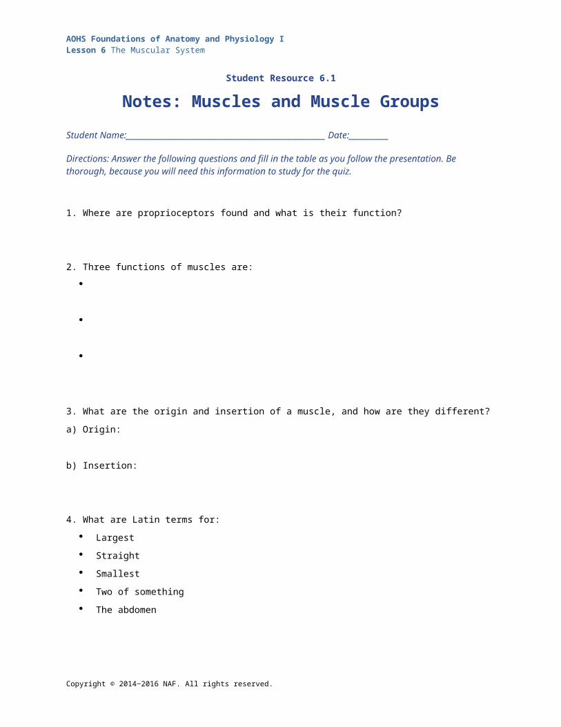
AOHS Foundations of Anatomy and Physiology ILesson 6 The Muscular System
Student Resource 6.1
Notes: Muscles and Muscle Groups Student Name:_______________________________________________________ Date:___________
Directions: Answer the following questions and fill in the table as you follow the presentation. Be thorough, because you will need this information to study for the quiz.
1. Where are proprioceptors found and what is their function?
2. Three functions of muscles are:
3. What are the origin and insertion of a muscle, and how are they different?
a) Origin:
b) Insertion:
4. What are Latin terms for:
Largest
Straight
Smallest
Two of something
The abdomen
5. What is an antagonistic pair of muscles?
Copyright © 2014‒2016 NAF. All rights reserved.
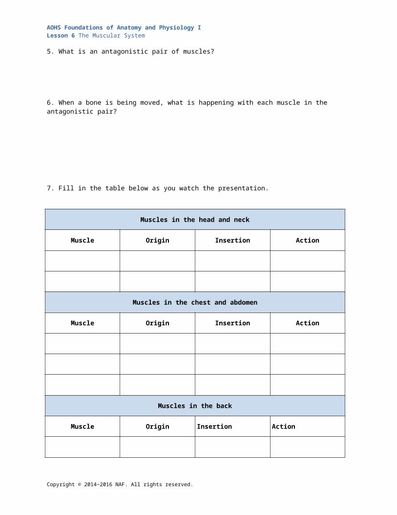
AOHS Foundations of Anatomy and Physiology ILesson 6 The Muscular System
6. When a bone is being moved, what is happening with each muscle in the antagonistic pair?
7. Fill in the table below as you watch the presentation.
Muscles in the head and neck
Muscle Origin Insertion Action
Muscles in the chest and abdomen
Muscle Origin Insertion Action
Muscles in the back
Muscle Origin Insertion Action
Copyright © 2014‒2016 NAF. All rights reserved.
AOHS Foundations of Anatomy and Physiology ILesson 6 The Muscular System
Muscles in the arms and legs
Muscle Origin Insertion Action
Copyright © 2014‒2016 NAF. All rights reserved.
AOHS Foundations of Anatomy and Physiology ILesson 6 The Muscular System
Student Resource 6.2
Reading: Muscles and Muscle Groups
Copyright © 2014‒2016 NAF. All rights reserved.

AOHS Foundations of Anatomy and Physiology ILesson 6 The Muscular System
Copyright © 2014‒2016 NAF. All rights reserved.
AOHS Foundations of Anatomy and Physiology ILesson 6 The Muscular System
Muscles provide the power for almost all of our body movements. But muscles don’t create movement on their own: all your skeletal muscles (with the exception of one) are attached to bones. When a muscle contracts—meaning it shortens—it pulls with it one of the bones it’s attached to. Flex your arm. That movement is the result of muscles in your arm (most visibly, your biceps) contracting. Your biceps are attached to your radius. When the muscle shortens, it pulls the bone with it. This is the mechanism behind all your movements. While it may sometimes seem like your muscles are pushing—for example, if you’re pushing a chair across the floor—it’s actually the contraction of muscles in your back and arms creating the force to move your body and push the chair. Altogether, we have over 600 muscles in our bodies but only 208 bones.
Copyright © 2014‒2016 NAF. All rights reserved.
AOHS Foundations of Anatomy and Physiology ILesson 6 The Muscular System
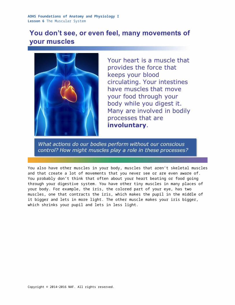
You also have other muscles in your body, muscles that aren’t skeletal muscles and that create a lot of movements that you never see or are even aware of. You probably don’t think that often about your heart beating or food going through your digestive system. You have other tiny muscles in many places of your body. For example, the iris, the colored part of your eye, has two muscles, one that contracts the iris, which makes the pupil in the middle of it bigger and lets in more light. The other muscle makes your iris bigger, which shrinks your pupil and lets in less light.
Copyright © 2014‒2016 NAF. All rights reserved.

AOHS Foundations of Anatomy and Physiology ILesson 6 The Muscular System
Your skeletal muscles are at work 24/7, making tiny adjustments all the time to adapt to new positions and to keep your posture and balance. Right now, you’re probably sitting, and muscles in your back and abdomen are working to keep you upright. Special sensors called proprioceptors send signals to your brain about where parts of your body are relative to each other, so that your muscles can work in unison to keep you steady. Thanks to these sensors (and maybe some training), you can kick your leg up and not fall over.
Copyright © 2014‒2016 NAF. All rights reserved.
AOHS Foundations of Anatomy and Physiology ILesson 6 The Muscular System
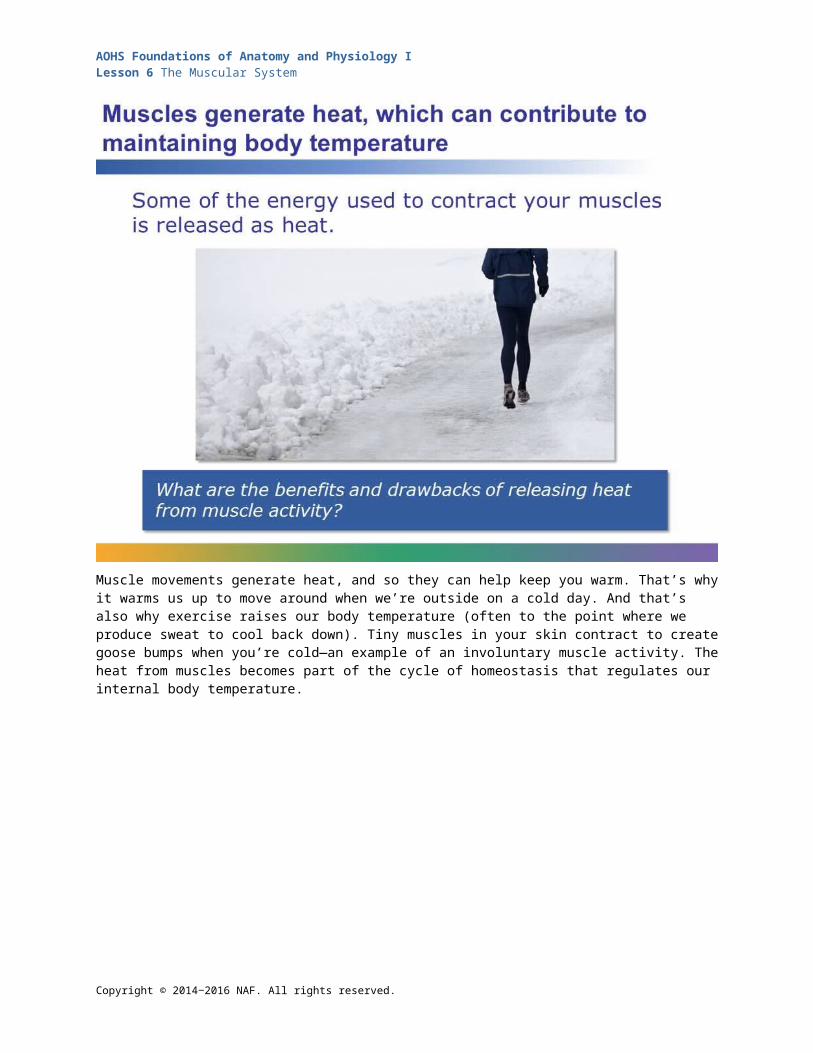
Muscle movements generate heat, and so they can help keep you warm. That’s why it warms us up to move around when we’re outside on a cold day. And that’s also why exercise raises our body temperature (often to the point where we produce sweat to cool back down). Tiny muscles in your skin contract to create goose bumps when you’re cold—an example of an involuntary muscle activity. The heat from muscles becomes part of the cycle of homeostasis that regulates our internal body temperature.
Copyright © 2014‒2016 NAF. All rights reserved.

AOHS Foundations of Anatomy and Physiology ILesson 6 The Muscular System
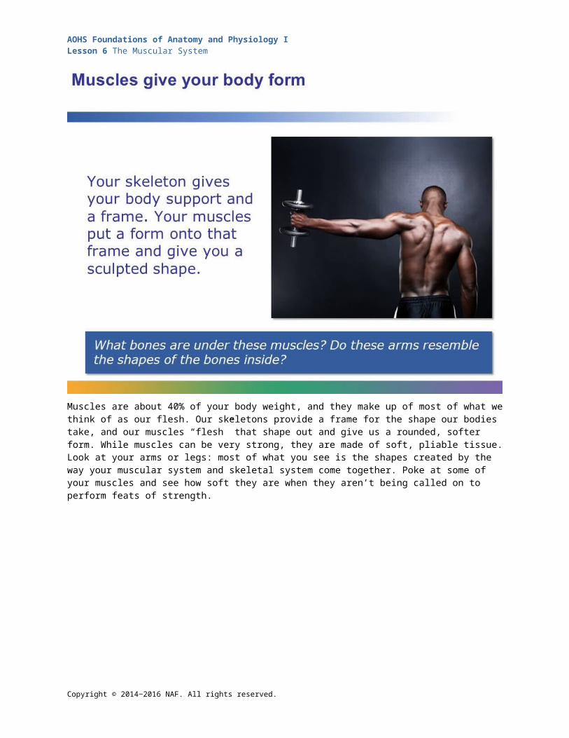
Muscles are about 40% of your body weight, and they make up of most of what we think of as our flesh. Our skeletons provide a frame for the shape our bodies take, and our muscles “flesh” that shape out and give us a rounded, softer form. While muscles can be very strong, they are made of soft, pliable tissue. Look at your arms or legs: most of what you see is the shapes created by the way your muscular system and skeletal system come together. Poke at some of your muscles and see how soft they are when they aren’t being called on to perform feats of strength.
Copyright © 2014‒2016 NAF. All rights reserved.
AOHS Foundations of Anatomy and Physiology ILesson 6 The Muscular System
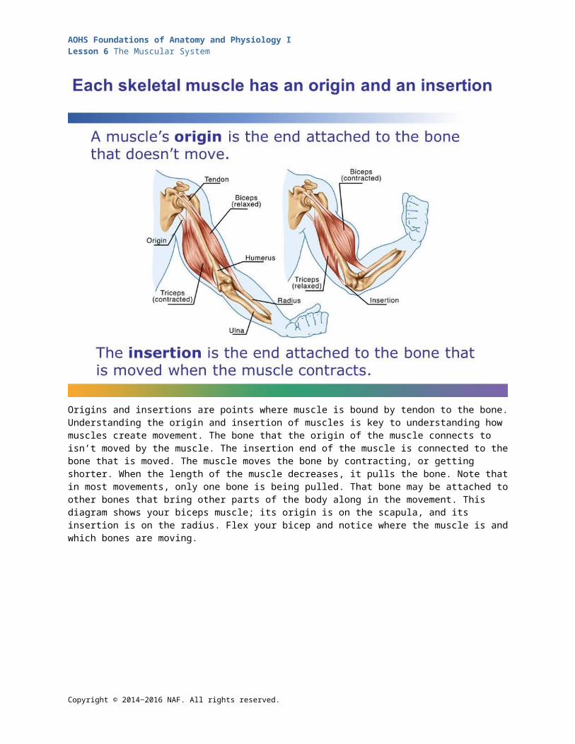
Origins and insertions are points where muscle is bound by tendon to the bone. Understanding the origin and insertion of muscles is key to understanding how muscles create movement. The bone that the origin of the muscle connects to isn’t moved by the muscle. The insertion end of the muscle is connected to the bone that is moved. The muscle moves the bone by contracting, or getting shorter. When the length of the muscle decreases, it pulls the bone. Note that in most movements, only one bone is being pulled. That bone may be attached to other bones that bring other parts of the body along in the movement. This diagram shows your biceps muscle; its origin is on the scapula, and its insertion is on the radius. Flex your bicep and notice where the muscle is and which bones are moving.
Copyright © 2014‒2016 NAF. All rights reserved.
AOHS Foundations of Anatomy and Physiology ILesson 6 The Muscular System
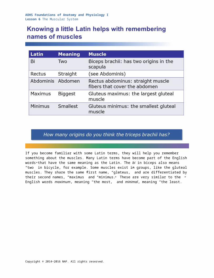
If you become familiar with some Latin terms, they will help you remember something about the muscles. Many Latin terms have become part of the English words that have the same meaning as the Latin. The bi in biceps also means “two” in bicycle, for example. Some muscles exist in groups, like the gluteal muscles. They share the same first name, “gluteus,” and are differentiated by their second names, “maximus” and “minimus.” These are very similar to the English words maximum, meaning “the most,” and minimal, meaning “the least.”
Copyright © 2014‒2016 NAF. All rights reserved.
AOHS Foundations of Anatomy and Physiology ILesson 6 The Muscular System
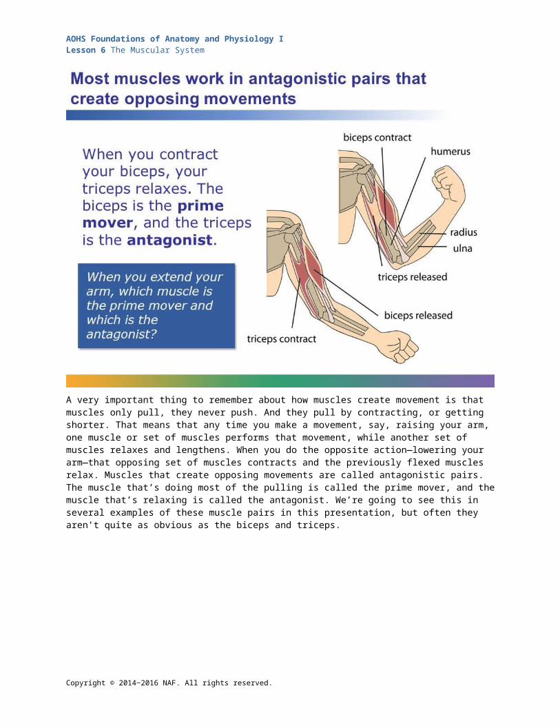
A very important thing to remember about how muscles create movement is that muscles only pull, they never push. And they pull by contracting, or getting shorter. That means that any time you make a movement, say, raising your arm, one muscle or set of muscles performs that movement, while another set of muscles relaxes and lengthens. When you do the opposite action—lowering your arm—that opposing set of muscles contracts and the previously flexed muscles relax. Muscles that create opposing movements are called antagonistic pairs. The muscle that’s doing most of the pulling is called the prime mover, and the muscle that’s relaxing is called the antagonist. We’re going to see this in several examples of these muscle pairs in this presentation, but often they aren’t quite as obvious as the biceps and triceps.
Copyright © 2014‒2016 NAF. All rights reserved.
AOHS Foundations of Anatomy and Physiology ILesson 6 The Muscular System
There are many, many more muscles in our bodies than we can learn in this course. We’re going to focus on major ones, starting at the head. The strongest muscle in your body, relative to its size, is the masseter, which you use when you bite down. When the masseter combines with other smaller, secondary muscles controlling your jaw, you can bite down on your molars with a force equal to about 200 pounds! The origin of the masseter is on your cheekbone, and the insertion is in your mandible. The sternocleidomastoid is the muscle that pulls your head toward your shoulders and rotates your head to the side. It has two origins, on the sternum and clavicle, and it inserts in the skull, behind the ear. The sternocleidomastoid lends a characteristic human shape to the neck, and so animators often include it in an alien or made-up creature—like the Na’vi in the movie Avatar, if they want that creature to look friendly. The muscles of our face also give us that “human” look. We have over 40 muscles in our faces, and all humans seem to make the same kind of expressions, no matter what environment they’ve grown up in.
Copyright © 2014‒2016 NAF. All rights reserved.
AOHS Foundations of Anatomy and Physiology ILesson 6 The Muscular System
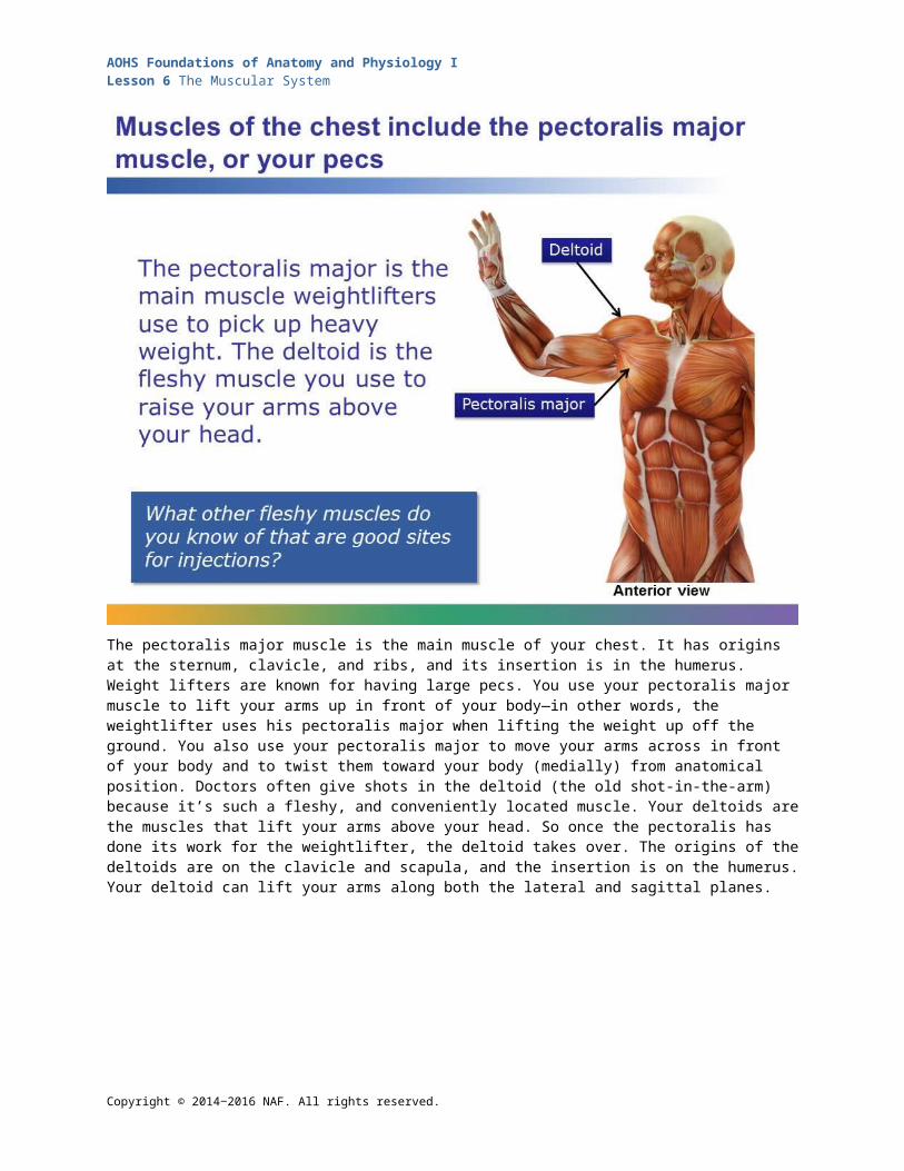
The pectoralis major muscle is the main muscle of your chest. It has origins at the sternum, clavicle, and ribs, and its insertion is in the humerus. Weight lifters are known for having large pecs. You use your pectoralis major muscle to lift your arms up in front of your body—in other words, the weightlifter uses his pectoralis major when lifting the weight up off the ground. You also use your pectoralis major to move your arms across in front of your body and to twist them toward your body (medially) from anatomical position. Doctors often give shots in the deltoid (the old shot-in-the-arm) because it’s such a fleshy, and conveniently located muscle. Your deltoids are the muscles that lift your arms above your head. So once the pectoralis has done its work for the weightlifter, the deltoid takes over. The origins of the deltoids are on the clavicle and scapula, and the insertion is on the humerus. Your deltoid can lift your arms along both the lateral and sagittal planes.
Copyright © 2014‒2016 NAF. All rights reserved.

AOHS Foundations of Anatomy and Physiology ILesson 6 The Muscular System
Your rectus abdominis is your abs, or your “six-pack.” It’s actually a pair of muscles that run vertically upward, with the origin at the pelvic girdle and the insertion at the ribs and sternum. When you exhale forcefully, you’re using your rectus abdominis. The rectus abdominis also helps stabilize your spine and hold you upright. Two other important abdominal muscles are the external and internal obliques. These are each pairs of muscles that wrap around your abdomen, and their muscle fibers are aligned at right angles to each other. Together, they allow you to twist and bend. More importantly―and more than your rectus abdominis―keeping these muscles strong helps prevent back injuries, because the obliques are the main support for your vertebral column.
Copyright © 2014‒2016 NAF. All rights reserved.
AOHS Foundations of Anatomy and Physiology ILesson 6 The Muscular System
Once you’ve lifted that weight, you have to put it down. The latissimus dorsi is the opposing muscle to the deltoids, and it contracts when you lower your arms from above your head. Raise your arms straight out to the side and then pull them back in so your palms are by your sides. When you raise them, you’re using your deltoids, and relaxing your latissiumus dorsi. When you pull them back down, you’re contracting your lats and relaxing your deltoids. And when you’re swimming the crawl stroke, your latissimus dorsi is hard at work bringing power to your stroke. You also use your latissiumus dorsi when you carry weight in your arms and when you uncross your arms. Its origin is along the spinal column and ribs, and the insertion is in the humerus. Your trapezius is one of the muscles opposing your pectorals. It originates at the back of the skull and along the vertebrae of your upper spine. Its insertion is in the scapula. Bend your elbows and bring them together in front of you; you’re using your pecs. Now move your bent arms back so they’re in line with your body laterally. Notice that you’re also pulling your shoulders back at the same time? You’re using your trapezius to do that. You use your trapezius when you stand up straight with your shoulders back or when you shrug your shoulders. Body builders work to strengthen these muscles just as much as they work out the muscles of the chest and shoulder.
Copyright © 2014‒2016 NAF. All rights reserved.

AOHS Foundations of Anatomy and Physiology ILesson 6 The Muscular System
If it’s arm curls you’re doing, then the biceps brachii and triceps brachii are the muscles you’re working on. The biceps brachii spans across the length of your humerus but isn’t attached to it; its origin is the scapula, and its insertion is the radius. It’s the muscle that bulges when you bend your elbow. The triceps brachii, as we discussed earlier, is the opposing muscle. It has three heads, thus its name, two on the scapula and one on the humerus. Its insertion is on the ulna. You’re using your triceps brachii quite a bit during the up part of a push up, and if you were to give someone a good, straight-on punch (which we know you wouldn’t), the force for that punch would come from you extending your elbow with your triceps brachii.
Copyright © 2014‒2016 NAF. All rights reserved.

AOHS Foundations of Anatomy and Physiology ILesson 6 The Muscular System
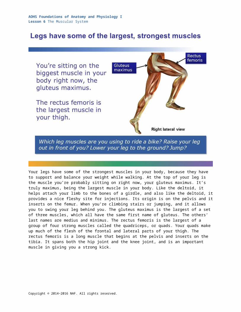
Your legs have some of the strongest muscles in your body, because they have to support and balance your weight while walking. At the top of your leg is the muscle you’re probably sitting on right now, your gluteus maximus. It’s truly maximus, being the largest muscle in your body. Like the deltoid, it helps attach your limb to the bones of a girdle, and also like the deltoid, it provides a nice fleshy site for injections. Its origin is on the pelvis and it inserts on the femur. When you’re climbing stairs or jumping, and it allows you to swing your leg behind you. The gluteus maximus is the largest of a set of three muscles, which all have the same first name of gluteus. The others’ last names are medius and minimus. The rectus femoris is the largest of a group of four strong muscles called the quadriceps, or quads. Your quads make up much of the flesh of the frontal and lateral parts of your thigh. The rectus femoris is a long muscle that begins at the pelvis and inserts on the tibia. It spans both the hip joint and the knee joint, and is an important muscle in giving you a strong kick.
Copyright © 2014‒2016 NAF. All rights reserved.

AOHS Foundations of Anatomy and Physiology ILesson 6 The Muscular System
The biceps femoris is one of the trio of muscles called the hamstrings that oppose the quads. The biceps femoris bends your knee. It originates on your pelvis and femur and inserts in your tibia. You use both your hamstrings and your quads while walking, but the muscle that really propels you forward is the gastrocnemius. The gastrocnemius is the shapely muscle you see on the calves of cyclists and runners. It arises on the femur and inserts at the base of the heel, connected to the Achilles tendon. You contract your gastrocnemius when you stand on your toes or when you flex your ankle when taking a step forward.
Copyright © 2014‒2016 NAF. All rights reserved.
AOHS Foundations of Anatomy and Physiology ILesson 6 The Muscular System
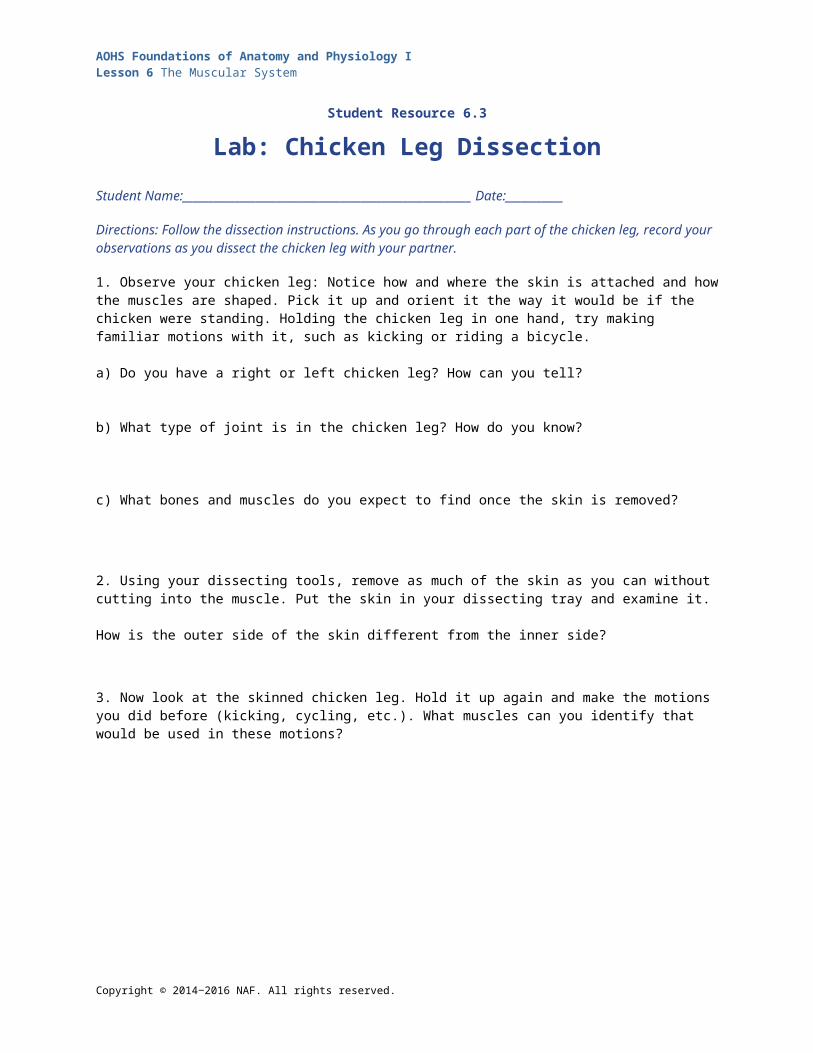
Student Resource 6.3
Lab: Chicken Leg DissectionStudent Name:_______________________________________________________ Date:___________
Directions: Follow the dissection instructions. As you go through each part of the chicken leg, record your observations as you dissect the chicken leg with your partner.
1. Observe your chicken leg: Notice how and where the skin is attached and how the muscles are shaped. Pick it up and orient it the way it would be if the chicken were standing. Holding the chicken leg in one hand, try making familiar motions with it, such as kicking or riding a bicycle.
a) Do you have a right or left chicken leg? How can you tell?
b) What type of joint is in the chicken leg? How do you know?
c) What bones and muscles do you expect to find once the skin is removed?
2. Using your dissecting tools, remove as much of the skin as you can without cutting into the muscle. Put the skin in your dissecting tray and examine it.
How is the outer side of the skin different from the inner side?
3. Now look at the skinned chicken leg. Hold it up again and make the motions you did before (kicking, cycling, etc.). What muscles can you identify that would be used in these motions?
Copyright © 2014‒2016 NAF. All rights reserved.

AOHS Foundations of Anatomy and Physiology ILesson 6 The Muscular System
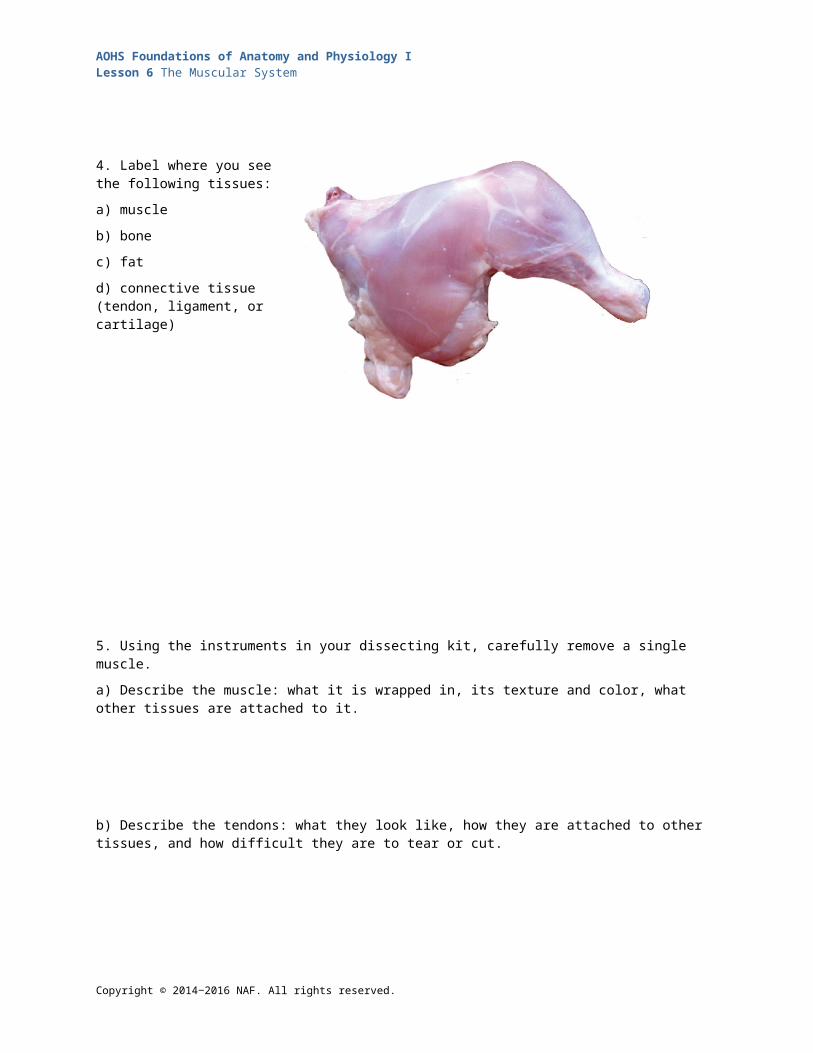
4. Label where you see the following tissues:
a) muscle
b) bone
c) fat
d) connective tissue (tendon, ligament, or cartilage)
5. Using the instruments in your dissecting kit, carefully remove a single muscle.
a) Describe the muscle: what it is wrapped in, its texture and color, what other tissues are attached to it.
b) Describe the tendons: what they look like, how they are attached to other tissues, and how difficult they are to tear or cut.
c) How many different muscles do you see? How difficult was it to separate them from one another?
6. Did you find other tissues that you think might be nerves or blood vessels? Describe what you found, where it is located, and what clues you see as to what it might be.
Copyright © 2014‒2016 NAF. All rights reserved.
AOHS Foundations of Anatomy and Physiology ILesson 6 The Muscular System
7. Next, remove as much muscle tissue as you can from the bones. Again, hold up what’s left of the chicken leg and make the motions you made earlier, noting how the motion looks in the knee joint.
On this picture of a chicken knee, label the bones, ligaments, cartilage and other tissues that you can recognize, and showing how the shapes of the bones fit together.
8. Cut the bones apart at the knee joint, and break one of the bones open. Describe what you see inside the bone.
9. What does the periosteum look and feel like?
10. Compare what it was like to cut the muscle tendon and to cut the ligaments (remember, ligaments connect bone to bone, and they are found in the joints).
Copyright © 2014‒2016 NAF. All rights reserved.
AOHS Foundations of Anatomy and Physiology ILesson 6 The Muscular System
Student Resource 6.4
Reading: How Muscles Work
Copyright © 2014‒2016 NAF. All rights reserved.

AOHS Foundations of Anatomy and Physiology ILesson 6 The Muscular System
Copyright © 2014‒2016 NAF. All rights reserved.

AOHS Foundations of Anatomy and Physiology ILesson 6 The Muscular System
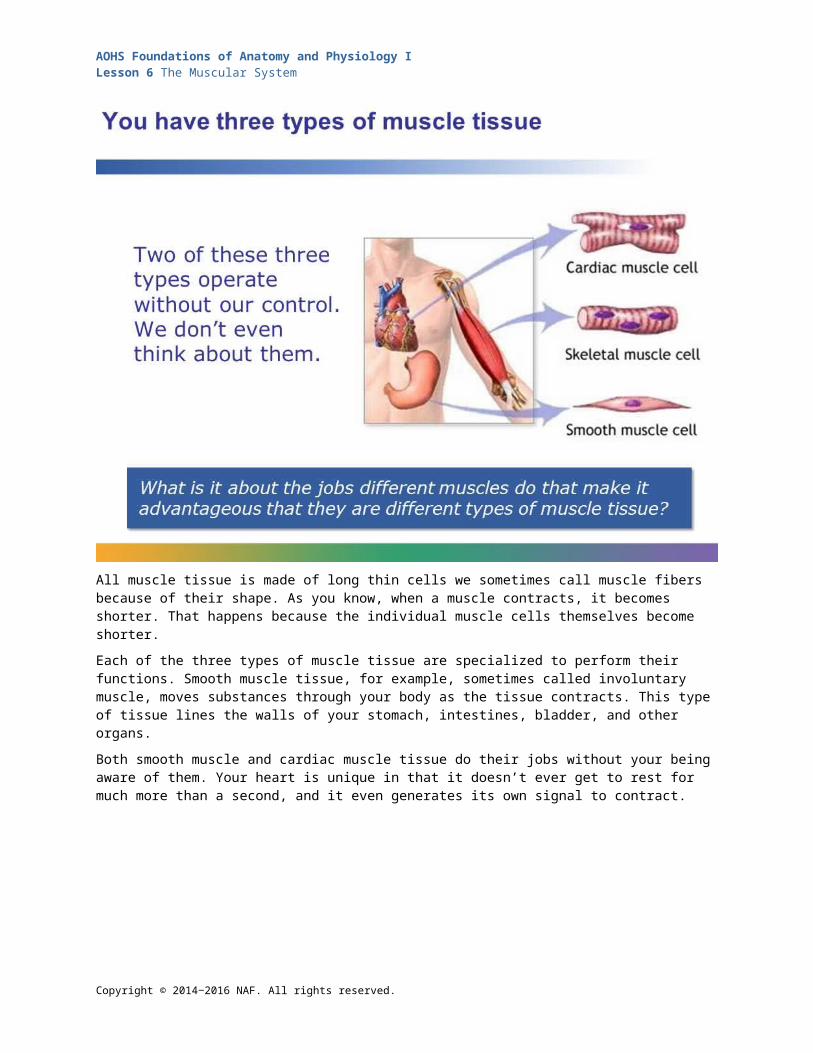
All muscle tissue is made of long thin cells we sometimes call muscle fibers because of their shape. As you know, when a muscle contracts, it becomes shorter. That happens because the individual muscle cells themselves become shorter.
Each of the three types of muscle tissue are specialized to perform their functions. Smooth muscle tissue, for example, sometimes called involuntary muscle, moves substances through your body as the tissue contracts. This type of tissue lines the walls of your stomach, intestines, bladder, and other organs.
Both smooth muscle and cardiac muscle tissue do their jobs without your being aware of them. Your heart is unique in that it doesn’t ever get to rest for much more than a second, and it even generates its own signal to contract.
Copyright © 2014‒2016 NAF. All rights reserved.

AOHS Foundations of Anatomy and Physiology ILesson 6 The Muscular System
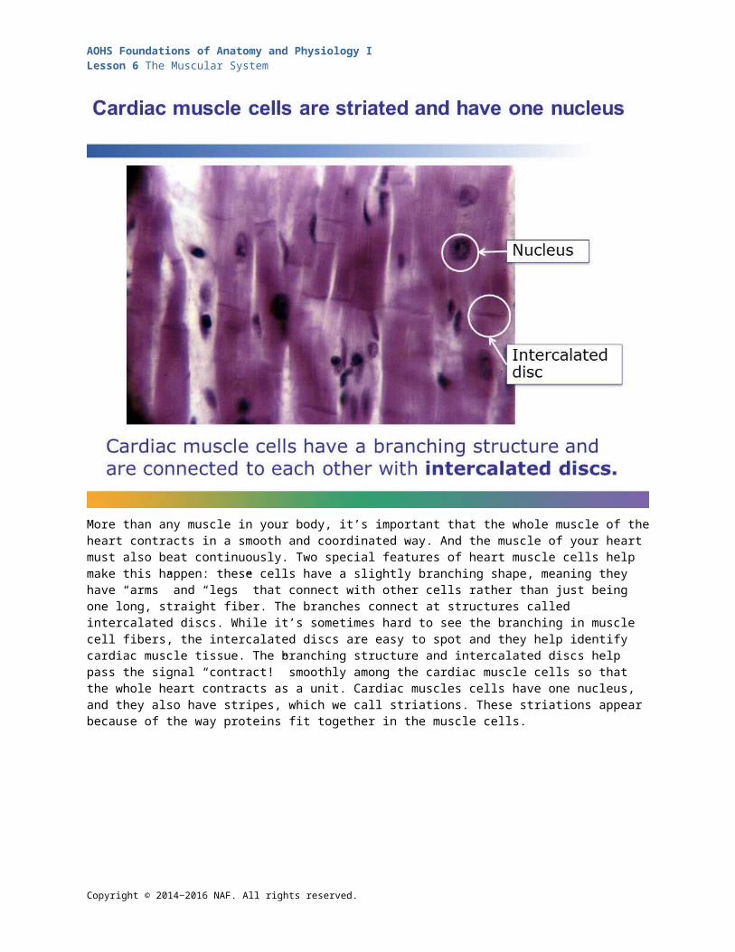
More than any muscle in your body, it’s important that the whole muscle of the heart contracts in a smooth and coordinated way. And the muscle of your heart must also beat continuously. Two special features of heart muscle cells help make this happen: these cells have a slightly branching shape, meaning they have “arms” and “legs” that connect with other cells rather than just being one long, straight fiber. The branches connect at structures called intercalated discs. While it’s sometimes hard to see the branching in muscle cell fibers, the intercalated discs are easy to spot and they help identify cardiac muscle tissue. The branching structure and intercalated discs help pass the signal “contract!” smoothly among the cardiac muscle cells so that the whole heart contracts as a unit. Cardiac muscles cells have one nucleus, and they also have stripes, which we call striations. These striations appear because of the way proteins fit together in the muscle cells.
Copyright © 2014‒2016 NAF. All rights reserved.
AOHS Foundations of Anatomy and Physiology ILesson 6 The Muscular System
Smooth muscle is also sometimes called involuntary muscle, because, like cardiac muscle, we can’t consciously control the workings of it. Smooth muscle cells are thick in the middle, with tapered ends, and these cells come together in sheets that make up the walls of hollow organs like your stomach, intestines, and bladder. They also compose our sphincters (circular muscles that often control the openings and closings of organs); many of the sphincters you are familiar with, like the urinary sphincter, hold the contents in at the end of your hollow organs. Some sphincters control what goes into your body and how blood circulates. The uterus is also made of smooth muscle. Smooth muscle is the champion at sustaining contraction: some smooth muscle organs, like your sphincters, are in contraction almost all the time. Other organs, like your stomach and intestines, go through long, sustained contractions. Smooth muscle can remain in contraction much longer than skeletal muscle. One of the only times we become aware of its action is when an expectant mother is about to give birth; she becomes very aware of smooth muscle contraction!
Copyright © 2014‒2016 NAF. All rights reserved.
AOHS Foundations of Anatomy and Physiology ILesson 6 The Muscular System
Skeletal muscle cells are truly fiber-like: think of the texture of a steak or chicken breast when you cut into it. That’s why muscle cells are often called muscle fibers. The fibers of the muscle all run the same direction, and have a thread-like shape. Skeletal muscle cells are easy to recognize because they have many nuclei and are striated. This tissue is sometimes called voluntary muscle, because we can (usually) control its contractions: if you want to jump, you contract muscles in your legs and you jump. While you aren’t conscious of the contractions themselves, you are conscious of the movement being generated. Individually, skeletal muscle fibers are wispy and fragile. The strength of a muscle comes from how the fibers are packaged, in a complex system of bundles.
Copyright © 2014‒2016 NAF. All rights reserved.
AOHS Foundations of Anatomy and Physiology ILesson 6 The Muscular System
An important key to the strength of our muscles is the way they’re packaged. A skeletal muscle is made of bundles of muscle cells that are then bundled together again. Both the small and large bundles are held together with tough connective tissue. Each individual muscle cell is wrapped in a thin connective tissue, and these tissue-wrapped cells are then bound together in bunches that are held together by another wrapping of connective tissue. Then these bunches of muscle fibers are bound up into a larger package, which is the muscle itself, and wrapped up in one last wrapper called the epimysium. At the ends of muscles, the epimysium blends into the rope-like structure of the tendons similar to the way that the periosteum and tendons blend together. In most skeletal muscles, there are thousands of fibers bound together in this way, which allows them to work together as a unit to exert force.
Copyright © 2014‒2016 NAF. All rights reserved.

AOHS Foundations of Anatomy and Physiology ILesson 6 The Muscular System
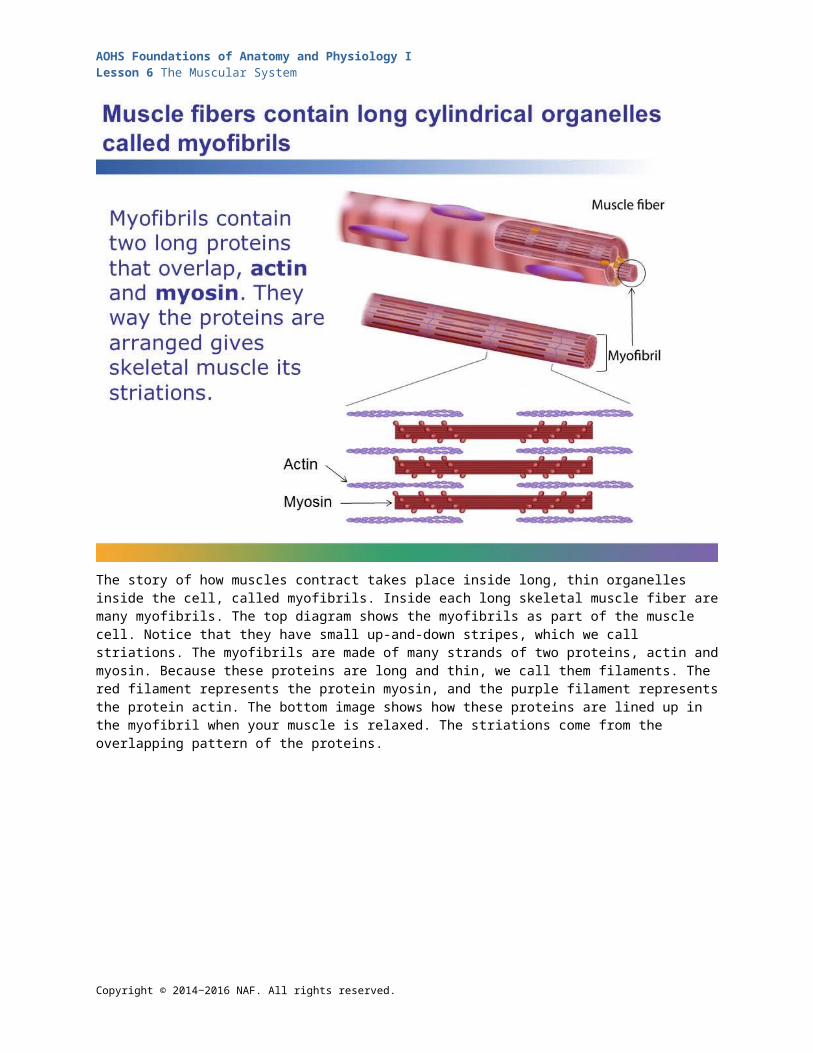
The story of how muscles contract takes place inside long, thin organelles inside the cell, called myofibrils. Inside each long skeletal muscle fiber are many myofibrils. The top diagram shows the myofibrils as part of the muscle cell. Notice that they have small up-and-down stripes, which we call striations. The myofibrils are made of many strands of two proteins, actin and myosin. Because these proteins are long and thin, we call them filaments. The red filament represents the protein myosin, and the purple filament represents the protein actin. The bottom image shows how these proteins are lined up in the myofibril when your muscle is relaxed. The striations come from the overlapping pattern of the proteins.
Copyright © 2014‒2016 NAF. All rights reserved.

AOHS Foundations of Anatomy and Physiology ILesson 6 The Muscular System
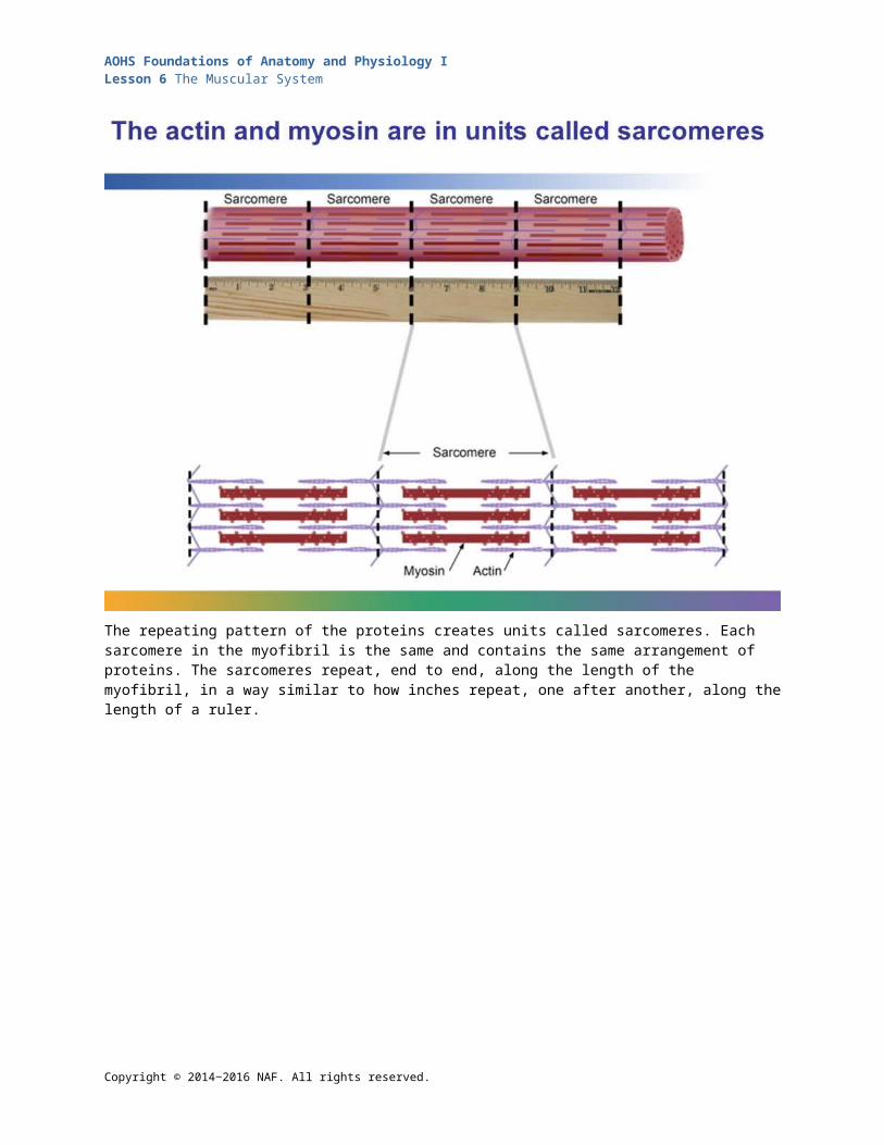
The repeating pattern of the proteins creates units called sarcomeres. Each sarcomere in the myofibril is the same and contains the same arrangement of proteins. The sarcomeres repeat, end to end, along the length of the myofibril, in a way similar to how inches repeat, one after another, along the length of a ruler.
Copyright © 2014‒2016 NAF. All rights reserved.

AOHS Foundations of Anatomy and Physiology ILesson 6 The Muscular System
But unlike the inches in a ruler, the sarcomeres can get shorter, which is what happens when a muscle cell contracts. The sarcomeres get shorter because the myosin latches onto the actin and sort of “rows” its way along it, toward the end of the sarcomere (where you see the purple vertical zigzag line), pulling the actin molecules together. If you compare the two diagrams, you can see that the length of the sarcomere shrinks as the proteins move past each other. This contraction happens in all the myofibrils of the cell at the same time, so that that whole cell itself gets shorter. This process requires a lot of energy; when you’ve been exercising and you get hungry and need fuel, it’s because your myosin molecules have been so busy “rowing.”
Copyright © 2014‒2016 NAF. All rights reserved.
AOHS Foundations of Anatomy and Physiology ILesson 6 The Muscular System
When you want to make a movement, like raising your hand, your muscle needs to receive a signal to do that. Often the signal starts in your brain, but if the movement is a reflex, like pulling your hand off a hot burner, the signal may start in your spinal cord. Either way, the signal travels down a neuron that makes connections to individual muscle cells. The part of the neuron that delivers the signal to the muscle cells is called the axon. This system, taken together, is called a motor unit. Note that one axon connects to many individual muscle cells.
Copyright © 2014‒2016 NAF. All rights reserved.
AOHS Foundations of Anatomy and Physiology ILesson 6 The Muscular System
When the signal gets to the end of the neuron, it causes changes at what’s called the neuromuscular junction. The neuromuscular junction is a kind of synapse, a place where a signal from the nervous system is passed from one cell to another. It’s important to note here is that the neuron and the muscle cell aren’t directly connected. There’s a tiny space between them called the synaptic cleft. What we’re going to see now is a mechanism that’s similar to how all neurons pass signals, whether they are communicating with a muscle to make you move or with another neuron to create a thought or perception. In all synapses, when the signal reaches the end of an axon, it causes the axon to release a chemical called a neurotransmitter into the synaptic cleft. Neurotransmitters diffuse across that tiny space between the two cells, and when they reach the other side, they create changes in the membrane of the other cell.
Copyright © 2014‒2016 NAF. All rights reserved.

AOHS Foundations of Anatomy and Physiology ILesson 6 The Muscular System
There are many different neurotransmitters in your body, and they have different functions. The neurotransmitter that causes muscle contraction is called acetylcholine. When acetylcholine has crossed the synapse, it binds with receptor proteins on the surface of the muscle cell membrane.
Copyright © 2014‒2016 NAF. All rights reserved.

AOHS Foundations of Anatomy and Physiology ILesson 6 The Muscular System
The effect of acetylcholine on the cell membrane is that it opens up ion channels. Because there is more sodium outside the cell than inside, and more potassium inside than outside, these ions can now cross the membrane. With the ion channels open, lots of sodium rushes into the cell, putting more positively charged ions inside of it. Some potassium goes out of the cell, but not as much, so, for a moment, there’s a higher positive charge inside the cell than out. This change in charge is called depolarization.
Copyright © 2014‒2016 NAF. All rights reserved.

AOHS Foundations of Anatomy and Physiology ILesson 6 The Muscular System
The “charge swap” of depolarization produces an imbalance of charge; there are now more positive ions inside the cell and fewer outside. We call this current the action potential, because it’s what’s going to make the cell “act.” The action potential travels through the length of the muscle cell, spreading the depolarization. The depolarization sets off a series of events that make the myosin filaments in the myofibrils bind with and “row” along the actin, pulling the protein filaments past each other. This shortens the sarcomeres and the muscle cell as a whole. Because muscle cells can be very long, it can take billions of contracting sarcomeres to move one muscle.
Copyright © 2014‒2016 NAF. All rights reserved.

AOHS Foundations of Anatomy and Physiology ILesson 6 The Muscular System
A single large muscle, like your biceps, can exert enormous force when all its cells are at work. But you don’t need nearly as much force to lift a pen as you need, for example, to lift a heavy couch. And it would be pretty inefficient if your muscles used more energy than they needed all the time. The amount of strength behind a muscle’s action depends on how many cells of that muscle contract. Remember, every cell that contracts does its job to the fullest. When you’re picking up a pen, only a fraction of the number of muscle fibers kick in versus when you pick up a heavy sofa. This is one way your body can fine-tune your movements.
Copyright © 2014‒2016 NAF. All rights reserved.

AOHS Foundations of Anatomy and Physiology ILesson 6 The Muscular System
Student Resource 6.5
Notes: How Muscles WorkDirections: Take notes as you follow the presentation How Muscles Work, answer all of the questions, and label the diagrams. Be thorough, because you will need this information to study for the quiz.
Check the boxes that apply: Cardiac Smooth Skeletal
Striated?
Multinucleated?
Voluntary?
Note other features and why they are important
Copyright © 2014‒2016 NAF. All rights reserved.

AOHS Foundations of Anatomy and Physiology ILesson 6 The Muscular System
1. Draw a diagram of how muscles are bundled together, and label an individual muscle cell.
2. Why is this arrangement inside the muscle important?
3. What is the “wrapper” on the outside of the muscle and how is it similar to the periosteum?
4.
a) Label the proteins actin and myosin.
b) In the myofibril, show where one sarcomere begins and ends. Also draw lines from the protein diagram to the myofibril to show where the protein structure fits into the myofibril.
c) Draw brackets to show how the length of the sarcomere is different when the myofibril is relaxed and when it is contracted.
Copyright © 2014‒2016 NAF. All rights reserved.

AOHS Foundations of Anatomy and Physiology ILesson 6 The Muscular System
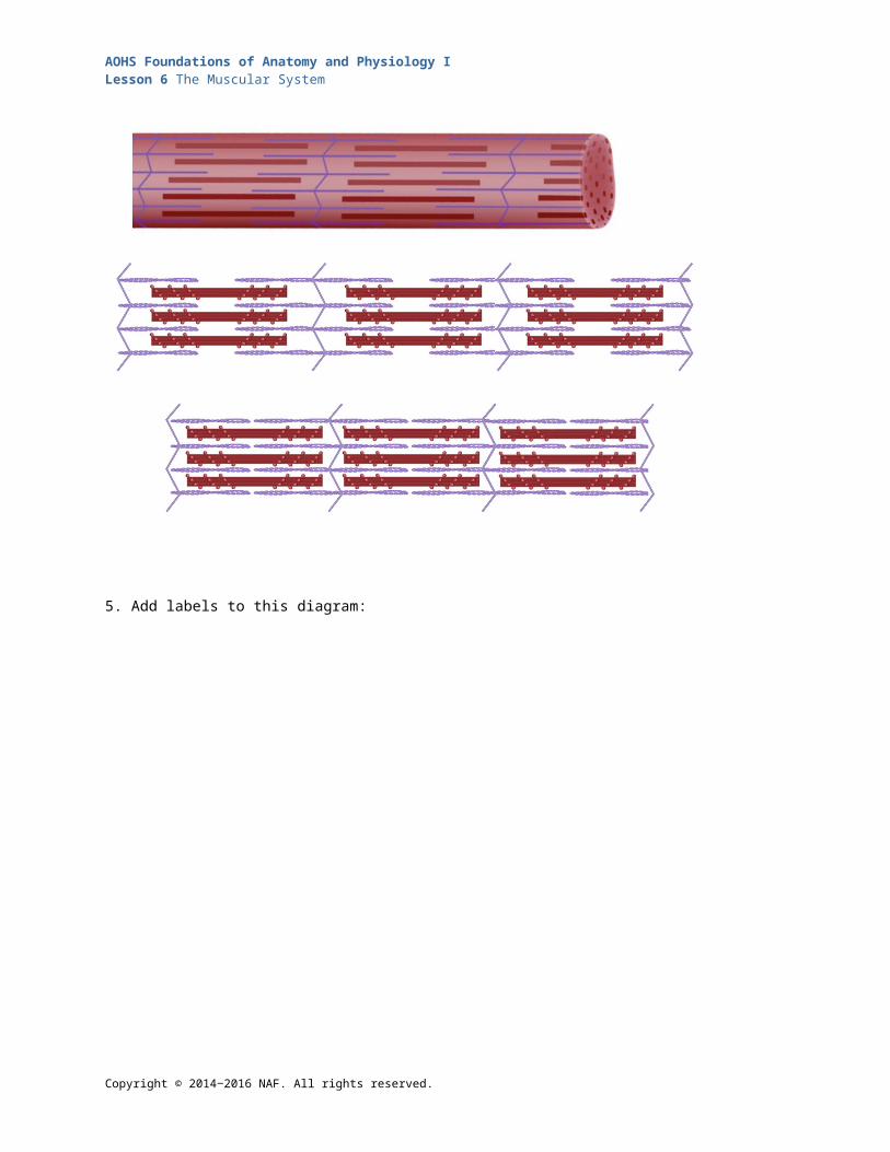
5. Add labels to this diagram:
6. Explain what an axon, synapse, and neurotransmitter are. How do they function together?
7. What happens to the cell membrane when more sodium enters the cell than potassium leaving the cell?
8. What is the electrical current that causes the cell to contract?
9. What adjustments do your muscles make when you have to pick up a heavy object and then move a light one?
Copyright © 2014‒2016 NAF. All rights reserved.

AOHS Foundations of Anatomy and Physiology ILesson 6 The Muscular System
Student Resource 6.6
Reading: The Impact of Exercise Student Names:_______________________________________________________ Date:___________
Directions: As you read, make notes in the margins every time you see information to add to one of the following topics, which are written on the chart papers that your teacher has posted.
1. Benefits of exercise
2. Examples of endurance exercise/activities
3. Examples of resistance exercise/activities
4. Effects of inactivity
Sprinting up a flight of stairs leaves some of us gasping for breath. At the top, while waiting for our thighs to stop burning, we might say, “Wow, I need to get in shape!” What does “getting in shape” mean in terms of your muscles? Why is it easier to get up the stairs once you’ve gotten in shape? And why is it good to be in shape in the first place? Regular exercise changes your muscles—including your heart—in ways that affect your whole body and your overall health.
Different Activities Have Different Effects on Your MusclesDifferent kinds of activities strengthen and improve your muscles in different ways, so it’s important to get a mix of exercise in your day. You’re doing resistance activities when you’re working against a heavy object or the force of gravity. Lifting weights, picking up trays of dishes, and pushing a lawn mower are all resistance activities. Many resistance exercises are also weight-bearing exercises, so they help build your bones, too. Just as weight-bearing activity promotes new bone cells, resistance activities spark your muscle cells to create more myofibrils. What that means is that when your brain sends a signal to your muscle cells to contract, there are many more sarcomeres that will be part of that contraction. The contraction is stronger, without your having to use any additional muscle cells to do it. A good trick of efficiency!
Resistance activities also increase the amount of connective tissue your muscles have, which makes them able to withstand the greater force you can now exert with them. That additional tissue, along with the cells making new myofibrils, results in your muscles getting bigger and more defined. Body builders get their sometimes-exaggerated muscles by doing resistance exercises.
Resistance exercise doesn’t help you run long distances or dance all night, though. To gain that kind of stamina, you need endurance activities. Endurance exercise increases blood flow to your muscles and makes them more efficient at making energy and storing oxygen. That means you don’t get tired as quickly and so can run farther or dance longer. Often people can keep at endurance activity like walking or cycling for hours. And while endurance exercises won’t give you the bulging biceps of a champion weight lifter, they have beautifying effects on almost every other part of your body, inside and out.
Muscle ToneAt the gym, when we refer to muscle tone, we’re actually thinking about the size and strength of the muscles. A medical professional thinks about muscle tone differently, as a measure of how contracted our muscles are at rest. The amount of real muscle tone (as opposed to “gym strength”) is determined by our genetics and doesn’t change when we work out. What does change when you repeatedly exercise the same muscles is the actual muscles and muscle cells.
Copyright © 2014‒2016 NAF. All rights reserved.

AOHS Foundations of Anatomy and Physiology ILesson 6 The Muscular System
Endurance Exercise Affects Many Body SystemsRemember that all the systems in your body are connected, and that what affects one system usually affects many others. In the case of your muscles, they’re loaded with blood vessels, nerves, and connective tissue, and they are fed and fueled by oxygen from your lungs and nutrients from your food. So while your body is building muscle, it’s also improving the function of all the systems that work on your skeletal muscles. For starters, your heart—also a muscle, remember—gets exercised as well, and also gets larger. A bigger heart can pump more blood with each heartbeat and so doesn’t have to work as hard, another good efficiency trick. A more efficient heart gets more blood going through your blood vessels, too, and a more effective circulatory system usually means lower blood pressure and cholesterol. Endurance exercise also enhances your digestive system’s ability to make fuel and nutrients from your food and your circulatory system’s ability to get rid of waste products. Your lungs also become better at taking in oxygen and getting rid of carbon dioxide as you exercise. Endurance exercise even helps your brain and mood by releasing neurotransmitters call endorphins that can lift your mood. Beyond that, exercise can actually stimulate your brain to make new neurons, and seeing yourself get stronger and be able to do more things can make you feel better about yourself physically and emotionally.
Inactivity Also Affects Your Body Systems As a growing teen, it can be difficult to believe that you’re doing yourself harm by not exercising. After all, you feel fine. In the long term, though, you will feel the effects and they will not be pretty. The World Health Organization estimates that inactivity is the world’s fourth leading risk factor for mortality after high blood pressure, smoking, and high blood glucose levels. A factor for mortality is a professional way of saying that it’s something that we know contributes to people dying.
The most immediate and visible affect of not enough exercise is muscle atrophy. If you’ve ever broken a bone and had a cast, you’ve experienced what can happen to your muscles if you don’t use them: they lose their shapeliness and strength. They are actually losing myofibrils; in other words, you lose the stuff that gives you strength. And with that effect comes many others, like a cascade: your heart becomes weaker and less efficient, your body is less proficient at digestion and ferrying out toxins, you don’t breathe as well, your blood vessels don’t get oxygen and nutrients to your muscles and other body parts as efficiently, and your blood pressure and cholesterol may increase.
People who aren’t active also suffer from more depression and low self-esteem. And the fact that exercise is harder when you’re not used to doing it, especially if your muscles have atrophied, can be a discouragement. But anyone can increase their activity level in very simple ways that provide enormous health benefits.
Easy Ways to Increase Activity LevelsOf course working out at the gym or riding your bike are perfect examples of healthy activity. But there are lots of other ways for you to get your muscles going. For example, you can park your car on the far side of a parking lot or a block away from your friend’s house, and enjoy a brisk walk. Take the stairs instead of the elevator, especially if you’re going down. Get up from your TV-watching chair or couch during commercials and stretch your muscles, run in place, or do some jumping jacks.
You don’t need a fancy weight set to build up strength; it’s easy to do resistance exercises just by resisting the force of gravity. Move your arms up and down, hold them straight out, or try doing some leg lifts. Do push ups or lift weights using heavy objects like cans of soup. You can work your abdominal muscles by contracting them while you’re waiting in line to buy movie tickets, and no one will even know you’re exercising!
How do you know if an activity counts towards exercise? Here are some indications that you’re benefiting your body with an activity:
The muscle or group of muscles feels tired from the activity
Copyright © 2014‒2016 NAF. All rights reserved.
AOHS Foundations of Anatomy and Physiology ILesson 6 The Muscular System
Your pulse is faster and harder (your heart rate increases)
You sweat
You’re out of breath
Suggestions for Activities and Forms of ExerciseBelow, you’ll find a chart of different kinds of activities. Any of them are better than being a couch potato!
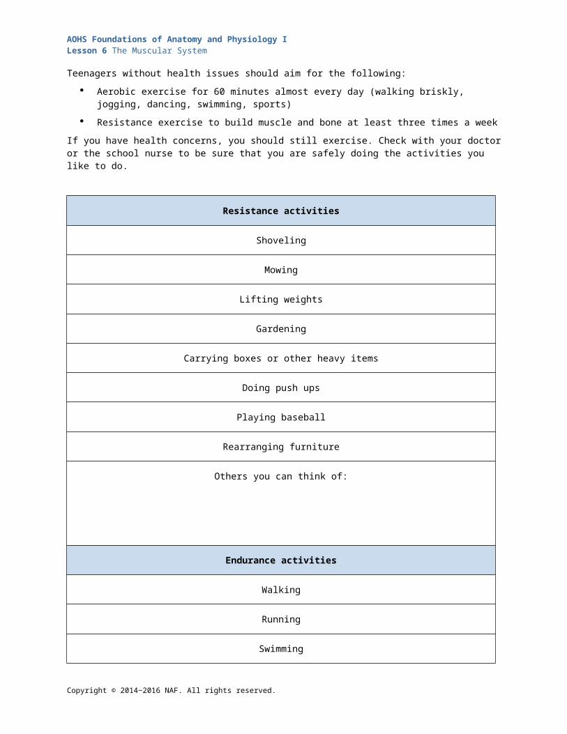
Teenagers without health issues should aim for the following:
Aerobic exercise for 60 minutes almost every day (walking briskly, jogging, dancing, swimming, sports)
Resistance exercise to build muscle and bone at least three times a week
If you have health concerns, you should still exercise. Check with your doctor or the school nurse to be sure that you are safely doing the activities you like to do.
Resistance activities
Shoveling
Mowing
Lifting weights
Gardening
Carrying boxes or other heavy items
Doing push ups
Playing baseball
Rearranging furniture
Others you can think of:
Endurance activities
Walking
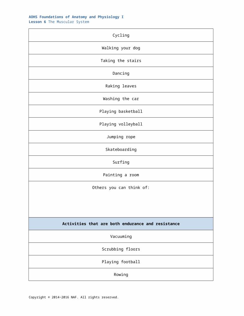
Running
Copyright © 2014‒2016 NAF. All rights reserved.

AOHS Foundations of Anatomy and Physiology ILesson 6 The Muscular System
Swimming
Cycling
Walking your dog
Taking the stairs
Dancing
Raking leaves
Washing the car
Playing basketball
Playing volleyball
Jumping rope
Skateboarding
Surfing
Painting a room
Others you can think of:
Activities that are both endurance and resistance
Vacuuming
Scrubbing floors
Playing football
Rowing
Tennis
Copyright © 2014‒2016 NAF. All rights reserved.

AOHS Foundations of Anatomy and Physiology ILesson 6 The Muscular System
Skiing
Snowboarding
Ice skating
Doing home improvement jobs like installing a new door
Others you can think of:
Activities that aren’t technically considered exercise but are better than inactivity
Marching in place (try it while watching TV or talking on the phone)
Pushing a stroller
Ironing
Cooking
Building a snowman
Housecleaning, like doing dishes or cleaning the bathtub
Making small house repairs
Playing exercise-based video games, like those for Wii Fit
Others you can think of:
Copyright © 2014‒2016 NAF. All rights reserved.

AOHS Foundations of Anatomy and Physiology ILesson 6 The Muscular System
Student Resource 6.7
Venn Diagram: Favorite ExercisesDirections: Think about which activities or exercises you already incorporate into your regular schedule. Add them to the diagram below according to whether you think they are resistance activities, endurance activities, or both. If you do simple household activities, list those in the chart below the diagram. Then answer the questions that follow.
Copyright © 2014‒2016 NAF. All rights reserved.
AOHS Foundations of Anatomy and Physiology ILesson 6 The Muscular System
Activities I do that aren’t technically exercising but are better than inactivity
Copyright © 2014‒2016 NAF. All rights reserved.
RESISTANCE
ENDURANCE
BOTH
AOHS Foundations of Anatomy and Physiology ILesson 6 The Muscular System
Favorite exercises and activities I already do
Activity/Exercise How often? How long?
Ways to increase doing more of my favorite exercises and activities
Activity/Exercise How to increase
Activities or exercises I would like to start doing
Activity/Exercise How to start
Copyright © 2014‒2016 NAF. All rights reserved.
AOHS Foundations of Anatomy and Physiology ILesson 6 The Muscular System
Copyright © 2014‒2016 NAF. All rights reserved.

AOHS Foundations of Anatomy and Physiology ILesson 6 The Muscular System
Student Resource 6.8
Reading: Conditions of the Muscular SystemStudent Name:_______________________________________________________ Date:___________
Directions: Read the section below that your teacher has assigned to you and your group. Practice summarizing your section with your group. See Student Resource 6.9, Summaries: Conditions of the Muscular System, for an example of what a good summary looks like. When you change groups, you will be the expert on the section you have read, and you will need to explain it so that your new group members fully understand the facts.
Part A: Delayed Onset Muscle Soreness (DOMS)Exercise today, be sore tomorrow: You’ve probably experienced for yourself how exercise affects your muscles if you haven’t been using them for a while. If you’re muscles are sore a day or two after exerting yourself, you’ve got what’s called delayed onset muscle soreness, or DOMS.
DOMS can happen to anyone exercising, whether they’re a pro or a complete beginner, but it’s more common when a person is starting a new activity. The pain associated with DOMS is caused by tiny, microscopic tears in the muscle tissue or connective tissue, and the inflammation that can follow. It’s perfectly normal to experience these microtears, and they’re considered part of the processes of conditioning your body when you’re getting used to a new activity. Once you’ve done an activity a number of times, your body adapts, and you don’t feel the soreness anymore.
Doctors and researchers aren’t entirely sure what happens when your muscles adapt, but one theory is that your motor units learn that they need to stimulate more muscle fibers so that your muscles contract more strongly, which will reduce the strain on them.
Stretching before your activity can help reduce the pain you might get from DOMS later on. If you’ve been working up a sweat, you can also do some cool-down activities after exercising. If you’re feeling the ache the next day, you could try some anti-inflammatory pain relievers. And if you’re starting with a new activity, take it easy and work your way into it. You’ll find it doesn’t take long before you can put more effort in without having DOMS effects.
DOMS can be very mild or it can be painful enough to make it difficult to walk or make other motions. The pain involved depends on the extent of the damage and the inflammation. Personal trainers may sometimes say that DOMS is “good pain.” While there’s no clear evidence that there’s a benefit to DOMS, it is clear that the pain associated with taking on new activities isn’t harmful. In fact, it usually subsides after just a session or two of activity, and ultimately, taking on new activities and exercise has enormous benefits.
Part B: Using SteroidsThere’s nothing more disappointing to a sports fan than to find out their admired athlete’s victories may have been assisted by “doping.” Home run hitter Barry Bonds, champion cyclist Lance Armstrong, and many others have admitted to using anabolic steroids, commonly known as “juice,” to make themselves stronger and faster.
Anabolic steroids were first developed in the 1950s as a way to treat certain muscle-wasting diseases such as multiple sclerosis and conditions like anemia. The drugs are relatives of testosterone, the naturally occurring hormone that is responsible for the increases in muscle and bone mass—and the masculine appearance—that boys gain as they grow to become men. It didn’t take athletes long to come up with the notion that these drugs, which lend strength to patients who have a medical need for them,
Copyright © 2014‒2016 NAF. All rights reserved.
AOHS Foundations of Anatomy and Physiology ILesson 6 The Muscular System
would also enhance athletic ability and the chance to win. By the 1960s, steroid use had begun spreading among professional athletes.
By now, we’ve all heard a great deal about the use of steroids, so much so that their use has also spread to teenagers and college students looking for an edge. Some teens buy the drugs illegally online, and others try over-the-counter substances that promise “performance enhancement.” Athletes claim that the drugs make them stronger, keep them from getting winded, and make their muscles larger.
Steroids, which are technically known as anabolic-androgenic steroids, do add protein to muscle fibers and can encourage the growth of new myofibrils. They can indeed increase strength and muscle mass, which is why they are useful medically. But whether they provide much benefit for athletes like runners and swimmers remains in question, because steroids work mostly on the type of muscle used in resistance activities rather than endurance.
Although the impact of steroids is mixed, their side effects are clear: they can give you a bloated face, liver damage, and high blood pressure, and they can stunt your growth. Steroids can cause substances in your body to cling to the insides of your blood vessels, blocking them and leading to heart attacks and strokes. In young men, steroids are responsible for hair loss, infertility, enlarged breasts, and shriveled testicles. Young women might find themselves with increased body and facial hair, a deepened voice, and bald patches on their heads. The drugs can also make a person aggressive and manic, and a third of all steroid users have been found to develop psychiatric problems.
As you can see, the impact steroids have on a person’s body go a lot farther than just increasing the size of some muscles, and the side effects can be more than physical. Steroids can even affect your image as an athlete among your friends: a victory doesn’t feel real if it’s helped along by artificial enhancers.
Part C: Muscular Dystrophy (MD)In February 2013, Natalie Jones drew thousands of followers on Facebook with the story of her son Mitchell’s muscular dystrophy (MD). As the disease withered Mitchell’s muscles—including his heart—and sent him in and out of the hospital, Natalie shared their experience with the world.
Mitchell was diagnosed as a young boy with MD, an inherited disorder that makes muscles susceptible to damage. There are nine kinds of MD; some affect children and others aren’t apparent until adulthood. Mitchell’s type of MD, called Duchenne muscular dystrophy, or DMD, is the most common form, occurring in 1 out of every 3,500 births. It affects primarily boys, and symptoms usually start to appear between ages 3 and 5.
A boy with DMD will likely first start to experience muscle weakness in his hips, thighs, and pelvic area. Often these boys have larger-than-normal calves, and poor balance and coordination. His arm muscles will also become weak, making it hard to raise his arms. By the time he reaches his teenage years, a boy with DMD will usually begin to experience the effects of the disorder on his heart and on the muscles that help him breathe—this is what began to happen to Mitchell in early 2013. As the effects of DMD progress, the degeneration of the heart becomes life threatening.
DMD, like all other forms of MD, is caused by flaws in the person’s DNA. In the case of DMD, having this flaw means the boy’s body isn’t able to produce a protein called dystrophin (DIS-tro-fin). Your body uses dystrophin to make the strong connective tissue that binds and bundles the muscle cells together. Without it, your muscles aren’t protected against the strain put on them when you use them. So, for a boy with DMD, just using his muscles to move can damage them and make them weaker.
DMD occurs almost exclusively in boys because the gene that codes for dystrophin is found on the X chromosome. Every child inherits one X chromosome from his or her mother. From their father, girls inherit another X chromosome, and boys inherit a Y chromosome. The reason girls rarely have DMD is that even if they have a flaw in one X chromosome, they’re like to be able to make up for it with their other X chromosome. Boys don’t have that protection, so if they inherit the flawed gene—which they would have gotten from their mother—they will have the disorder.
Copyright © 2014‒2016 NAF. All rights reserved.
AOHS Foundations of Anatomy and Physiology ILesson 6 The Muscular System
There is no cure for any of the forms of muscular dystrophy, but there are many treatments that can help prolong the life of someone with the disorder. Corticosteroids (which are different from anabolic steroids) can keep the muscle deterioration at bay for some time. A patient with MD might also get a pacemaker put into their heart to help keep their heartbeats steady and strong. Researchers are looking at whether some drugs commonly used to treat high blood pressure can also help slow the deterioration of heart muscle. And doctors usually pair MD patients with a physical therapist to help them learn and practice exercises that keep their muscles strong without subjecting them to too much strain.
In the past, many boys with DMD didn’t live much longer than Mitchell has been alive. But thanks to treatments like these, many boys with DMD grow up to lead full adult lives, into their 40s and even 50s.
Part D: TendonitisYou know you’ve been texting a bit too much when you start to feel some pain in your thumbs. “Twitter tendonitis” is becoming more common, so if you start to feel a twinge while texting, don’t ignore it.
As you know, tendons are the thick, fibrous cords that connect muscles to bones. On the muscle end, tendons form from the epimysium, and on the bone end, they arise from the periosteum. Like other connective tissues, tendons can get strained by overuse, and when they are damaged or irritated, they become inflamed. We call the painful inflammation of a tendon tendonitis.
You have hundreds of tendons all over your body, but some seem more susceptible to tendonitis because we use those muscles for activities that repeat the same motions over and over. Many people get tendonitis in their wrists from typing at their computers. Tennis players can get tennis elbow and runners, jumpers, and basketball players need to be careful to avoid tendonitis in their knees and in their Achilles tendon, which connects the gastrocnemius to the heel bone.
A simple test can reveal whether you have a bit of inflammation in your texting thumbs that could be, or lead to, tendonitis. Put your forefinger at the base of the thumb on your other hand. Put your other thumb behind it and give a squeeze, pressing your forefinger into your palm. If you’re a little tender (and most of us are), that’s a sign to ease up on your texting so that you don’t suffer long-term consequences.
A doctor will use some similar measures to diagnose tendonitis; one of the most common symptoms is tenderness near the injured tendon and a dull ache when you move the bones that tendon is attached to. The usual treatment is to avoid using the affected muscle, ice it, and give it time to heal. Tendonitis can persist for a few days to a few months. If you don’t treat it right and keep doing what you’ve been doing, you may damage your tendon and require a replacement. So, in the long term, it’s a good idea to find out ways you can accomplish the same tasks but to switch positions occasionally so that you don’t repeatedly strain the tendon.
The best approach is to do what you can to prevent tendonitis, because it can be rather debilitating. When you’re exercising, make sure to do warm up and stretch beforehand. Don’t overdo your exercises if you feel pain. When you’re texting, take breaks to stretch your hands and fingers (something to keep you busy while you’re waiting for your friend to text you back). Don’t text with your palms or thumbs touching each other. There are even apps you can download that will remind you to take a tendon-relief break. When you’re typing at your computer, keep your wrists relaxed. If you’re writing a long report, make sure your keyboard is in line with your elbows and that your chair is the right height to allow you to sit up straight and see the screen. Most of all, when you start to feel pain, pay attention to it, and change how you’re doing things. It’s easier to prevent tendonitis than to wait for your tendons to heal.
Copyright © 2014‒2016 NAF. All rights reserved.

AOHS Foundations of Anatomy and Physiology ILesson 6 The Muscular System
Student Resource 6.9
Summaries: Conditions of the Muscular System Student Name:_______________________________________________________ Date:___________
Directions: In the appropriate section below, record the information you learn about each reading topic from your team members. Remember to fill out the section for the reading you are presenting on.
Here is an example of a summary for arthritis, which you read about for the skeletal system.
Arthritis is a painful inflammation of the joints. Arthritis also makes it difficult to move the affected joints. It is most often caused by wear and tear of the cartilage that helps our bones glide past each other. It happens over time. There are many different kinds of arthritis, such as osteoarthritis, rheumatoid arthritis, and gout. Each requires a different form of treatment, but all of them are helped with anti-inflammatory medications and painkillers.
Part 1: Delayed Onset Muscle Soreness (DOMS)
What causes DOMS?
What are the symptoms of DOMS?
What are some ways you can avoid or minimize DOMS when you start a new activity?
Explain why DOMS is not considered something to be concerned about and what someone might expect when they start a new activity.
Copyright © 2014‒2016 NAF. All rights reserved.

AOHS Foundations of Anatomy and Physiology ILesson 6 The Muscular System
Part 2: Using Steroids
What are anabolic steroids?
What effects do anabolic steroids have that might appeal to athletes?
What are some side effects of anabolic steroid use? What side effects can men experience? What side effects can women experience?
Copyright © 2014‒2016 NAF. All rights reserved.
AOHS Foundations of Anatomy and Physiology ILesson 6 The Muscular System
Part 3: Muscular Dystrophy (MD)
What is MD?
What is the most common form of MD?
What is the progression of symptoms for a person with this form of MD?
Why is this form of MD so much more common among boys?
What are some things MD patients can do to prolong the use of their muscles?
Copyright © 2014‒2016 NAF. All rights reserved.
AOHS Foundations of Anatomy and Physiology ILesson 6 The Muscular System
Part 4: Tendonitis
What is tendonitis?
What are the symptoms of tendonitis?
What are some measures you can take to treat your tendonitis?
What are some ways you can prevent tendonitis in the first place, especially while texting or typing at your computer?
Copyright © 2014‒2016 NAF. All rights reserved.