Transcriptomic analysis of the interaction between Helianthus annuus and its obligate parasite
Upload
griselda-barrettCategory
view
220download
0
VIRUSES

WHAT IS A VIRUS?
Viruses are not alive
A virus in an obligate intracellular parasite
Requires host cell to reproduce
Can be seen at magnifications provided by the electron microscope (they are microscopic)
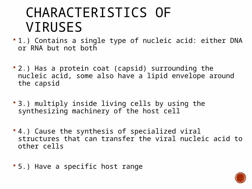
CHARACTERISTICS OF VIRUSES
1.) Contains a single type of nucleic acid: either DNA or RNA but not both
2.) Has a protein coat (capsid) surrounding the nucleic acid, some also have a lipid envelope around the capsid
3.) multiply inside living cells by using the synthesizing machinery of the host cell
4.) Cause the synthesis of specialized viral structures that can transfer the viral nucleic acid to other cells
5.) Have a specific host range

SIZE OF A VIRUS Usually much smaller than bacteria
must be smaller than the cells they infect: 20-14,000nm in length

SIZE CONTINUED

STRUCTURE OF VIRUSES
Virion = infectious viral particle: completely assembled with a protein coat surrounding the nucleic acid
All viruses are made of at least 2 parts
Inner core of nucleic acid
Enclosed in protein capsid
* Some also contain lipoprotein envelope

STRUCTURE OF VIRUSES 1.) Nucleic Acids:
Either DNA or RNA, but not both
Single or Double Stranded (SS or DS) if RNA, it can be plus sense strand (has codons) or
minus/antisense (need to make complement sense strand for translation)
If DNA- usually double stranded
Linear or circular
Genome is SMALL Only a few genes (most have 6-10 genes)

STRUCTURE OF VIRUSES CONTINUED 2. Capsid – protein coat (protein shell)
Surrounds the nucleic acid protects the virion in the external environment Aids in transfer between host cells Composed of subunits called capsomeres some capsids have protein-carbohydrate pointed
projections called pentons if pentons are present they are used for attachment to
the host cell

STRUCTURE OF VIRUSES *3. Envelope (not all viruses)
Function is to protect the virion some viruses have an envelope around the capsid consisting of
lipids, proteins and carbohydrates (cell membrane like) with envelope = enveloped virus
the envelope may be coded for by the virus or taken from the host cell plasma membrane
some envelopes have carbohydrate-protein complexes called spikes which are used for attachment to the host cell
if a virus does not have an envelope it is called a non-enveloped virus, “naked”
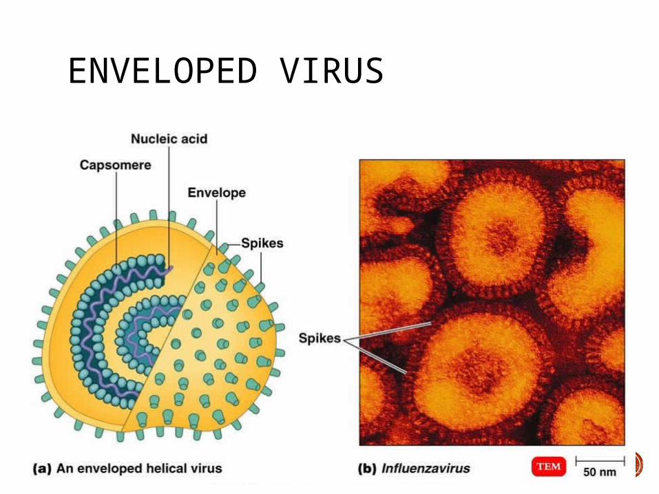
ENVELOPED VIRUS
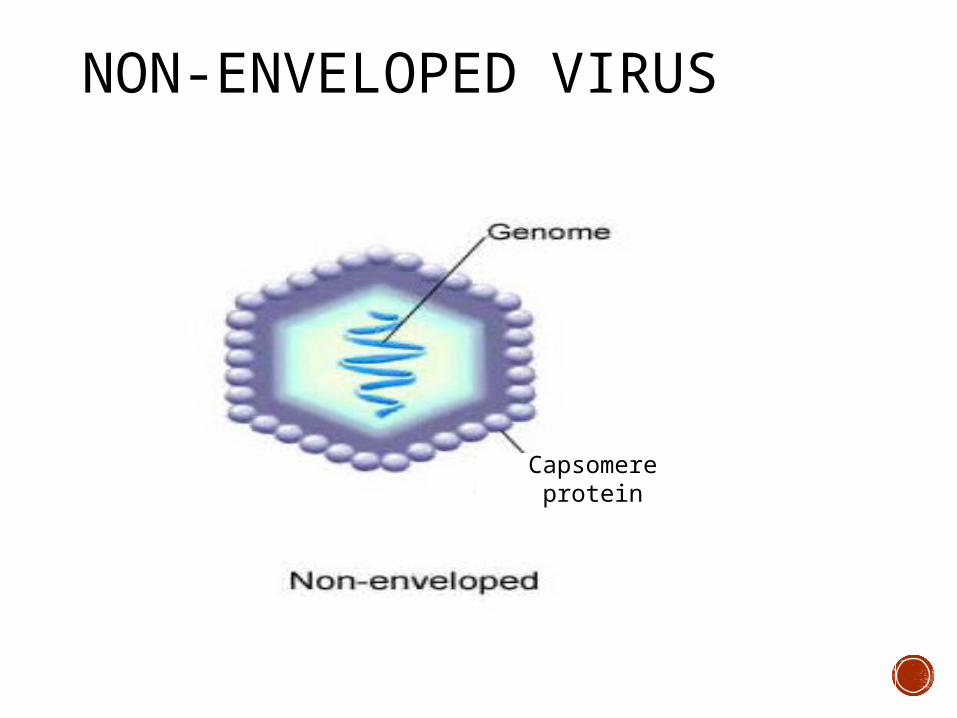
NON-ENVELOPED VIRUS
Capsomere protein
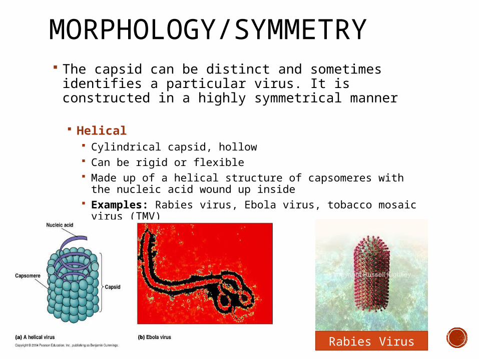
MORPHOLOGY/SYMMETRY The capsid can be distinct and sometimes identifies a
particular virus. It is constructed in a highly symmetrical manner
Helical Cylindrical capsid, hollow Can be rigid or flexible Made up of a helical structure of capsomeres with the nucleic
acid wound up inside Examples: Rabies virus, Ebola virus, tobacco mosaic virus
(TMV)
Rabies Virus

MORPHOLOGY/SYMMETRY
Polyhedral Most are icosahedrons (icosohedral)
20 equilateral triangle faces and made from capsomeres 12 corners made form capsomeres called pentons which
contain 5 protomers each Appear spherical Examples: Adenovirus, Polio virus
Polio virus

MORPHOLOGY/SYMMETRY Complex
Several types of symmetry in one virus Unique shape Examples:
Bacteriophage: capsid and accessory structure
Pox virus: no clear capsid, just several protein layers around the nucleic acid
Glass sculpture of pox virus
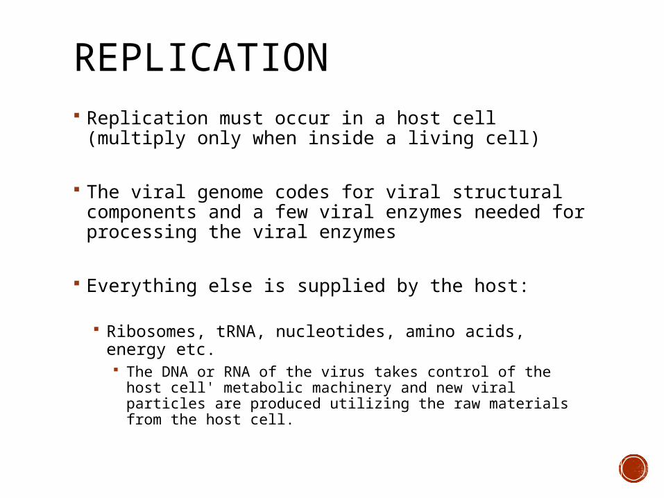
REPLICATION Replication must occur in a host cell (multiply only
when inside a living cell)
The viral genome codes for viral structural components and a few viral enzymes needed for processing the viral enzymes
Everything else is supplied by the host:
Ribosomes, tRNA, nucleotides, amino acids, energy etc. The DNA or RNA of the virus takes control of the host cell'
metabolic machinery and new viral particles are produced utilizing the raw materials from the host cell.

REPLICATION Replication of viruses is studied in great detail in
bacteriophages Bacteriophages are viruses that infect a specific bacteria
Two possible types of infection cycles:
1.) Lytic cycle (virulent) Ends with the lysis and death of the host bacterial wall
2.) Lysogenic cycle Host cell remains alive, but carries the virus in its genome
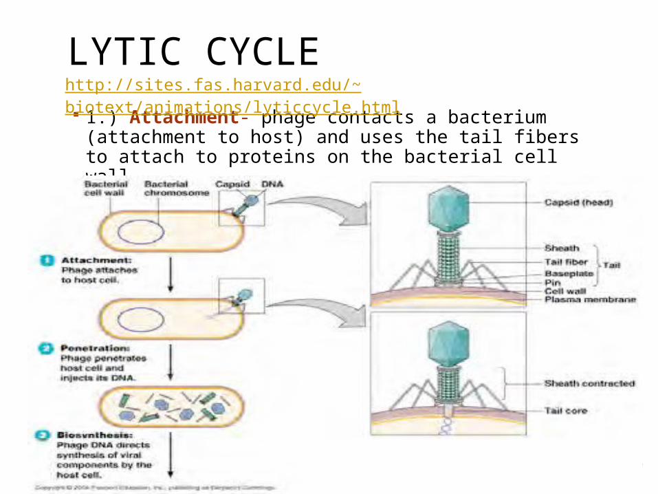
LYTIC CYCLE 1.) Attachment- phage contacts a bacterium
(attachment to host) and uses the tail fibers to attach to proteins on the bacterial cell wall
http://sites.fas.harvard.edu/~biotext/animations/lyticcycle.html

LYTIC CYCLE 2.) Penetration/Entry- the phage injects its DNA into
the bacterium The phage tail releases lysozyme to break down the
bacterial cell wall The sheath contracts to drive the tail core through the
weakened cell wall and plasma membrane The DNA is injected into the bacterium through the tail core
Uncoating- During or before penetration
3.) Synthesis of new virus particles (Multiplication) Once inside, host protein synthesis is stopped Virus has host make proteins and nucleic acid Virus directs viral nucleic acid replication and transcriptions
and translation of viral genes (host’s cell transcription stops) This results in a pool of viral genomes and capsid parts

LYTIC CYCLE 4.) Assembly
“eclipse period” – the time of viral entry The bacteriophage DNA and capsid spontaneously
assemble into complete virons 5-10 hrs DNA viruses 2-10 hrs RNA viruses

LYTIC CYCLE 5.) Lysis- release of virus and death of host cell
A single virus can give rise to up to 1000 new virus particles from on host cell
Virions will leave bacteria (host) Lysozyme encoded by viral genes causes the cell wall to
break down The bacteria lyses releasing the virions
Cycle will then repeat with new phages
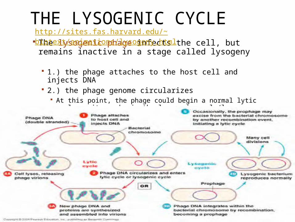
THE LYSOGENIC CYCLE The lysogenic phage infects the cell, but remains
inactive in a stage called lysogeny
1.) the phage attaches to the host cell and injects DNA 2.) the phage genome circularizes
At this point, the phage could begin a normal lytic cycle or it can begin the lysogenic cycle/lysogeny
http://sites.fas.harvard.edu/~biotext/animations/lysogeny.html

THE LYSOGENIC CYCLE
Latency- “dormant” state- unpredictability Viral DNA/RNA integrated into DNA of host = hidden
DNA=provirus
Can be reactivated in the future Factors that influence: stress, other viral infections, UV light
Example: fever blisters, chicken pox, HIV 2+ years
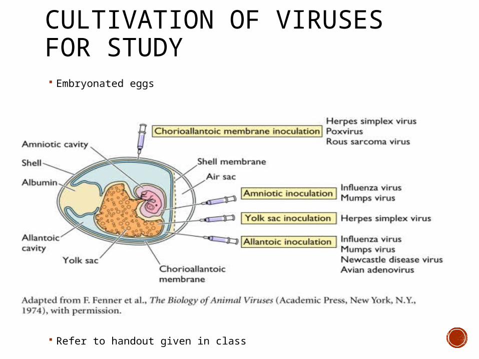
CULTIVATION OF VIRUSES FOR STUDY Embryonated eggs
Refer to handout given in class

CELL CULTURES
Refer to handout given in class

ANIMAL MODELS
Refer to handout given in class