Three major functions: › 1. Receive sensory input Gather info by monitoring changes or stimuli...
84
Nervous System The Brains of the Operation
-
Upload
claud-lambert -
Category
Documents
-
view
216 -
download
0
Transcript of Three major functions: › 1. Receive sensory input Gather info by monitoring changes or stimuli...
Nervous SystemThe Brains of the Operation

A.Functions Three major functions:
› 1. Receive sensory input Gather info by monitoring changes or
stimuli from inside & outside body› 2. integration of input
Process & interpret sensory input & determine action
› 3. motor output Carry out response decided by integration
usually by muscles (movement) or glands (secretion)
Remember this from Intro Unit?
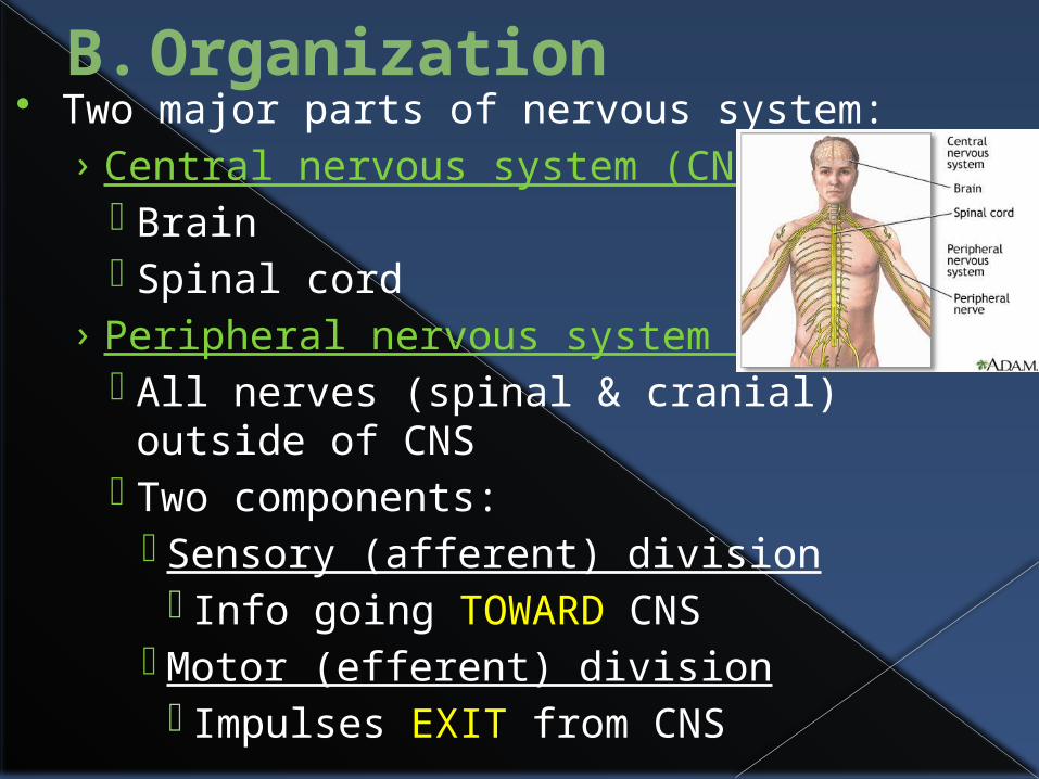
B. Organization Two major parts of nervous system:
› Central nervous system (CNS) Brain Spinal cord
› Peripheral nervous system (PNS) All nerves (spinal & cranial) outside of
CNS Two components:
Sensory (afferent) division Info going TOWARD CNS
Motor (efferent) division Impulses EXIT from CNS

Two subdivisions of motor (efferent): Somatic nervous system – voluntary
Conscious control of skeletal muscles Autonomic nervous system - involuntary Controls smooth/cardiac muscle & glands

Two subdivisions of autonomic that often bring about opposite effects: Parasympathetic – stimulate rest & digest activities Ex: stimulate flow of saliva
Sympathetic – stimulate flight or fight activities Ex: inhibit flow of saliva
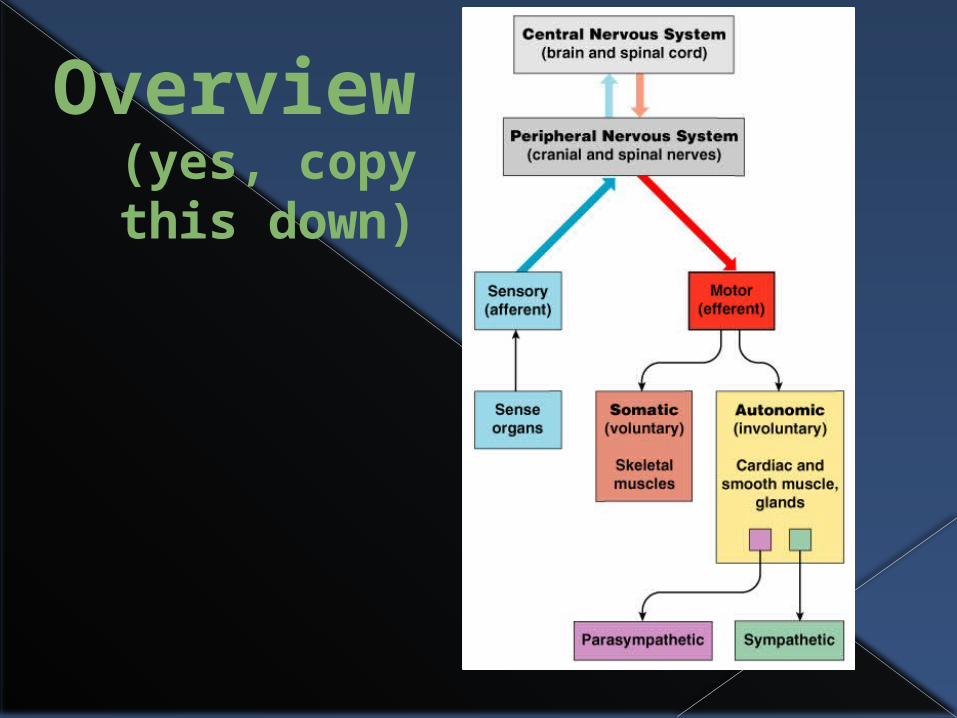
Overview(yes, copy this down)
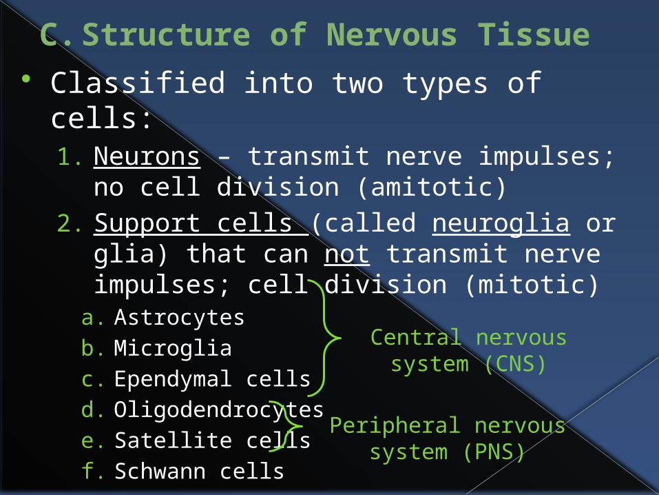
C. Structure of Nervous Tissue Classified into two types of cells:
1. Neurons – transmit nerve impulses; no cell division (amitotic)
2. Support cells (called neuroglia or glia) that can not transmit nerve impulses; cell division (mitotic)
a. Astrocytesb. Microgliac. Ependymal cellsd. Oligodendrocytese. Satellite cellsf. Schwann cells
Central nervous system (CNS)
Peripheral nervous system (PNS)

1. Neurons Also known as nerve cells Structure allows them to
receive & transmit messages or impulses
All have same basic structures› Cell body – usual cell organelles including nucleus
except no centrioles (no mitosis)› Processes/fibers – arms that extend to/from body
› To body = dendrites (1-100s)› From body = axon (only 1)
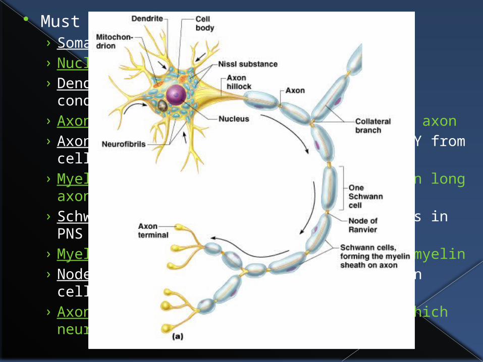
Must know cell parts:› Soma - cell body› Nucleus – metabolic center of cell› Dendrite(s) – one or more processes that conducts
impulses TOWARD cell body› Axon hillock – where cell branches out to axon› Axon – process that conducts impulses AWAY from
cell body› Myelin – whitish, fatty substance found on long
axons in CNS; speeds up transmission rate › Schwann cells – cells that myelinate axons in PNS › Myelin sheath – membranes wrapped around
myelin› Nodes of Ranvier – gaps in between Schwann cells› Axon terminal – branched end of axon in which
neurotransmitters are stored in vesicles

Large concentrations of cell bodies in CNS are in clusters & called nuclei
Small concentrations are called ganglia (pl) or ganglion (sing) – found in CNS & PNS
Bundles of nerve fibers in CNS called tracts, but in PNS are called nerves› Myelinated nerve fibers/tracts in CNS
called white matter › Unmyelinated fibers and cell bodies called
gray matter

Three types of neurons based on function or direction of nerve impulse:› Sensory (afferent) neurons
Carry impulses from sensory receptors TOWARD CNS Nerve endings – pain & temperature
receptors Meissner’s corpuscle – touch receptor Pacinian corpuscle – deep pressure receptor Proprioceptors – stretch or tension in
tendons & muscles› Motor (efferent) neurons
Carry impulses FROM CNS to organs or muscles› Association neurons/interneurons
Connect motor & sensory neurons
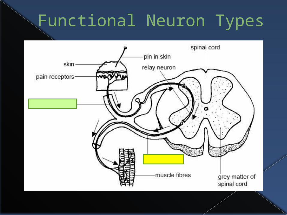
Functional Neuron Types
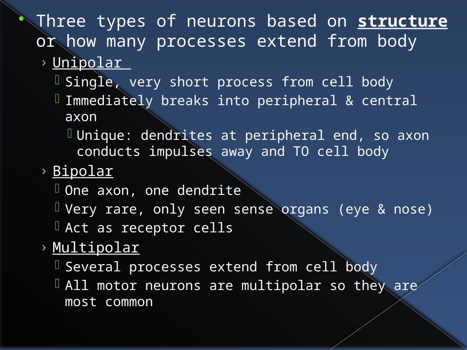
Three types of neurons based on structure or how many processes extend from body› Unipolar
Single, very short process from cell body Immediately breaks into peripheral & central axon
Unique: dendrites at peripheral end, so axon conducts impulses away and TO cell body
› Bipolar One axon, one dendrite Very rare, only seen sense organs (eye & nose) Act as receptor cells
› Multipolar Several processes extend from cell body All motor neurons are multipolar so they are most
common

Structural Neuron Types

2. Support Cells2a. Astrocytes
Star-shaped Account for ~ 50% of
neural tissue Form living barrier
between capillaries & neurons therefore make exchanges between them› Help protect neurons from harmful
substances› Pick up extra ions› Recapture released neurotransmitters

2b. Microglia Spiderlike phagocytes (cell eaters)
› Dispose of debris like dead brain cells & bacteria

2c. Ependymal Cells Covered with cilia Line cavity of brain & spinal cord
› Beating of cilia help circulate cerebrospinal fluid that fills brain & spinal cord cavity
› Forms protective cushion around CNS

2d. Oligodendrocyte Wrap flat extensions tightly around
nerve fibers› Produces fatty insulating covering of axons
called myelin sheaths in CNS
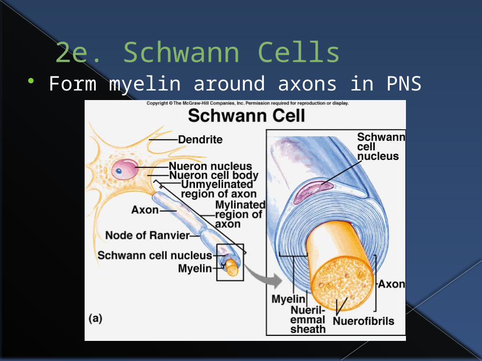
2e. Schwann Cells Form myelin around axons in PNS

2f. Satellite Cells Protective, cushioning cell body in PNS

Nerve Cells of CNS

D. Nerve Impulse Recall a neuron has two distinct properties that
differentiate it from any other cell in the human body:› Irritability - ability to respond to stimuli & convert it
to a nerve impulse› conductivity - ability to transmit an impulse to
other neurons, muscles, or glands Most CNS neurons receive chemical stimulus at
plasma membrane (everywhere on neuron), transmits it as electrical signal along axon, & ends as chemical signal at axon terminals
Most PNS neurons (sensory organs) receive stimulus as light (eyes), sound waves (ears), pressure (touch), chemicals (taste), or chemicals (smell)

plasma membrane is where nerve impulse begins
Plasma membrane at rest is polarized› fewer positive ions (K+) are inside cell than
positive ions (Na+) outside cell› More negative (Cl-) ions inside cell than
outside
Na+ Na+Na+ Na+Na+
Na+
K+K+K+ K+
K+
--
-
-- -
K+
K+
K+
K+- - -
-
-
-
--
Na+ Na+Na+
Na+Na+ Na+
Na+ Na+ Na+Na+ Na+ Na+
Na+ Na+ Na+
-
--
-
--
-
- - -- -
-
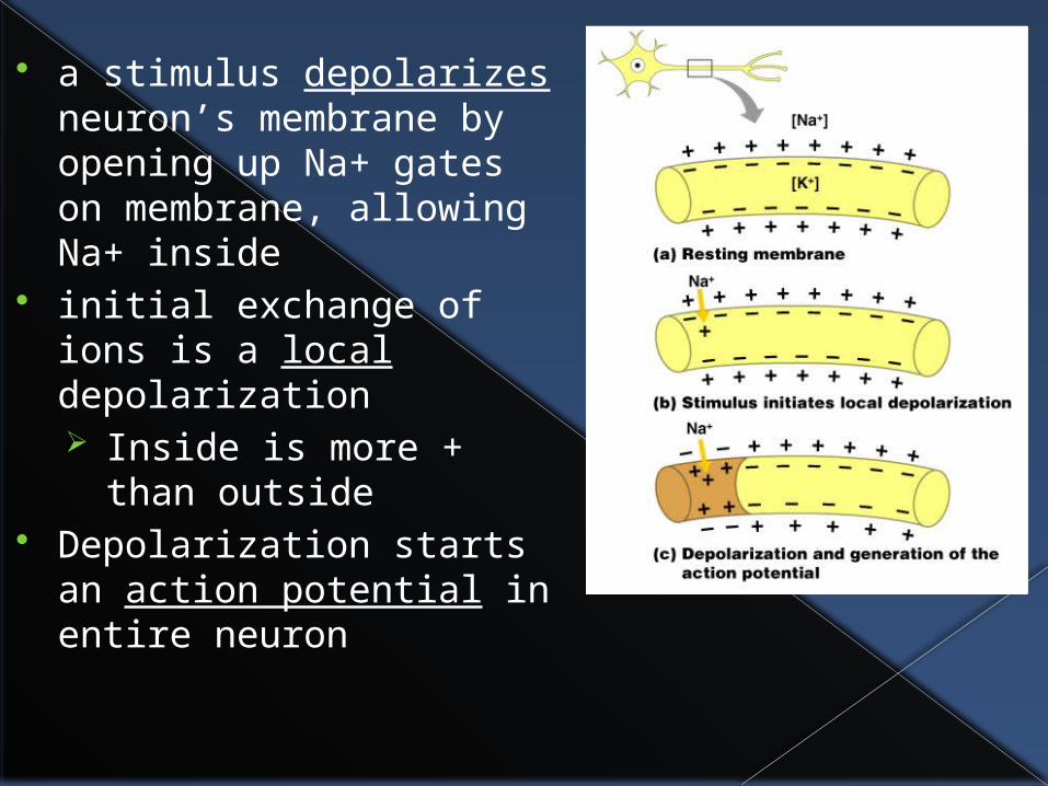
a stimulus depolarizes neuron’s membrane by opening up Na+ gates on membrane, allowing Na+ inside
initial exchange of ions is a local depolarization Inside is more + than
outside Depolarization starts an
action potential in entire neuron

Once action potential (nerve impulse) starts, it’s propagated over entire axon (all or nothing principle)
K+ ions rush out of the neuron after Na+ ions rush in, which repolarizes the membrane
Na+/K+ pump on membrane restores original configuration by shoving Na+ back out and allowing K+ back in› requires ATP
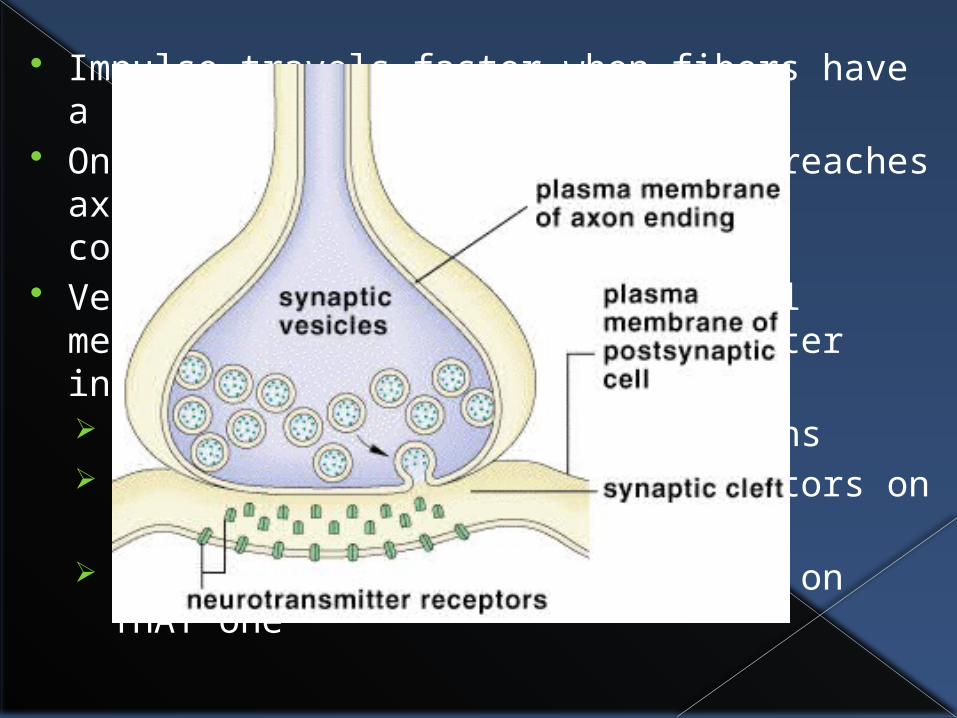
Impulse travels faster when fibers have a myelin sheath
Once electrical action potential reaches axon terminals, excites vesicles containing neurotransmitters
Vesicles move toward axon terminal membrane & releases neurotransmitter into synaptic cleft Neurons NEVER touch other neurons Neurotransmitters bind to receptors on
neighboring neuron’s dendrites New action potential will start on THAT one


Recap
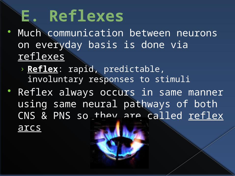
E. Reflexes Much communication between neurons
on everyday basis is done via reflexes› Reflex: rapid, predictable, involuntary
responses to stimuli Reflex always occurs in same manner
using same neural pathways of both CNS & PNS so they are called reflex arcs

Two types of reflexes:› Somatic: stimulates skeletal muscles
Ex: pull hand away from hot object, blinking when air burst aimed at eyes
› Autonomic: regulate smooth & cardiac muscles, & glands Ex: secretion of saliva, change in pupil
size

Reflex arcs have at least 5 elements involved in same arc or pattern:1. Sensory receptor – react to stimulus2. Sensory neuron – connect receptor & CNS3. Integration center (brain) – connect neurons4. Motor neuron – connect CNS & effector5. Effector organ – muscle/gland to be
stimulated

patellar (knee-jerk) reflex is simplest type of reflex – two neurons involved
Withdrawal reflex (remove from painful stimulus) is more complicated – three neurons involved utilizing association neuron

F. Brain Structure & Functions
Average adult brain weighs 3 lbs Divided into 4 regions:
1. Cerebrum – largest region, broken into left & right hemispheres
2. Diencephalon – interbrain atop brain stem3. Brain stem – stalk on which brain sits,
connects to spinal cord4. Cerebellum – bulbous projection at occipital
region, broken into two hemispheres

1. Cerebrum Made of two
hemispheres together called cerebrum
Encloses other three parts of brain
Entire surface made of peaks and valleys› Gyrus (gyri) – peaks of
ridges› Sulcus (sulci) – shallow
valleys› Fissures – deep grooves
separating large regions
Function of cerebrum is vast› speech, memory, logical & emotional
response, consciousness, interpretation of sensation, voluntary movement
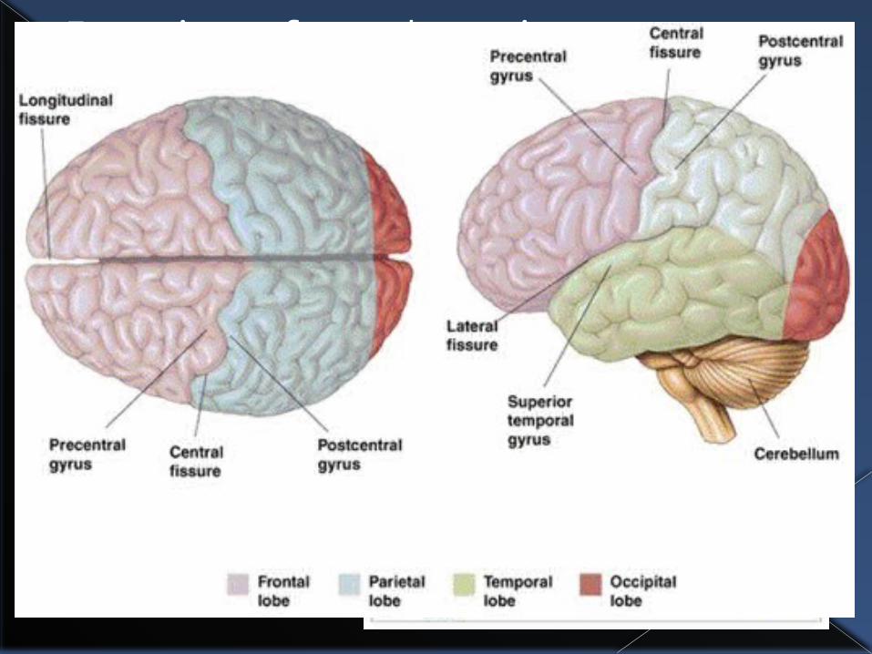
Sulci & fissures divide cerebrum into lobes (named after cranial bones)› Parietal lobe› Frontal lobe› Temporal lobe› Occipital lobe

Parietal Lobe› Somatic sensory area located just posterior
to central sulcus receives & interprets impulses from body’s sensory receptors (NOT special senses) Pain, cold, light touch

Spatial map depicting region on body where senses come from and how much brain power is devoted to them is called sensory homunculus
Model depiction showing areas of body given more
brain “power” than others

› Sensory pathways are crossed pathways, meaning left side of brain receives impulses from right side of body & vice versa Itch on right hand interpreted on left side of
somatic sensory area.

Occipital lobe› Visual area located in posterior part
Temporal lobe› Auditory area bordering lateral sulcus› Olfactory (smell) area deep inside

Frontal lobe› Contains primary motor area, just anterior to
central sulcus, which allows us control of skeletal muscles
› Spatial map region calledmotor homunculus
› Broca’s area – located inleft hemisphere gives ability to speak
› Higher intellectual reason› Socially acceptable
behavior› Language comprehension

Sensory & Motor Areas of Cerebral Hemisphere


Two layers of cerebral hemisphere:› Gray matter (cerebral cortex)
Outermost layer made out of cell bodies of neurons (no myelin)
Ridges allow greater surface area, increasing amount of neurons
Several islands of gray matter that jut inward called basal ganglia
› White matter Deeper cerebral layer made from fiber tracts
(bundles of nerve fibers) Major tract called corpus callosum connects
right & left cerebral hemisphere


2. Diencephalon AKA interbrain, made of 3 areas:
› Thalamus – relay station for sensory impulses going up to sensory cortex Get rough idea if sensation will be pleasant or
unpleasant – sensory cortex figures it out› Hypothalamus – regulates body temperature,
water balance (thirst), metabolism (appetite), sex, pain, pleasure, pituitary gland Pituitary gland is attached & secretes
hormones› Epithalamus – pineal gland(secretes
hormones) & choroid plexus (knots of capillaries that form cerebrospinal fluid)

Diencephalon

3. Brain Stem Made of 3 structures:
› Midbrain – reflex centers for vision & hearing
› Pons – fiber tracts that control breathing› Medulla oblongata – control heart rate,
blood pressure, breathing, swallowing, vomiting
Many small gray matter areas that control breathing, blood pressure
Running along length is reticular formation which regulates consciousness, awake/sleep cycles› Damage here results in permanent
unconsciousness or coma

4. Cerebellum Two hemispheres & wrinkly (convoluted)
surface› Outer cortex is gray matter while inner region
is white matter called arbor vitae (tree of life) Provides timing for muscle activity,
controls balance & equilibrium› Constantly monitors body position & makes
adjustments to keep balance
StructureSubdivision
Function
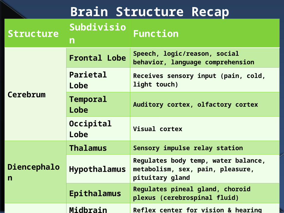
Cerebrum
Frontal Lobe Speech, logic/reason, social behavior, language comprehension
Parietal Lobe
Receives sensory input (pain, cold, light touch)
Temporal Lobe
Auditory cortex, olfactory cortex
Occipital Lobe
Visual cortex
Diencephalon
Thalamus Sensory impulse relay station
Hypothalamus
Regulates body temp, water balance, metabolism, sex, pain, pleasure, pituitary gland
Epithalamus Regulates pineal gland, choroid plexus (cerebrospinal fluid)
Brain Stem
Midbrain Reflex center for vision & hearing
Pons breathing
Medulla oblongata
Controls heart rate, blood pressure, breathing, swallowing, vomiting
Cerebellum none Muscle coordination, balance, equilibrium
Brain Structure Recap

G. Protection of CNS As nervous tissue is very soft and delicate,
injury to irreplaceable neurons can be catastrophic
Three methods of protection:› Bony skull & vertebral column› Membranes› Cerebrospinal fluid

Membranes Three connective tissue membranes called
meninges cover & protect CNS› Top: Dura mater (“tough
mother”) Periosteal layer (touches
skull) Meningeal layer
› Middle: Arachnoid mater (“spider mother”) Looks like a cobweb
› Bottom: Pia mater (“gentle mother”) Clings gently but tightly to
brain surface

CSF (Cerebrospinal Fluid) Watery broth similar to blood plasma Constantly formed by choroid plexuses
› Little protein, lots of vitamin C, lots of ions Always circulating among ventricles,
canals, & aqueducts in brain› Spinal tap removes CSF from lumbar area

Brain can not handle tiniest fluctuations of chemicals (all kinds) as other organs can
As result, neurons are kept separated from blood borne substances by “blood-brain barrier” which is composed of least permeable capillaries in human body› Only water, glucose, essential amino acids, fats,
respiratory gases, and fat-soluble alcohols, nicotine, caffeine, and anesthetics can pass
› Metabolic wastes (urea), toxins, proteins, most drugs are prevented
› Nonessential amino acids & K, are always pumped from brain


The other component of CNS, it’s a two-way conduction pathway from PNS & brain composed of neurons with long axons
Reflex center where reflexes are determined
H. Spinal Cord

17” long spinal cord is continuation of brain stem ending at L2
Starting at L3, branched into 31 pairs of spinal nerves exit vertebral column called cauda equina (horse’s tail)
Covered by meninges for protection

Gray matter of spinal cord resembles butterfly› Posterior projections = posterior/dorsal horns› Anterior projections = ventral/anterior horns› Gray matter surrounds central canal which
contains CSF› Spinal (nerve) fibers entering spinal cord
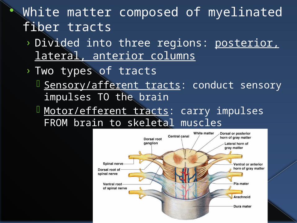
White matter composed of myelinated fiber tracts› Divided into three regions: posterior, lateral,
anterior columns› Two types of tracts
Sensory/afferent tracts: conduct sensory impulses TO the brain
Motor/efferent tracts: carry impulses FROM brain to skeletal muscles

I. Peripheral Nervous System (PNS) Consists of nerves &
scattered groups of ganglia found outside CNS
Nerve is bundle of neuron fibers not in CNS› Neuron fibers (processes)
surrounded by endoneurium
› Groups of fibers bound by perineurium
› Whole bundles called fascicles
› Fascicles bound together by epineurium

Nerves carrying both sensory & motor fibers called mixed nerves› All spinal nerves are
mixed› Sensory (afferent)
nerves – toward CNS
› Motor (efferent) nerves – away from CNS
Cranial nerves – 12 pairs that serve head and neck
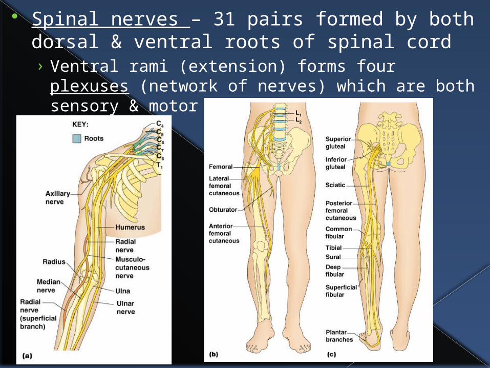
Spinal nerves – 31 pairs formed by both dorsal & ventral roots of spinal cord› Ventral rami (extension) forms four plexuses
(network of nerves) which are both sensory & motor

Three nerves to know:› Sciatic nerve
part of sacral plexus Largest nerve in
body Serves lower trunk &
posterior thigh/leg Inflammation or
damage causes sciatica

› Median nerve Part of brachial
plexus Allows flexion of
forearm & some hand muscles
Pressure on nerve from tendon causes carpal tunnel syndrome Inability to pick up small objects, fine motor control

› VII Facial nerve 7th cranial nerve Serves muscles for facial expression, salivary
& lacrimal (tear ducts) glands, taste buds Weakening or paralysis causes Bell’s palsy

J. Autonomic Nervous System Involuntary motor branch of PNS that controls smooth muscles, cardiac muscles, glands
Information from CNS activates nerves that release neurotransmitters which then signal appropriate muscle/gland

Recall two divisions of ANS that have opposite effects:› Sympathetic – extreme situations (fear,
exercise, rage)› Parasympathetic – rest & conserve energy
Three neurotransmitters in ANS:› Acetylcholine – both sympathetic &
parasympathetic › Epinephrine – sympathetic division› Norepinephrine – sympathetic division
K. Development Formation
› Nervous system formed during first 4 weeks of embryonic development
› Maternal infection or poor health habits may cause permanent damage Measles causes deafness Smoking decreases oxygen causing low
birth weight, others Drugs (OTCs & illegal) can permanently
damage

Maturation› Last areas of CNS to mature is
hypothalamus Preemies have problems controlling body temperature
› Throughout childhood, no neurons grow but in fact become myelinated, allowing neuromuscular control

Aging› Brain at maximum weight as young adult› Next 60+ years, neurons get damaged &
die Other unused pathways can take over &
be developed› Sympathetic nervous system becomes
less efficient› Premature shrinking of brain occurs when
individuals accelerate normal process with lifestyle Boxers, alcoholics, drug abusers

L. Diseases/Injuries Huntington’s Disease
› Dominant genetic disease (one dominant allele needed) for 50% chance of acquiring it
› Strikes in middle age (around 50) › Massive degeneration of basal nuclei then
cerebral cortex› Initial symptoms are wild, jerky movements
termed chorea (Latin for dance)› Usually fatal within 15 years of onset› Treated with neurotransmitter (dopamine)
blockers

Parkinson’s Disease› Degeneration of dopamine-releasing
neurons in substantia nigra (in midbrain) so basal nuclei dopamine targets becomes overactive, causing tremors
› Treatment with L-dopa drugs helps some symptoms, but after more neurons are affected, it is ineffective
› Newer (albeit controversial) treatments include transplanting embryonic substantia nigra tissue, or genetically engineered (stem cells), or cells from fetal pigs

Alzheimer’s› Progressive degenerative disease that
results in dementia (mental deterioration)› Nearly 50% of all people in nursing homes
have Alzheimer’s› Begins with short-term memory loss, short
attention span, disorientation, loss of language
› Result of shortage of acetylcholine & structural changes in brain (areas of cognition & memory)
› Microscopy of tissue shows abnormally large deposits of protein
› About 5-15% of people over 65 will get this
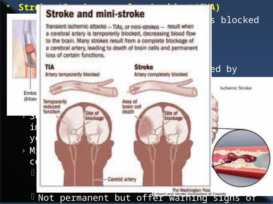
Stroke (Cerebrovascular Accident/CVA)› Blood circulation to brain area is blocked resulting in
tissue death Blood clot Ruptured blood vessel
› Area of tissue is initially located by looking at patient’s symptoms Left cerebral hemisphere results in aphasia
(language impairment)› Severe strokes kill 2/3 people almost immediately,
and remaining 1/3 die within 3 years› Mild strokes do not cut off blood flow completely
Called temporary brain ischemia or transient ischemic attack (TIA)
Not permanent but offer warning signs of CVA later
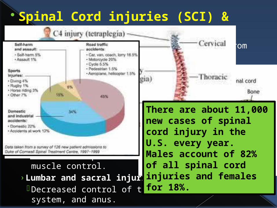
Spinal Cord injuries (SCI) & Paralysis› Any damage to the spinal cord resulting from crushing
or severing.› Cervical injuries
Cervical (neck) injuries usually result in full or partial tetra/quadriplegia.
› Thoracic injuries Injuries at or below the thoracic spinal levels result in
paraplegia. T1 to T8 : inability to control the abdominal
muscles. T9 to T12 : partial loss of trunk and abdominal
muscle control.› Lumbar and sacral injuries
Decreased control of the legs and hips, urinary system, and anus.
There are about 11,000 new cases of spinal cord injury in the U.S. every year. Males account of 82% of all spinal cord injuries and females for 18%.

Multiple Sclerosis› Autoimmune disease in which
myelin sheaths around axon fibers in CNS are gradually destroyed by own immune system
› Myelin converts to hardened sheaths called scleroses
› Lack of insulation leads to inability to control muscles
› Treatment today includes hormone-like substance called interferon
› Will result in complete inability to function

Meningitis› Inflammation of
meninges due to viruses or bacteria
› Can be life threatening since can spread to nervous tissue of CNS
› Diagnosed by spinal tap to look at CSF

M. Diagnosing Problems EEG (electroencephalogram)
› Assess electric activity of brain impulses› Many electrodes are placed on scalp and
measurement of activity pattern is recorded
› Used to diagnose epileptic lesions, tumors
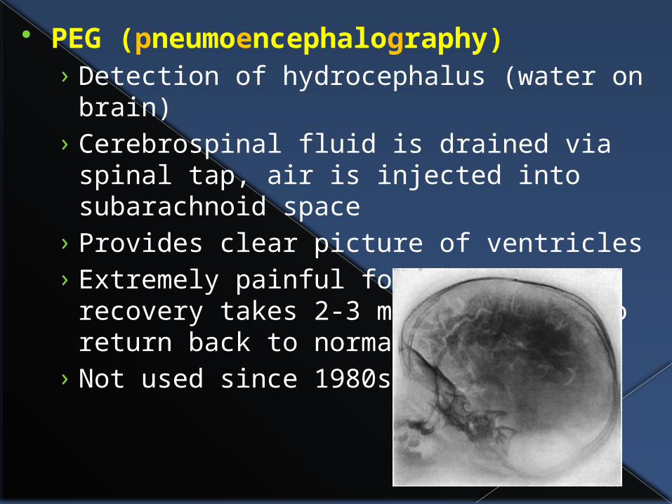
PEG (pneumoencephalography)› Detection of hydrocephalus (water on
brain)› Cerebrospinal fluid is drained via spinal tap,
air is injected into subarachnoid space› Provides clear picture of ventricles› Extremely painful for patients – recovery
takes 2-3 months for CF to return back to normal
› Not used since 1980s
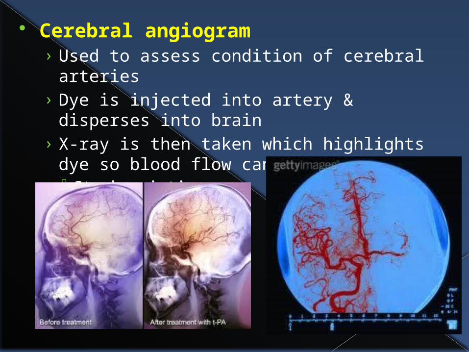
Cerebral angiogram› Used to assess condition of cerebral
arteries› Dye is injected into artery & disperses into
brain› X-ray is then taken which highlights dye so
blood flow can be assessed Stroke victims

Computed Tomography (CT or CAT) scan› Used to see tumors, lesions, MS or Alzheimer’s
plaques, infarcts (dead brain tissue)› Important in mapping brain prior to surgery

MRI scan (Magnetic Resonance Imagery)› Used to see tumors, lesions, MS or
Alzheimer’s plaques, infarcts (dead brain tissue)
› Similar to CT scan but 3D image capabilities

PET scan (Positron Emission Tomography)› Used to determine sugar (glucose) uptake/usage of
cells Faster growing cells (cancer) use sugar faster Active brain areas also use sugar faster
Alzheimer, Parkinson, epilepsy, tumors, dementia› Patient drinks glucose solution, areas of fast uptake
show up on image