-Schwann: neurons in PNS -Satellite cells: ganglia of PNS · -The human basal forebrain nucleus...
8
Neurohistology Location of cells -Schwann: neurons in PNS -Satellite cells: ganglia of PNS -Astrocyte: prominent in grey matter near synapses -Oligodendrocyte: prominent in white matter -Microglia: distributed in CNS -Ependymal: line ventricles -Meninges (Pia, Arachnoid, Dura): membranes, which surround the brain •Grey matter -Cell bodies, dendrites and axon of neurons -Synapses -Astrocytes -Many capillaries •White matter -Axons -Oligodendroglia -Not many astrocytes (fibrous) or capillaries •Common stains used for visualising neurons ▪Haematoxylin and Eosin -Grey matter – distribution can be defined but not connections -Cell nuclei the size, numbers, density and distribution of neurons can be approximated and seen -Glial and other cells are also stained (glia in particular cannot be readily differentiated)
-
Upload
truongduong -
Category
Documents
-
view
213 -
download
0
Transcript of -Schwann: neurons in PNS -Satellite cells: ganglia of PNS · -The human basal forebrain nucleus...

Neurohistology
Location of cells
-Schwann: neurons in PNS
-Satellite cells: ganglia of PNS
-Astrocyte: prominent in grey matter near synapses
-Oligodendrocyte: prominent in white matter
-Microglia: distributed in CNS
-Ependymal: line ventricles
-Meninges (Pia, Arachnoid, Dura): membranes, which surround the brain
•Grey matter
-Cell bodies, dendrites and axon of neurons
-Synapses
-Astrocytes
-Many capillaries
•White matter
-Axons
-Oligodendroglia
-Not many astrocytes (fibrous) or capillaries
•Common stains used for visualising neurons
▪Haematoxylin and Eosin
-Grey matter – distribution can be defined but not connections
-Cell nuclei the size, numbers, density and distribution of neurons can be approximated and seen
-Glial and other cells are also stained (glia in particular cannot be readily differentiated)

▪Nissl - toluidine blue and cresyl violet (basophilic stain)
-Nissl stains colour acidic components (DNA, RNA) and thus the nucleus and rER (DNA, RNA in nucleus/nucleolus and
RNA in ribosomes/ rER)
-The nuclei and rER of all cell types of the brain (microglia, macroglia, endothelial cells) are stained but neuronal
somata are more prominent due to rich Nissl bodies.
-Distinguishing neurons from other cell types (glia, and blood vessel-related cells)
-Grey matter - organisation
-More detail on cell body (particularly nucleus, nucleolus, Nissl substance), location, density
-Measuring their number, size and visualizing their organization (nuclei or layers, for example)
-Differentiate axon from dendrite and cell body – you may see extensions of soma merging into dendrite (where
protein synthesis occurs) but NOT dendritic arbor or axon.
▪Reduced silver
-Grey and white matter
-Clearly show axons, dendrites, chase pathways/ connection
-The extent to which axons can be traced is limited by the section thickness and process density

▪Golgi
-Stains only select neurons = isolated (not obscured by dense fibres) cells completely
-Labels an entire cell to enable morphological comparison of different cell types
-Enables thick sections to be looked at
-Clear cell body shape but not intracellular structures
-Clear dendritic arbor and even the fine dendritic spines (The dendritic spine is a protrusion of the dendrite which is
the receptive side of the synapse)
-Can show synaptic spines and glial cells
-The yellow hue and silver grains in high power shots

▪Weigert/ Myelin staining
-White matter
-Membrane of oligodendrocytes
-Myelin staining shows location of underlying axons
▪Microinjection/ Cell filling
By using micropipettes marker substances can be injected into individual neurons. The marker travels throughout
the cytoplasm and can reveal the appearance of the cell in particular the cell processes much in the way the Golgi
stain does. Microinjection is more selective and prior to staining the cells can be recorded from or stimulated and a
correlation made between cell shape, location and behaviour.
-A single neuron is labelled
-The entire morphology of the cell is clear: soma, dendrites and axons
-Nucleus is not distinguished
▪Immunostaining and Histochemistry
Neurons and glial cells contain proteins such as structural and receptor proteins, enzymes, nucleic acids and
transmitter substances (e.g. synaptotagmin, microtubules, neurofilaments, substance P receptors, glial fibril acid
protein) that can act as antigens or take part in chemical reactions. Some of these proteins and molecules are unique
to particular groups of cells. They can be visualized by reacting them with antibodies or in chemical reactions and

attaching other molecules (e.g. ones which fluoresce) to them which allow them to be seen under the microscope.
Several antibodies and reactions can be used on the one preparation so that subpopulations and correlations
between populations can be made.
-Astrocytes (Glia limitans, astrocytes, brain capillaries)
-Microglia (+ endothelial cells)
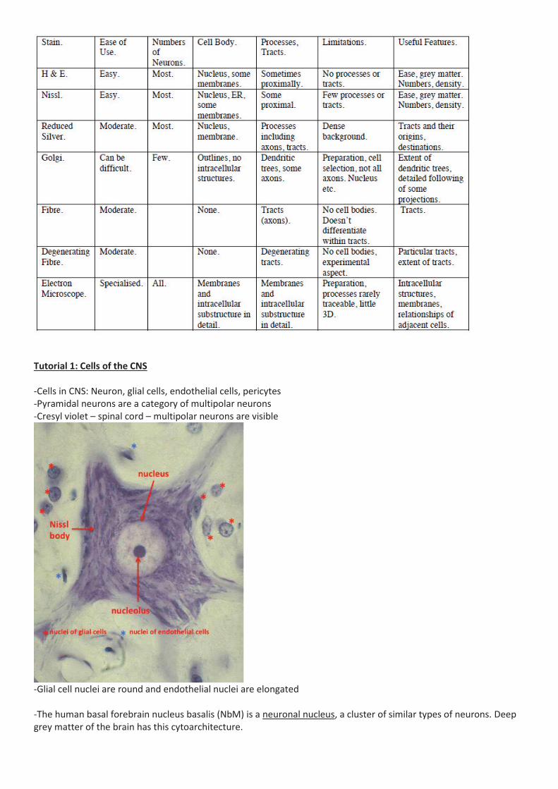
Tutorial 1: Cells of the CNS
-Cells in CNS: Neuron, glial cells, endothelial cells, pericytes
-Pyramidal neurons are a category of multipolar neurons
-Cresyl violet – spinal cord – multipolar neurons are visible
-Glial cell nuclei are round and endothelial nuclei are elongated
-The human basal forebrain nucleus basalis (NbM) is a neuronal nucleus, a cluster of similar types of neurons. Deep
grey matter of the brain has this cytoarchitecture.

The cerebral cortex is arranged in layers called laminae.

The cerebral cortical laminae have cytoarchitectural difference in neuronal size and layer thickness. The 6 cerebral
cortical laminae are numbered sequentially (I-VI) from the outermost layer I under glia limitans to layer VI which
abuts the underlying white matter. Subcortical white matter - sits inside the cerebral cortical mantle. It contains
axons exiting and entering the cortical layers.
Layer I -Thin layer
-No – few neuronal cell bodies
-High density of astrocytes
-Molecular layer contains the processes (apical dendrites of pyramidal neurons and horizontally-
oriented axons) of neurons
Layer II
Layer III
-Medium-sized, mainly pyramidal neurons
-Difficult to distinguish from one another
Layer IV -Small neurons
Layer V -Large, pyramidal neurons
Layer VI -Medium-sized neurons
-Grey matter = cell bodies: large blue dots/ triangles, nuclei of glia and endothelial cells: tiny blue dots
-White matter = nuclei of oligodendrocytes: tiny blue dots, NO neurons
-In Golgi stain, the visible cells are not the only cells. There are neurons and glia other than those seen. The visible
neurons aren’t the only neurons as Golgi stain only visualises a select subset of neurons (unobscured).
-Phagocytosis = A cell engulfs foreign particles or cells via internalization by the PM. Internalised material is
processed and digested in lysosomes.
-Endothelial cells make up small blood vessels (capillaries). These are joined by tight junctions, instead of being
fenestrated (not sealed by tight junctions and more permissive). This prohibits any diffusion of ion and forms the
blood brain barrier.