
بسم الله الرحمن الرحيم. Interpretation in·ter·pre·ta·tion DR.Tajuddin...
66
م ي ح ر ل ا ن م ح ر ل ه ا ل ل م ا س ب م ي ح ر ل ا ن م ح ر ل ه ا ل ل م ا س ب
-
Upload
dorthy-riley -
Category
Documents
-
view
221 -
download
2
Transcript of بسم الله الرحمن الرحيم. Interpretation in·ter·pre·ta·tion DR.Tajuddin...

الرحيم الرحمن الله الرحيم بسم الرحمن الله بسم
Interpretation
in·ter·pre·ta·tion
DR.Tajuddin Malabarey
Associate professor
365-March 2011


Interpretation• Something that serves to explain or clarify:
Clarification, Explanation, Illumination, Illustration.
• The act or process of explaining the meaning of something.

Interpretation
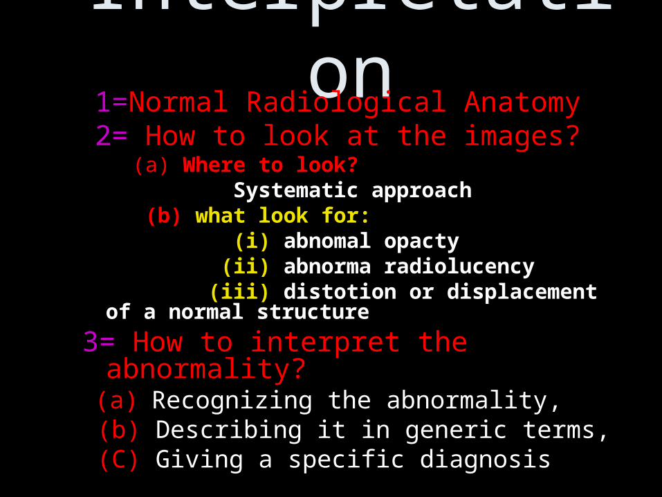
Interpretation 1=Normal Radiological Anatomy 2= How to look at the images? (a) Where to look? Systematic approach (b) what look for: (i) abnomal opacty (ii) abnorma radiolucency (iii) distotion or displacement of a normal
structure
3= How to interpret the abnormality? (a) Recognizing the abnormality, (b) Describing it in generic terms, (C) Giving a specific diagnosis

• Normal radiological image of certain age and sex is a mental image that must be developed
Normal Radiological Anatomy

• By developing a systematic approch to examine the radiological image
• Advantages:• Minimizes the chance of missing an
abnormality• Makes complex images easier to read
with practice• Builds up a mental databank of what is
normal
How to build up a normal mental image
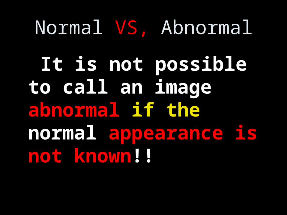
Normal VS, Abnormal
It is not possible to call an image abnormal if the normal appearance is not known!!


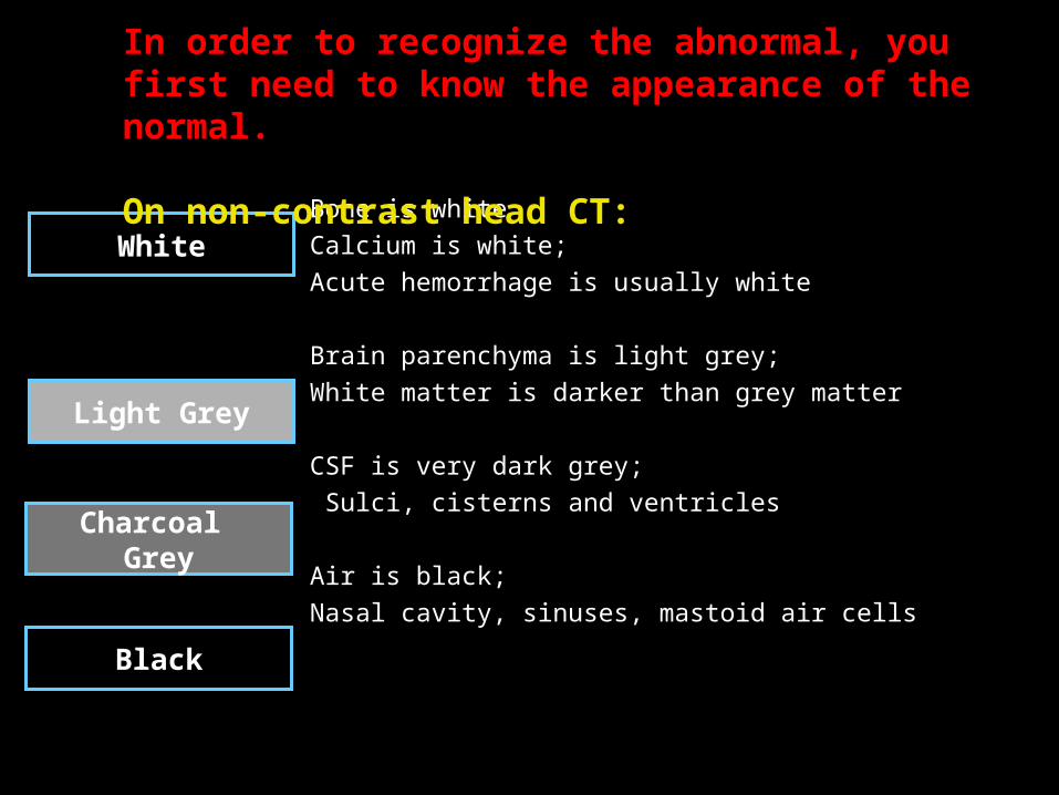
Bone is white
Calcium is white;
Acute hemorrhage is usually white
Brain parenchyma is light grey;
White matter is darker than grey matter
CSF is very dark grey;
Sulci, cisterns and ventricles
Air is black;
Nasal cavity, sinuses, mastoid air cells
White
Light Grey
Charcoal Grey
Black
In order to recognize the abnormal, you first need to know the appearance of the normal.
On non-contrast head CT:
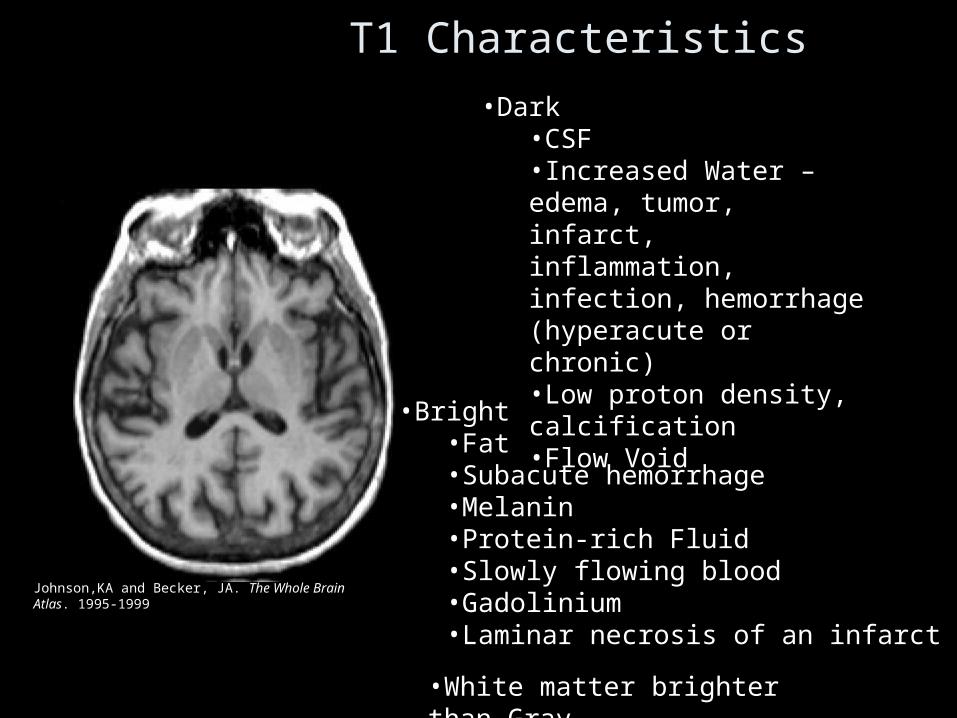
T1 Characteristics
•Dark•CSF•Increased Water – edema, tumor, infarct, inflammation, infection, hemorrhage (hyperacute or chronic)•Low proton density, calcification•Flow Void
•Bright•Fat•Subacute hemorrhage•Melanin•Protein-rich Fluid•Slowly flowing blood•Gadolinium•Laminar necrosis of an infarct
•White matter brighter than Gray
Johnson,KA and Becker, JA. The Whole Brain Atlas. 1995-1999
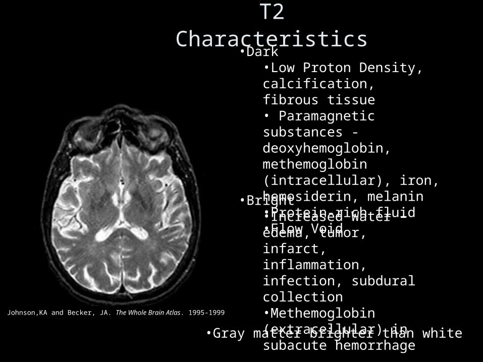
T2 Characteristics•Dark
•Low Proton Density, calcification, fibrous tissue• Paramagnetic substances - deoxyhemoglobin, methemoglobin (intracellular), iron, hemosiderin, melanin •Protein-rich fluid•Flow Void
•Bright•Increased Water – edema, tumor, infarct, inflammation, infection, subdural collection•Methemoglobin (extracellular) in subacute hemorrhage
•Gray matter brighter than white
Johnson,KA and Becker, JA. The Whole Brain Atlas. 1995-1999

Interpretation• Learn Normal Radiological
Anatomy
• How to look at the images?
• How to interpret the abnormality?
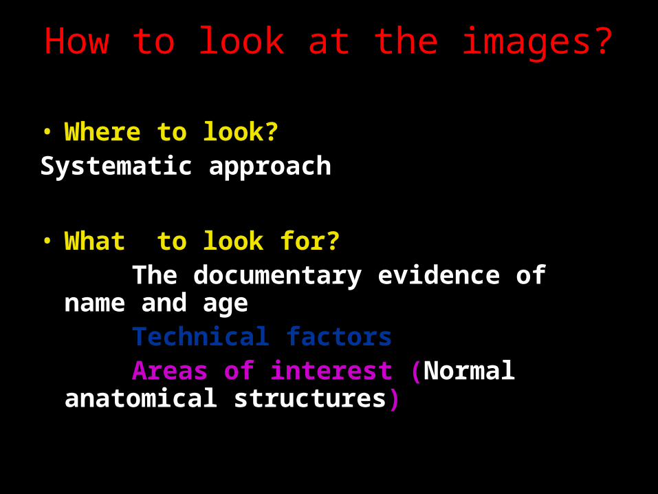
How to look at the images?
• Where to look?Systematic approach
• What to look for? The documentary evidence of name and
age Technical factors Areas of interest (Normal anatomical
structures)
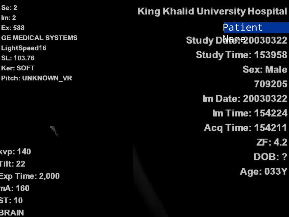
Patient Name
• What to look for?
3.Areas of interest (Normal anatomical structures)

Systematic Approach to reading a Head CT
I. Check Brain Parenchyma• Check grey/white differentiation• Gyri• Look for blood• Surgeons need to know . . . (size of hematoma, extent of
midline shift, herniation)
II. Check CSF spaces: Ventricles, Cisterns and Sulci• CSF spaces (ventricles and cisterns)
– size, symmetry, midline shift– herniation
• Subfalcine – cingulate gyrus crosses falx• Transtentorial – temporal lobe into tentorial notch • Cerebellar – cerebellum into foramen magnum
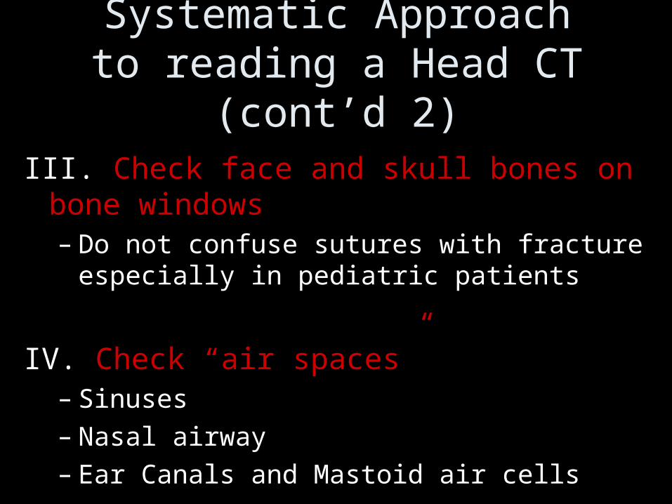
Systematic Approach to reading a Head CT (cont’d 2)
III. Check face and skull bones on bone windows– Do not confuse sutures with fracture especially in
pediatric patients
IV. Check “air spaces”– Sinuses– Nasal airway– Ear Canals and Mastoid air cells

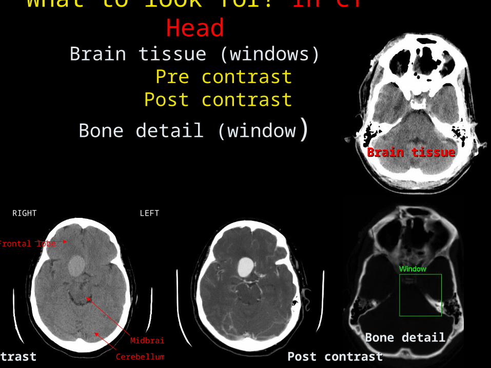
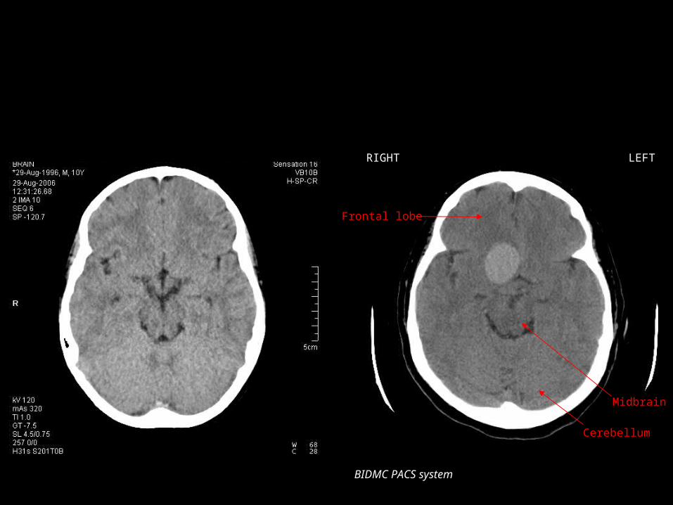
What to look for? In CT HeadBrain tissue (windows)
Pre contrast Post contrast
Bone detail (window)
Frontal lobe
Midbrain
Cerebellum
RIGHT LEFT
Pre contrast Post contrastPre contrast Post contrast
Bone detailBone detail
Brain tissueBrain tissue

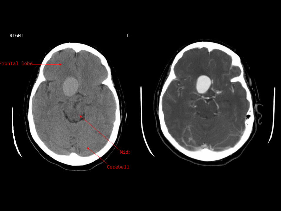
what look for: (i) abnomal opacty
(ii) abnorma radiolucency (iii) distotion or displacement of a normal structure
Frontal lobe
Midbrain
Cerebellum
RIGHT LEFT
Normal
abnomal opactyabnomal opacty
abnorma radiolucencyabnorma radiolucency
distotion or displacement of a normal structuredistotion or displacement of a normal structure
3= How to interpret the abnormality?
(a) Recognizing the abnormality,
(b) Describing it in generic terms,
(C) Giving a specific diagnosis
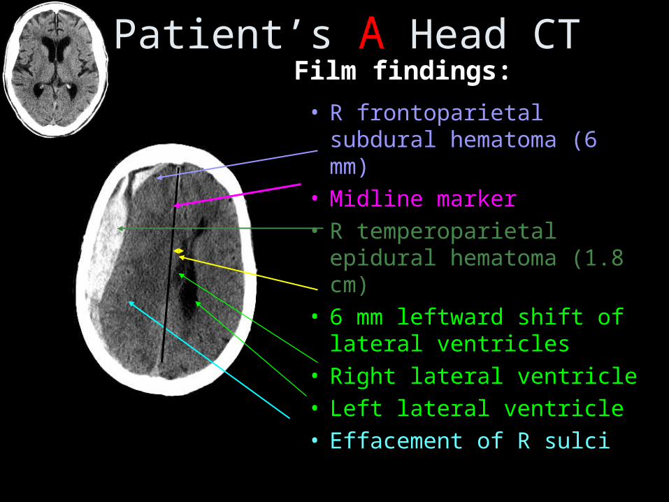
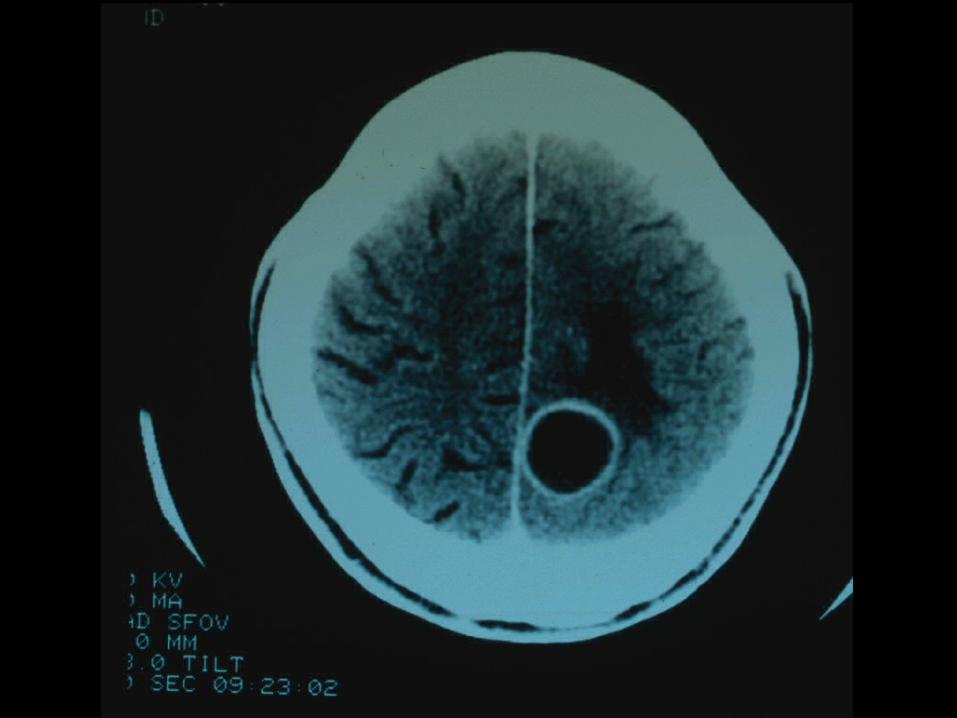
Patient’s A Head CT
• R frontoparietal subdural hematoma (6 mm)
• Midline marker• R temperoparietal epidural
hematoma (1.8 cm)• 6 mm leftward shift of
lateral ventricles• Right lateral ventricle• Left lateral ventricle• Effacement of R sulciBIDMC
Film findings:

Parenchymal Parenchymal HemorrhageHemorrhage
Subarachnoid Subarachnoid HemorrhageHemorrhage
Subdural Subdural HematomaHematoma
EpiduralEpidural

Patient B
• 57yr old woman
• History of migraines
• Presents with persistent headache– several months duration– different from her usual headache
Need to rule out intracranial abnormality
BIDMC PACS system
Frontal lobe
Midbrain
Cerebellum
RIGHT LEFT

Patient’s B Head CT (no contrast)
BIDMC PACS system
Frontal lobe
Midbrain
Cerebellum
RIGHT LEFTFilm Findings:• Spherical mass • Smooth margined• High attenuation• Slight mass effect• Located just
anterior to the Circle of Willis
• No acute hemorrhage, edema, infarct
Frontal lobe
Midbrain
Cerebellum
RIGHT LEFT
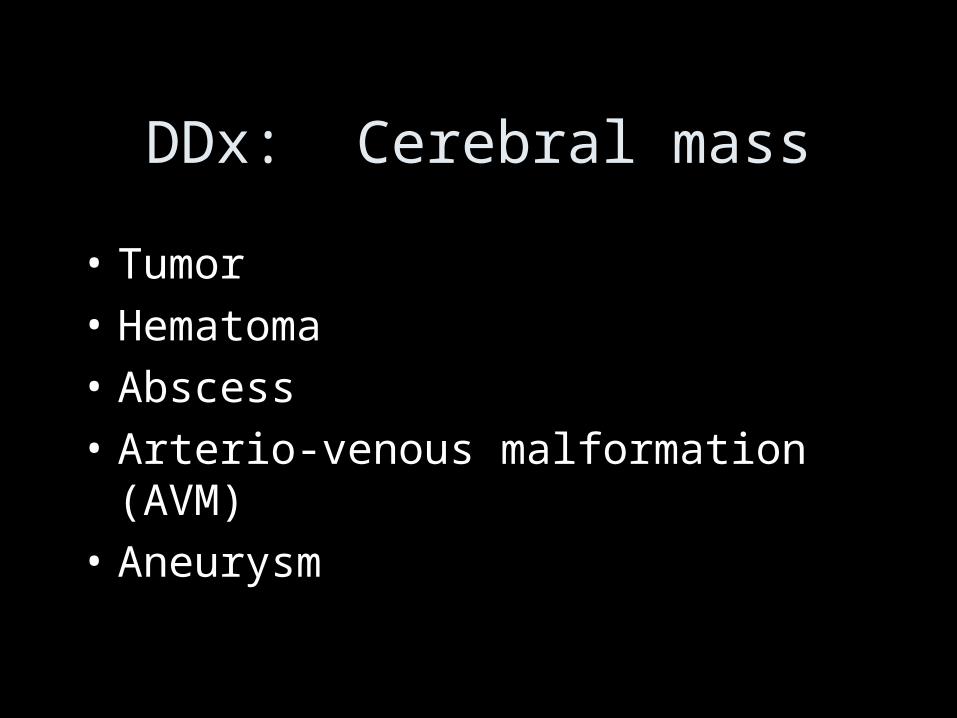
DDx: Cerebral mass
• Tumor
• Hematoma
• Abscess
• Arterio-venous malformation (AVM)
• Aneurysm
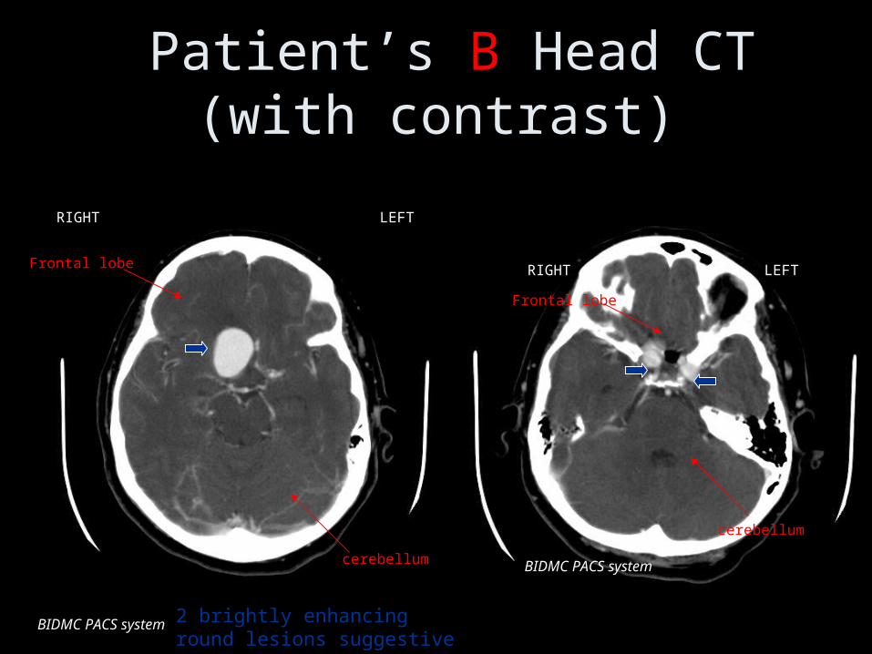
Patient’s B Head CT (with contrast)
BIDMC PACS system
RIGHT LEFT
BIDMC PACS system
Frontal lobe
cerebellum
Frontal lobe
cerebellum
RIGHT LEFT
2 brightly enhancing round lesions suggestive of cerebral aneurysms

Lets review the anatomy of the Circle of Willis
http://www.strokecenter.org/education/ais_vessels/ais048.html
• Communicating system of vessels that supplies blood to the brain
• Anterior portion fed by the internal carotid arteries
• Posterior portion fed by the vertebral arteries

Patients B Axial MR (T2 sequence)
BIDMC PACS system BIDMC PACS system
RIGHT LEFTRIGHT LEFT
T2 sequence: CSF is bright (“high signal”)
Round lesions with flow void confirmed
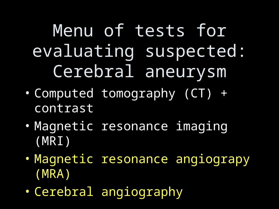
Menu of tests for evaluating suspected: Cerebral
aneurysm• Computed tomography (CT) + contrast
• Magnetic resonance imaging (MRI)
• Magnetic resonance angiograpy (MRA)
• Cerebral angiography

MRA - Circle of Willis
BIDMC PACS system
http://www.strokecenter.org/education/ais_vessels/ais048.html
b
Internal carotid artery aneurysms
asi
la
r
vertebral arteries
internalcarotid
internalcarotid
ACA
MCA
PCA
RIGHT LEFT
Our PatientAnatomic Diagram

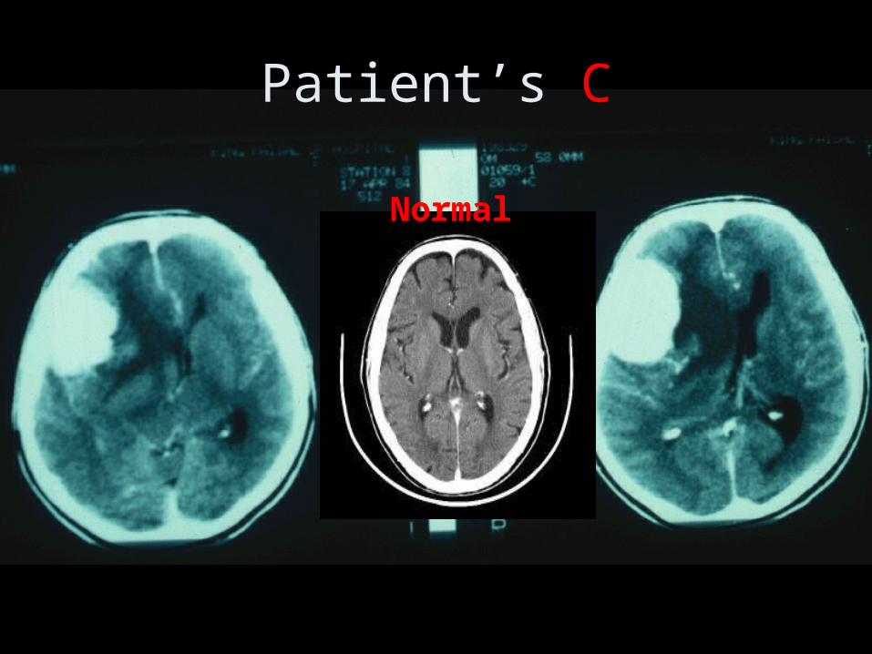
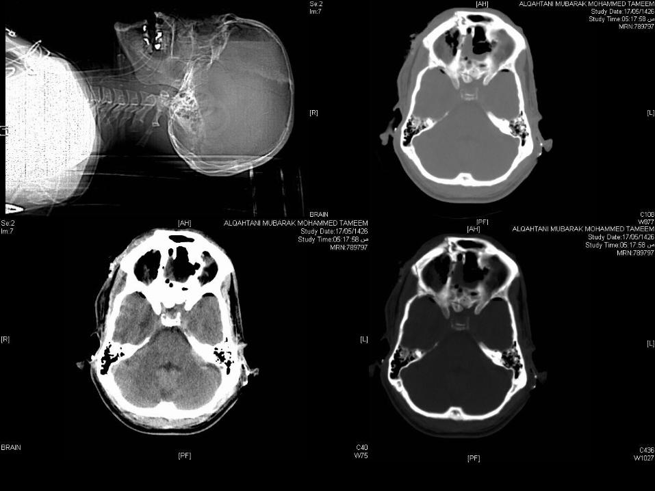
Patient C
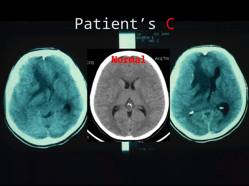
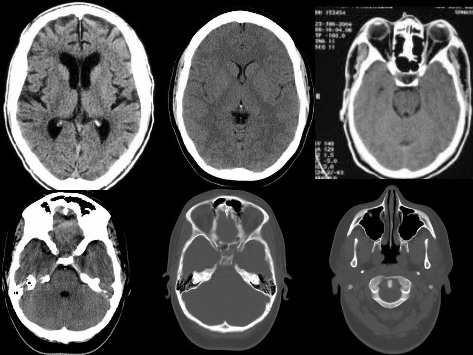
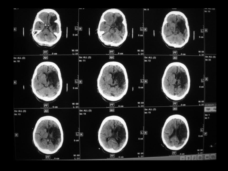
Patient’s C
Normal
Patient’s C
Normal
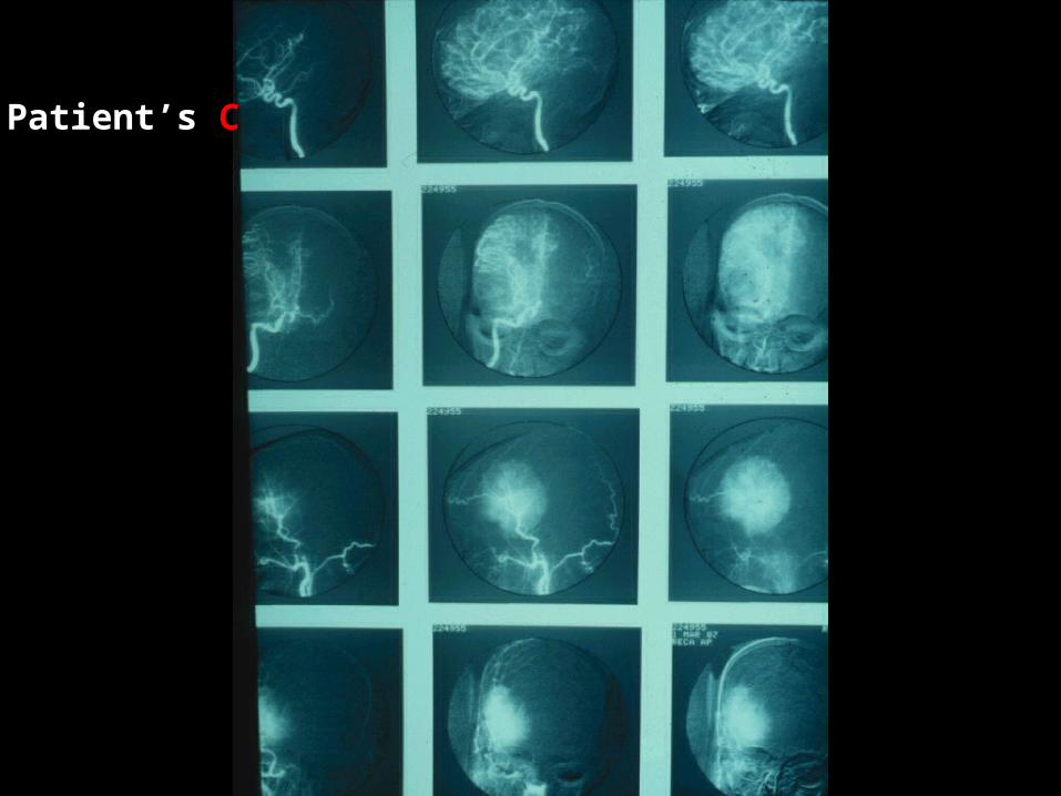
Patient’s C
Interpretation•Features of several diseases,
• trauma
•and non-trauma
CNS Trauma Imaging

SELLA SELLA TURCICATURCICA
CORONAL CORONAL SUTURESUTURE
GROOVE GROOVE FOR MCAFOR MCA
EXT.AUD EXT.AUD MEATUSMEATUS ORBITAL ORBITAL
GROOVEGROOVE
Normal Linear fracture
Epidural H Depressed fracture

Orbital Fracturesblow-out
NORMAL WATERS VIEWMedial/Inferior orbital wall blow-outMedial/Inferior orbital wall blow-out

Orbital Fracturesblow-outNORMAL WATERS VIEW Medial/Inferior orbital wall blow-outMedial/Inferior orbital wall blow-out
Axial CTCoronal CT

Medial/Inferior orbital wall Medial/Inferior orbital wall blow-outblow-out
Depressed right orbital floorDepressed right orbital floorOpacification of right Opacification of right maxillary sinusmaxillary sinusOpacification of right Opacification of right ethmoid sinusethmoid sinus
““Hanging tear drop”: Hanging tear drop”: herniation of orbital fat into herniation of orbital fat into maxillary sinus (not seenmaxillary sinus (not seen
here)here)
Orbital Fracturesblow-out

Interpretation
Non-trauma

Extra-axial vs Intra-axial
Meningioma Glioma
(external to pia) (beneath pia)

Supra-tentorial vs Infra-tentorial
Glioma Medulloblastoma

Nonenhanced computed tomography scan shows a hyperdense mass resulted in midline shift to the right aspect in the left frontal lobe
CECT shows a homogeneous enhancing mass located in the left frontal lobe.
DSA, Left external carotid artery injection shows early stain of the mass
DSA: Left external carotid artery shows delayed stain of the mass

Hydrocephalus versus Cerebral Atrophy
Definition
:Hydrocephalus is abnormal accumulation of intracranial fluid resulting from structural or functional block to normal flow of CSF,
cerebral atrophy is parenchymal volume loss

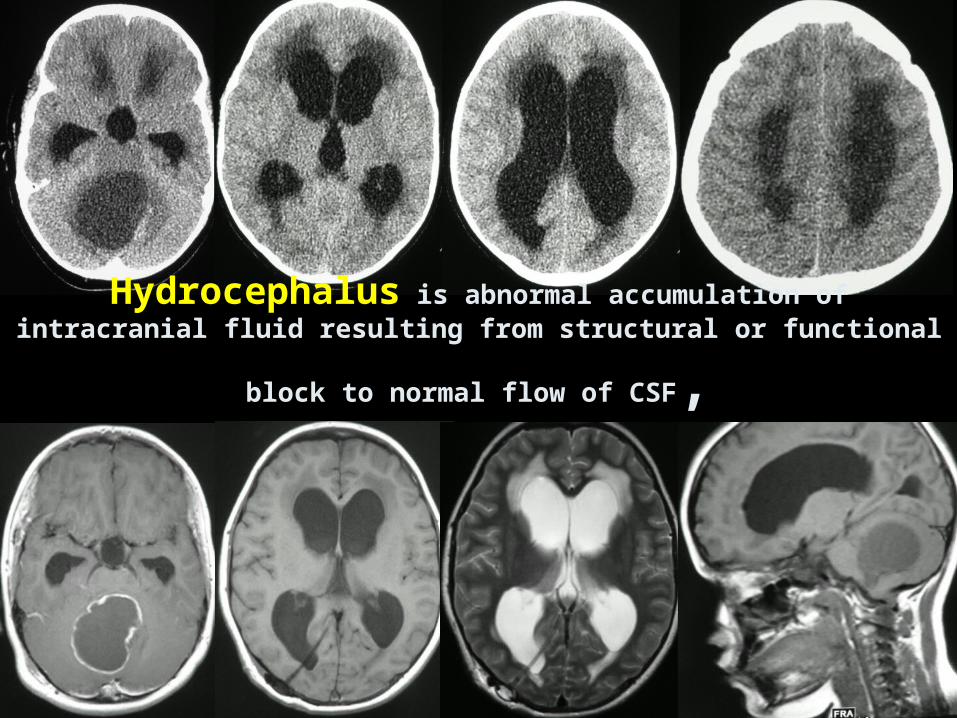
Hydrocephalus is abnormal accumulation of intracranial fluid
resulting from structural or functional block to normal flow of CSF,

cerebral atrophy is parenchymal volume loss
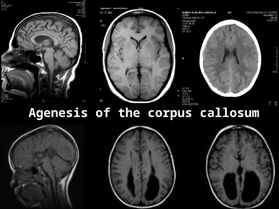
Agenesis of the corpus callosum

Patterns of edema
Edema: Increase in tissue water
CT - decreased density
MR - T1W - decreased signal
MR - T2W - increased signal
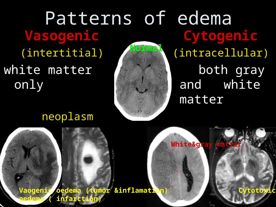
Patterns of edemaVasogenic
(intertitial)
white matter only
neoplasm
abscess
Cytogenic(intracellular)
both gray and white matter
infarction
Normal
Vaogenic oedema (tumor &inflamation) Cytotoxic oedema ( infarction)
White matter
White&gray matter


CONCLUSION KNOW YOUR Normal Radiological Anatomy
look for: (i) abnormal opacity (ii) abnormal radiolucency (iii) distortion or displacement of a normal structure
Interpret the abnormality: (a) Recognizing the abnormality, (b) Describing it in generic terms, (C) Giving a differential diagnosis
Give a specific diagnosis
KNOW your radiological terminology

BEST LUCK
HOPE THIS WAS
HELPFUL