© Imperial College LondonPage 1 Creating a Winning CV Careers Advisory Service .
date post
22-Dec-2015Category
view
214download
0
© Imperial College LondonPage 1
Detecting gold nanoparticles in electron
microscope images
John Cupitt, Exp. Med. & Tox.
16 November 2009

© Imperial College LondonPage 2
Technique
Use gold nanoparticles to label receptors, signalling proteins and lipids
Use up to three particle sizes to label different features
Image sections of cell membrane with transmission electron microscopy
Particle positions reveal information about membrane function

© Imperial College LondonPage 3
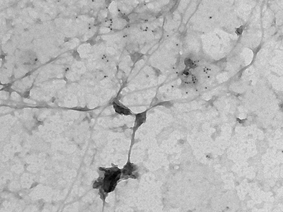
TEM imaging of cell membrane fragments
Then fix lightly, label with gold particles, fix securely, stain, dry and image with TEM

© Imperial College LondonPage 4
Problem
Gold nanoparticles are in the 3 – 15nm range, as few as 100 atoms
Image sections of cell membrane ~2000nm across containing several hundred particles
Particles therefore appear rather small: 4 - 12 pixels across
Standard blob detectors are designed for larger features and have poor accuracy at this scale
Only 50% accuracy with most systems

© Imperial College LondonPage 5

© Imperial College LondonPage 6

© Imperial College LondonPage 7

© Imperial College LondonPage 8

© Imperial College LondonPage 9
Characteristics
Approximately Gaussian profile7 – 13 pixels acrossApproximately circularAmong the darkest objects in the image

© Imperial College LondonPage 10
Our new detector
Gaussian ring divided into octants, convolve with each octant, take minimum response, compare to centre average

© Imperial College LondonPage 11
Results
On the test image, with a little tuning, we get 99+% accuracySearching the whole 2,500 x 2,500 pixel image takes about 2 secondsNeed to test on more images, add some better tuning

© Imperial College LondonPage 12
© Imperial College LondonPage 13

© Imperial College LondonPage 14
Credits
Nicolas Robidoux, Department of Mathematics and Computer Science, Laurentian University, designed the detector with help from Chantal Racette, an undergraduate studentThe NIH/NIGMS Center for the Spatiotemporal Modeling of Cell Signaling provided images and the gold standard, as well as funding for Chantal Racette's work.