Contents...Contents Full papers and Abstracts related to the subject of the thesis I. Abbreviations...
48
Contents Full papers and Abstracts related to the subject of the thesis............................................... I. Abbreviations………………………………………………………………………………..IV Introduction .............................................................................................................................. 1 Aims ......................................................................................................................................... 12 Materials and Methods ........................................................................................................... 13 Results ..................................................................................................................................... 22 Discussion ............................................................................................................................... 31 Summary ................................................................................................................................. 35 Acknowledgements ................................................................................................................. 37 References ............................................................................................................................... 38 Annex ......................................................................................Hiba! A könyvjelző nem létezik.
Transcript of Contents...Contents Full papers and Abstracts related to the subject of the thesis I. Abbreviations...

Contents Full papers and Abstracts related to the subject of the thesis............................................... I.
Abbreviations………………………………………………………………………………..IV
Introduction ..............................................................................................................................1
Aims.........................................................................................................................................12
Materials and Methods ...........................................................................................................13
Results .....................................................................................................................................22
Discussion ...............................................................................................................................31
Summary .................................................................................................................................35
Acknowledgements .................................................................................................................37
References ...............................................................................................................................38
Annex ......................................................................................Hiba! A könyvjelző nem létezik.

I IV
Abbreviations
AIDS acquired immunodeficiency syndrome
AV anchoring villus
BSA bovine serum albumin
CT cytotrophoblast
DAPI 4`,6-diamino-2-phenylindole
DC dendritic cell
DMEM Dulbecco's Modified Eagle Medium
DMSO dimethyl sulfoxide
E early
FcRc neonatal Fc receptor
FCS foetal calf serum
FITC fluorescein isothiocyanate
FV floating villus
gB glycoprotein B
HBSS Hanks` Balanced Salt Solution
HEPES hydroxyethylpiperazine-N’-2-ethanesulfonic acid
hCG human chorionic gonadotropin
HCMV human cytomegalovirus
HIV human immunodeficieny virus
IL Interleukin
IE immediate early
IgG immunoglobulin
L late
Mab monoclonal antibody
M/DC macrophage/dendritic cell
MOI multiplicity of infection
NK natural killer cell
ORFs open reading frames
PCR polymerase chain reaction
PBS phosphate buffered saline
PFU plaque forming unit
PMN polymorphonuclear leukocyte
ST syncytiotrophoblast

1
Introduction Human Cytomegalovirus
Human cytomegalovirus (HCMV), the largest and structurally most complex member
of the Herpesviridae family, is a ubiquitous virus that infects almost all humans at some time
in their lives (Figure 1). It has been classified as a betaherpesvirus on the basis of several
biochemical criteria such as the genome size, guanosine and cytosine contents, the slow
replicative cycle, and the restricted in vitro and in vivo tropism [1]. Although HCMV has
been shown to infect a broad spectrum of cells in vivo, the only cells that are fully
permissive for HCMV replication in vitro are human fibroblast. All known strains of HCMV
are genetically homologous, but none seem to be genetically identical, unless they are
obtained from epidemiologically related cases [2, 3]. The genetic and immunologic
variability and the differences in in vitro growth characteristics are well documented and
strain differences may affect HCMV virulence. Comparative genetic analysis of HCMV
strains is primarily limited by the size and complexity of the viral genome, which comprises
220–240 kB pairs of linear double-stranded DNA, depending upon the strain [4]. The
massive HCMV genome is approximately 50% larger than the genome of herpes simplex.
Figure 1.: Human Cytomegalovirus morphology. http://www.virology.net/Big_Virology/BVDNAherpes.html

2
Many of the capsid proteins appear to share structural, functional, and even antigenic
similarities with capsid proteins from other human herpesviruses. Outside the capsid, but
beneath the envelope, is tegument or matrix (Figure 1). The tegument of HCMV is the most
complex and heterogeneous structure in the virion. Proteins within the tegument are
characteristically phosphorylated and in many cases serve regulatory functions for virus
replication. Some tegument proteins appear to have a primary role in maintenance of the
structural integrity of the virion. However, the tegument contains the most immunogenic
proteins of the virions, including the immunodominant targets of T lymphocyte responses
and antibody responses [1]. The lipid-containing envelope is composed of virion
glycoproteins and host-derived membrane lipids (Figure 1). The envelope of HCMV is
thought to play an essential role in the initial steps of virus-host cell interactions and host
immune responses against virion componenst are thought to be important in protective
immunity. Since the envelope glycoprotein B (gB) of HCMV has been implicated in host
cell entry, cell-to-cell viral transmission and fusion of infected cells in addition to being an
important target for both antibody- and cell- mediated immune response, it is a candidate as
a HCMV virulence factor [5]. It is encoded by the gene UL55. Certain regions of the gB
gene vary considerably between different virus strains. On the basis of the nucleotide
sequence coding for the variable region, HCMV strains can be classified into 4 gB genotypes
[6]. The complete DNA sequence of AD169 has been determined, but an additional 22 viral
genes have been found in low passage clinical isolates that are missing in AD169. The wild
type HCMV genome has been predicted to encode approximately 165 genes (Figure 2) [7].
HCMV genes encode for structural proteins, which are incorporated in the architecture of
virions (nucleocapsid, tegument or envelope). In addition to structural genes, the genome
displays regulatory open reading frames (ORFs), whose functions are associated with the
onset and progression of HCMV infection and replication through the immediate early (IE),
early (E) and late (L) phases [4, 8-10]. Most genes are highly conserved in sequence between
HCMV strains, but some are characterized by a striking degree of variability, as revealed by
examination of individual genes [4] and by whole genome comparisons [7, 11]. These
variable genes are generally predicted to encode membrane-associated or secreted proteins
[7]. The hypervariation is probably due to immune system selection on the scale of human
evolutionary history or longer, and the genotypes are maintained stably within and among
infected individuals [12-14]. Several hypervariable genes are located in the sequence at the
right end of UL that is absent from laboratory-adapted strains AD169 and Towne. Two
notable examples are UL146 and UL139, which are located 5.2 kbp apart and oriented

3
leftward (Figure 2). The UL146 gene encodes a chemokine, designated vCXC-1, that
induces calcium mobilization, chemotaxis and degranulation of neutrophils. High-affinity
vCXC-1 binding has been shown to be mediated via CXCR2, but not CXCR1. vCXC-1
exhibits a potency approaching that of human interleukin-8 (IL-8). As the first example of a
virus-encoded a chemokine, vCXC-1 may ensure the active recruitment of neutrophils
during cytomegalovirus infection, thereby providing for efficient dissemination during acute
infection and accounting for the prominence of this leukocyte subset in cytomegalovirus
disease [15]. The most variable gene in the vicinity of UL146 is UL139, which is predicted
to encode a type I membrane glycoprotein. Variability is concentrated in a region of the
ectodomain [7].

4
Figure 2.: Consensus genetic map of wild-type HCMV based on the Merlin genome [7].
5
Epidemiology of HCMV infection
Distribution and transmission HCMV infections are endemic and has no seasonal variation. Studies on the age-
related prevalence of infection with HCMV suggest that there may be three periods with
particularly high rates of acquisition of the virus: early childhood, adolescence and the
childbearing years. The prevalence of HCMV infection in the normal population varies
widely, between 40% and 90%, depending on the race, the gender, the age, hygienic
circumstances and the socioeconomic factors [16]. In general, HCMV is more prevalent
among people in low socioeconomic brackets living in crowded conditions and in people
living in undeveloped countries, with 80% of 3-year-old children and most adults in such
groups being infected with HCMV [16-18]. In Hungary, the seroprevalence of HCMV at the
age of 10 years is 72%, which increases to 96% by the age of 50 [19]. The level of immunity
among women of childbearing age, which is an important factor in determining the
incidence and significance of congenital and perinatal HCMV infections, varies widely
among different populations. The prospective studies of pregnant women in the United
States indicate that the rate of HCMV acquisition for childbearing-aged women of middle to
higher socioeconomic background is approximately 2% per year, whereas it is 6 % per year
among women of lower socioeconomic background [20-22].
Infection with HCMV can be classified as either primary or recurrent. Similar to
other herpesvirus infections, primary HCMV infection is followed by persistent and/or
recurrent infections, most often due to reactivation of a latent infection, residing in the host
throughout life. It is reactivated periodically during episodes of mild immunosuppression
caused by intercurrent infection, pregnancy or stress. Recurrent infections are fairly common
[1]. Intermittent excretion of the virus can be anticipated in a significant proportion of
seropositive adults. Reinfection by a new strain of HCMV has been documented in
immunocompromised individuals, women attending a clinic for sexually transmitted
diseases and healthy children attending a day-care center [23-26].
HCMV rarely causes symptoms in an immunocompetent host, and these can be
nonspecific symptoms, such as malaise, fever, sweats, aching muscles, atypical
lymphocytosis and mild hepatitis during the self-limiting primary infection [27]. However, it
can give rise to serious disease in an immunodeficient person, such as those with acquired
immunodeficiency syndrome (AIDS) [28] and those with organ and bone marrow transplants

6
[29], thereby causing severe morbidity and eventually mortality, or neonates, and especially
premature babies [1]. Reactivation is asymptomatic, except in severely
immunocompromised individuals [28, 29].
HCMV is not very contagious: spreading of the infection appears to require close or
intimate contact of either a nonsexual or a sexual nature with another person who is shedding
the virus in the bodily secretions. The virus is present in urine, oropharyngeal, cervical and
vaginal secretions, breast milk, semen and tears, and can be shed intermittently for years.
Spread of HCMV also occurs frequently in child care centers. Cohort studies indicate that
children in day care centers frequently transmit virus to each other. It is reasonable to expect
that approximately 50% of susceptible children between the ages of 1 and 3 years who attend
group day care will aquire HCMV from their playmates and become an important potential
source of infection for serosusceptible child-care personnel and parents, particulary women
of childbearing age [20]. Fomites may also play role in transmission because HCMV has
been shown to retain infectivity for hours on plastic surfaces and has been isolated from
randomly selected toys and surfaces in day-care center [22].
HCMV can also be transmitted vertically, from mother to fetus by three routes:
transplacental, intrapartum and human milk [21]. The materno-fetal transmission of the
HCMV is the leading cause of congenital viral infection, affecting 0.3-2% of newborns in
developed countries, and the transmission could well be higher in developing countries [30].
The prevalence displays a specific geographical pattern: it is 0.9% in Hungary [1, 31]. The
individual course of the infection may vary between asymptomatic virus shedding, abortion
or stillbirth, and a congenital syndrome. Between 10% and 15% of infants infected with
congenital HCMV exhibit the clinically apparent or symptomatic form of the disease,
characterized by petechiae, hepatomegaly, splenomegaly, jaundice, periventricular
calcifications, microcephaly, hearing impairment and chorioretinitis. The remaining 85% to
90% of infected infants are asymptomatic at birth, but 15% of them will develop delayed
sequelae, especially progressive hearing loss [32].
The natural history of HCMV during pregnancy is particulary complex and has not been
fully explained.
Intrapartum transmission of HCMV is related to local shedding of virus.
Approximately 10% of woman shed HCMV from the vagina or cervix; near the time of
delivery, rates of 2% to 28% have been reported [21]. If the virus is present in the maternal
genital tract at the time of delivery, the rate of transmission to newborn is around 50%.

7
The most common route for mother to infant transmission of HCMV is human milk
[1]. The virus was isolated from human milk more than 30 years ago, but the roles of milk
cells and cell-free virus in transmission have remained unclear. More recent studies using
polimerase chain reaction (PCR) to detect HCMV DNA in milk from seropositive mothers
reported a strong relationship between the presence of viral DNA in milk and transmission
of HCMV to the infant. Breastfeeding significantly influences the epidemiology of HCMV
infection [33].
HCMV infection in pregnancy The in utero transmission of HCMV can occur as a consequence of primary and
recurrent infections, with equal frequency during all three trimesters [34]. Primary infection
with HCMV occurs in 0.7-4.1% of pregnancies, with a mean reported rate of transmission to
the fetus of 40% (range 25-75%). In contrast to the high rate of transmission in primary
infection, that during recurrent infection is much lower (1-1.2%) [35-40]. Primary infection
occurring in the mother and as intrauterine transmission during the first 16 weeks of
pregnancy has a much greater clinical impact on the fetus than nonprimary infections and
infections occurring during the last trimester of pregnancy.
After primary maternal infection, the most likely sequence of events leading to
congenital HCMV infection is maternal viremia, placental infection, and hematogenous
dissemination to the fetus. Anatomical considerations argue that in utero infection involves
the passage of virus or virus-infected cells across the placenta into the fetal compartment
[41-43].
During the viremic phase, the virus circulates and disseminates, carried by leukocytes.
HCMV is thought to be transmitted when infected leukocytes cross the placental barrier to
reach the fetal circulation via the umbilical cord vessels [41, 42]. Other routes may also be
accessible to viral transmission. Infected leukocytes may reach the fetal endothelium
directly, through breaches of the syncytiotrophoblast (ST) layer of the placenta, particularly
in the last stages of gestation. Virus coated by specific antibody may cross this layer by
transcytosis and be released, still infectious, to the underlying cytotrophoblast (CT) [42].
Located at the boundary between the maternal circulation and the fetal mesenchyma, the ST
is a central component of the placental barrier, imposing physical and possibly
immunological constraints to the passage of microbial pathogens and/or maternal cells into
the fetal compartment [41]. An additional possibility is that the virus may ascend from the

8
vagina via the ruptured membranes, to reach the decidua or amniotic cells [44].
Consequently, infected amniotic cells may be ingested by the fetus, after which the virus
may replicate in the oropharynx and invade the fetal circulation to reach the target organs.
Despite the morbidity and mortality associated with prenatal HCMV infection, little
is known about how the virus infects the conceptus. Approximately 15% of women with
primary infections during early pregnancy abort spontaneously [35]. In this case, the
placenta, but not the fetus, shows evidence of infection, which suggests that placental
involvement is important in its own right and precedes virus transmission to the fetus [45-
47]. Later in pregnancy, HCMV infection causes premature delivery and (in 25% of affected
infants) intrauterine growth retardation, outcomes that are often associated with placental
pathology [48]. Numerous reports indicate that the placentas from these births also contain
viral proteins, suggesting that placental infection and virus transmission to the infant are
related causally [47, 49].
Although the pathogenesis of HCMV transmission to the fetus during pregnancy is
unknown, congenital HCMV infections are commonly associated with chronic villitis and
infection of the placenta. Thus, passage probably occurs through the placenta, which may
also act as a viral reservoir [50].
Placenta
The human placenta is composed of villi that float in maternal blood and also villi
within the uterine wall that anchor the placenta and attach the fetus to the mother (Figure 3).
The individual chorionic villus has a connective core that contains fetal blood vessels and
numerous macrophages (Hofbauer cells) that often lie under a thick basement membrane
[51].
Three major trophoblast populations can be identified during placentation: CT stem cells and
two differentiated derivate cell types: ST and extravillous CT [52]. The undifferentiated
trophoblastic stem cell of the placenta, the CT, is the first fetal cell type arising during
embryogenesis. It derives from external trophectodermal cell layer of the blastocyst, thus it
is extraembryonic in origin. It represents the earliest epithelium and forms a variety of
different structures e.g., placental villi and fetal membranes [53]. Just as undifferentiated
basal layer of the skin gives rise to differentiated keratinocytes, the CT stem cell of the
placenta undergoes multistep differentiation and finally gives rise to villous (non-invasive)
and extravillous (invasive) trophoblast cell populations [54, 55].

9
In the human placenta, proliferating CT stem cells are attached to an extensive
basement membrane that surrounds the stromal core of two types of chorionic villi (Figure
3). In floating villi, CTs differentiate by fusion to form an overlying layer of multinucleated
ST. These cells are in immediate contact with maternal arterial blood that bathes the floating
villi, and their primary function is to perform nutrient, waste and gas exchange between the
maternal and fetal circulations. In anchoring villi, beginning with the third post-ovulatory
week, a subset of CTs at the distal tips of the villi proliferate and differentiate by leaving
their basement membrane to form multilayered cell columns consisting of highly migratory,
non-polarized, invasive CTs, covered by a thin layer of syncytium. The cell columns expand
distally, and populate the decidualized endometrium and the first third of the myometrium,
thereby anchoring the villous tip to the uterine wall [56, 57]. The penetrating cells spread
over the maternal decidual cells, forming the cytotrophoblastic shell. Some of these deeply
invasive cells penetrate the inner walls of the maternal spiral arteries up to their myometrial
segments and, by replacing the endothelial lining and the smooth muscle cells, transform
them in maximally dilated inert tubes. This process is termed endovascular invasion [53, 58-
62]. This unusual invasive behavior has two important roles: (1) CT invasion phisically
anchors the placenta to the uterus, and (2) by replacing the endothelial lining of the spiral
arteries invasive extravillous CTs create the large-diameter, low-resistance vessels that carry
blood to the floating villi at the material-fetal interface.
The mechanisms by which human trophoblast cells influence the vertical
transmission of HCMV have not been well studied. The cellular organization of the
decidual-placental interface suggests potential routes by which HCMV reaches the placenta
[63]. The virus might disseminate from infected maternal blood cells to the decidua (Figure
3, site 1), interstitial and endovascular CTs in the uterine wall (site 2), CT columns of
anchoring villi (site 3), and/or floating villi (site 4). An endothelial cell-tropic HCMV strain
replicates in uterine microvascular endothelial cells and spreads to invasive CTs in vitro
[64], suggesting that hematogenous transmission occurs in utero.

10
Figure 3: Diagram of the placental (fetal)-decidual (uterine) interface near the end of the first trimester of
human pregnancy (10 weeks of gestational age). Sites proposed as routes of HCMV infection at the maternal–
fetal interface are numbered 1–4; FcRn, neonatal Fc receptor; AV, anchoring villus; FV, floating villus; DC,
dendritic cell; PMN, polymorphonuclear leukocyte; NK, natural killer cell; M/DC, macrophage/dendritic cell
[51].
Numerous investigations have indicated that placental cells can be productively infected by
HCMV [42, 64-67]. Experiments have been performed in vitro by using laboratory-adapted
strains and a high multiplicity of infection (MOI), which may not be entirely representative
of the behavior of wild strains. Placental trophoblastic cells closely resemble macrophages;
both macrophages and trophoblasts are invasive, form syncytia, and express CD14, Fc
receptors, multiple cytokines and receptors for many cytokines [68]. Placental macrophages
and trophoblastic cells are known to constitutively secrete a variety of cytokines and
prostaglandins, which play crucial roles in normal reproductive processes [69]. In the
placenta, proinflammatory cytokines, including IL-1, IL-6, IL-8 and TNF-α, and anti-
inflammatory cytokines such as IL-4 and IL-10, are produced by the trophoblast [70-74].
Intrauterine infections are associated with the expression of placental cytokines such as IL-1
and IL-8 [71]. IL-8 has been demonstrated to upregulate the replication of laboratory-
adapted HCMV strains in various cells, including the ST [75-77]. Furthermore, IL-8 could

11
attenuate the antiviral activities of interferon, particularly type I interferon. These
observations would imply that endogeneously produced IL-8 in the placental
microenvironment may play a regulatory role in the expression of HCMV, and by this
mechanism latent virus in the placenta develops into a fulminating infection.
Griesinger et al. evaluated the influence of bacteria that have been identified in
uterine
infections on cytokine expression of highly purified primary trophoblast cultures
[78]. Their results indicated that the potential of various bacteria to stimulate cytokine
production in these cells varies. E. coli and B. fragilis exhibited the highest potencies to
stimulate IL-8 expression.

12
Aims
sent study was designated to address the following aims:
im 1: To investigate the production of proinflammatory cytokines of ST cultures infected
im 2: To examine the interrelationship between the level of IL-8 secreted and the
im 3: To determine the distribution of the genotypes of the HCMV genes UL55, UL146
he preT
A
with clinical HCMV isolates.
A
replication of HCMV in epithelial cells.
A
and UL139 among HCMV strains, and to investigate the association between
polymorphisms within the genes UL55, UL146 and UL139, and the ability to induce IL-8
production.

13
Materials and Methods
Isolation of villous cytotrophoblasts
A pure population of villous CTs was separated from individual first-trimester (6 to
12 weeks) human placenta villi and differentiated as described previously [79]. Briefly,
trophoblastic tissue was obtained from legal terminations of pregnancies performed in
accordance with the Hungarian Abortion Law between 6 and 12 weeks of gestation. Ethical
approval for this study was granted by the Human Investigation Review Board of the
University of Szeged. Villous tissue from individual placentas was dissected manually rinsed
and minced in Hanks’ Balanced Salt Solution (HBSS) (pH=7.4) containing 200 U/ml
penicillin and 200 µg/ml streptomycin (both from Sigma, Budapest, Hungary). The minced
tissue was then incubated at 37°C four times for 20 minutes in HBSS containing 0.25%
trypsin (Sigma, Budapest, Hungary), 50 U/ml DNase I (Sigma), 4.2 mM magnesium sulfate
(Sigma), 25 mM N-2-hydroxyethylpiperazine-N’-2-ethanesulfonic acid (HEPES) (Sigma)
and antibiotics (200 U/ml penicillin and 200 µg/ml streptomycin). The supernatant
containing the dissociated mixed placental cells was collected, and the trypsin activity was
neutralized by addition of 10% Foetal Calf Serum (FCS) (Gibco, Life Technologies, Vienna,
Austria). The neutralized supernatant was centrifuged at 800×g for 10 minutes and the
resulting cell pellet was resuspended in Dulbecco's Modified Eagle Medium (DMEM)
containing 25 mM HEPES, 200 µg/ml streptomycin. The cell suspension obtained was
placed immediately into an incubator and was maintained at 37°C until the end of the entire
dissociation procedure. Concomitantly, the remaining villous tissue was subjected to another
20 min trypsinization step. At the end of the dissociation procedure the remaining villous
fragments were discarded. The four fractions of cell suspensions were pooled, filtered over a
100 µm nylon mesh to remove remaining villous fragments, centrifuged at 800×g for 10
minutes, and resuspended in 2-3 ml of the same medium without FCS. This cell suspension
was layered over a 5% to 70% preformed discontinuous percoll gradient, according to
method of Kliman et al [80]. The fraction containing the CTs was washed and resuspended
in DMEM. The purity of the isolated CTs was tested by immunochemical staining with
monoclonal mouse antibodies to human cytokeratins (clone: MNF 116; Dako A/S, Glostrup,
Denmark), which stain only the trophoblasts within the placental villi [81] and with
monoclonal mouse antibodies to vimentin (clone: V9; Dako A/S, Glostrup, Denmark). The

14
viability of the percoll-enriched cells was estimated by trypan-blue exclusion. The viability
was constantly >95%.
At this point the isolated cells were frozen (5-10x106 cells/ml) and stored in liquid N2
until needed. The percoll-purified cells were tested routinely for bacterial infection,
including mycoplasma, chlamydia and infection with fungi or HCMV. When required, the
cells were thawed and subjected to an immunomagnetic separation procedure to remove the
remaining contaminating cells [82].
Storage of cells in liquid nitrogen
The percoll-purified cells were suspended in cold FCS containing 10% dimetyl
sulfoxide (DMSO) (Sigma) to give 5-10×106 cells/ml. One ml aliquots of this cell
suspension in 1.8 ml criogenic vials (Corning Inc., N.Y., USA) were placed in insulated
cardboard containers and kept at -70°C overnight. Next day the tubes were rapidly
transferred to liquid nitrogen for long term storage. When required, a tube was removed from
liquid nitrogen and thawed in a water bath at 37°C. The thawed cells were diluted 1/10 in
DMEM containing 20% FCS and centrifuged at 400×g for 5 minutes. The cell pellet was
resuspended in fresh culture medium to required cell density.
Purification of cytotrophoblasts
Cells were incubated for 30 minutes at 4°C with 50 µl of a monoclonal anti-HLA-
ABC (clone W6/32) and 50 µl of monoclonal anti- HLA- DP, DQ, DR (clone CR3/43; both
from Dako A/S). All the primery antibodies were raised in mice and added undiluted to the
cell suspension. After incubation, the cells were washed with phosphate buffered saline
(PBS) (pH 7.4) containing 0.1% bovie serum albumin (BSA) (radioimmunoassay grade,
Sigma). Prior to separation, 100 µl of magnetic particles coated with goat anti-mouse
immunoglobulin (IgG) (Dynal AS, Oslo, Norway) were washed twice with 5 ml PBS
containing 0.1% BSA and stored at 4°C until used. The cell suspension was then incubated
with the prewashed magnetic particles at 4°C for 30 minutes with occasional gentle shaking.
At the end of the incubation the tube was clamped to a magnetic concentrator (Dynal
Laboratories, Oslo, Norway) for 5 minutes. The supernatant containing the purified CTs was
removed from the tube and centrifuged at 800×g for 10 minutes at room temperature. The
pelleted cells were resuspended in culture medium and plated as required.

15
The purity of the CT population was tested by cytokeratin and vimentin staining. The
CT preparations from first-trimester placentas used in this study were highly pure (>99).
Differentiation of cytotrophoblasts
For the in vitro differentiation of CTs, Keratinocyte-SFM (Gibco BRL, Karlruhe,
Germany) supplemented with 15% FCS was used. CTs were seeded in 24-well plates
containing 5x10⁵ cells/well. The epidermal growth factor present in the Keratinocyte-SFM
promotes the differentiation of villous CTs from first-trimester chorionic villi to form STs
[81]. The media were changed every 48 h. All experiments were carried out on 5-day-old ST
cultures.
A549 cells
Cells of the human A549 lung carcinoma cell line were cultivated in Eagle’s
minimum essential medium (MEM) supplemented with 10% FCS and antibiotics. The cells
were grown on glass coverslips in 24-well plates containing 2x10⁵ cells/ well for cytokine
production, immunofluorescence and virus replication studies.
Viruses
HCMV clinical isolates of different origins and laboratory strains Davis (ATCC
#VR-807), Toledo (ATCC), Towne (ATCC #VR-977), Merlin (ATCC #VR-1590), TB40/E
(ATCC) and AD169 (ATCC #VR-538) (American Type Culture Collection) were used.
(Table I.). Most strains were isolated in our laboratory from the urine of congenitally
infected neonates and mothers, and strains 5DV and E5 from the urine of adults. Strains S
and B were isolated by Dr. J. Schirm (Regional Health Laboratory, Groningen, The
Netherlands) from the urine of neonates. Strains 20 and 74 originated from the blood of renal
transplant recipients, strains 83 and 89 from congenital infection and were provided by
Professor C. Bruggeman (University Hospital Maastrich, The Netherlands). The clinical
isolates were passaged <5 times. The stock of HCMV laboratory strain Towne and clinical
isolates were propagated in confluent MRC-5 cells grown in RPMI medium supplemented
with 10% FCS and antibiotics. The infectivity titers were determined by plaque assay, with
the inoculation of confluent MRC-5 cultures in 24-well plates.
The clinical samples were obtained in accordance with local ethical guidelines.

16
Table I. HCMV isolates of different origins and laboratory-adapted strains
Straina Clinical sourceb Age/sexc Clinical detailsd
16/5 U/P Infant C 128V U/P Infant C E24 U/P Infant C 16/3 U/P Infant C
E34AV U/P Mother C+ 5DV U/P Adult CH E5 U/P Adult C+
E33AV U Mother C+ E33BV U/P Infant C E36BV2 U Infant C E36AV1 U Mother C+ E41BV3 U Infant C E41AV1 U Mother C+ E42BV1 U Infant C E42AV1 U Mother C+ E43BV1 U Infant C E43AV1 U Mother C E62BV1 U Infant C E62AV1 U Mother C+ E45BV1 U Infant C E59BV1 U Infant C E28BV1 U Infant C
16/2 U Infant C E34B/BV U 5m/M C+ E60BV1 U Infant C+ E55BV1 U Infant C+ E63BV1 U 10 w/F C+ E63AV1 U Mother C+ E49AV1 U Mother C+ E52BV1 U 14 m/M C+
20 B/P Adult R 74 B/P Adult R S U/P Infant C B U/P Infant C 83 U Infant C 89 U Infant C
Davis BP/P Infant C Toledo U/P Infant C Towne U/P Infant C Merlin U/P Infant C TB40/E TS/P Adult B AD169 BP/P Infant C+
a Virus strains; b U, urine; BP, biopsy; B, blood; TS, throat swab; P, passaged in cell culture; C Ages are in weeks (w) or months (m). Sexes: M, male; F, female; d C+, HCMV positive by PCR or IgM in serum; C, congenital; CH, chronic hemodialysis patient; R, renal transplant recipient; B bone marrow transplant recipient;

17
Hormone assay
The release of human chorionic gonadotropin (hCG) from the trophoblasts was
measured in the culture supernatant daily after seeding in culture. The microparticle enzyme
immunoassay kit used for hCG detection (WHO Matched Assay Reagents for Immunoassay
of Hormones). The sensitivity of the assay was 2 mIU/ml.
Determination of cytokine production
Five-day-old ST cultures were infected with the Towne strain or isolates of HCMV
(16/3, 128V and 20) at a MOI of 0.1. The MOI was calculated from the number of CTs
seeded for syncytium formation. The infected ST cultures were centrifuged at 300xg for 60
min at room temperature and then incubated for 2 h at 37 ºC. The STs were washed five
times with serum-free medium. After washing, fresh medium was added to the ST cultures.
Twenty-four-hour A549 cell cultures were infected with one or other of the laboratory-
adapted strains Towne or AD169 or each isolate of HCMV at an MOI of 0.1. The infected
cultures were centrifuged at 750xg for 60 min in a Heraeus Megafuge 1.0 (Osterode,
Germany) at room temperature and then incubated for 2 h at 37 ºC. The unabsorbed virus
was removed and the cells were washed five times with serum-free medium.
Supernatants of virus-infected and mock-infected cultures were collected at different
time intervals after infection and assayed for TNF-α, IL-1β, IL-6 and IL-8. Cytokine
concentrations were measured in each supernatant by using ELISA kits according to the
manufacturer’s technical guidelines. All ELISA kits were purchased from Biosource Europe
S.A., Nivelles, Belgium. Sensitivity of cytokine assay: the minimum detectable doses of
hTNF-α, hIL-1β, hIL-6 and hIL-8 were 1.7, 1, <2 and <5 pg/ml, respectively.
Immunofluorescence assay
After a 48 h incubation the HCMV-infected and mock-infected cultures were washed
twice with cold PBS and fixed with cold acetone:ethanol (1:1) for 20 min at –20ºC. The
fixed cells were stored at –20 ºC until the immunofluorescence assay was performed. The
nuclei of the cells were stained with 4’,6-diamidino-2-phenylindole (DAPI), and the HCMV
IE antigen was detected in the nuclei of cells by immunostaining using monoclonal antibody
(MAB810) (Chemicon International Inc., Temecula, CA, USA) and fluorescein

18
isothiocyanate (FITC)-conjugated rabbit anti-mouse IgG (Sigma, Budapest, Hungary).
Nuclei positive for IE antigen were counted under Leitz UV microscope.
Assay for virus replication in A549 cells
For analysis of the growth kinetics of HCMV strains of different IL-8-inducing
abilities in A549 cultures, cells were infected at an MOI of 0.1 with strain 128V, E5 or
Towne. During the adsorption period, the plates were first subjected to centrifugation at
room temperature for 60 min at 750xg in a Heraeus Megafuge 1.0 (Osterode, Germany) and
then incubated at 37 ºC for 2 h.
The inoculum was removed and the cells were washed 5 times with serum-free
medium to remove residual input infectivity completely. After washing, the cells were
overlayed with MEM with 1% FCS and antibiotics, and were harvested at zero time point.
After infection, culture supernatants were collected daily, and made cell-free by
centrifugation. Cell-associated virus was collected by 3 freeze-thaw cycles of infected cells
in medium. The cell-free and cell-associated virus-containing samples collected at the same
time were pooled. The virus titers were determined by plaque assay, with inoculation of
confluent MRC-5 cultures in 24-well plates. All assays were performed in triplicate.
Determination of the effect of exogenous IL-8 on IE antigen expression
A549 cells were incubated with different concentrations of recombinant human IL-8
(rhIL-8) (R&D Systems Europe Ltd.) for 1 h at 37 ºC. After the removal of the medium, the
cells were infected with various strains of HCMV at an MOI of 0.1. During the adsorption
period, the plates were first subjected to centrifugation at room temperature for 60 min at
750xg in a Heraeus Megafuge 1.0 (Osterode, Germany) and then incubated at 37 ºC for 2 h.
The inoculum was removed, the cells were washed five times with serum-free medium to
remove residual input infectivity completely, and were overlayed with MEM with 1% FCS
and antibiotics in the presence or absence of the various concentrations of IL-8. After 48 h
incubation, the cultures were washed twice with cold PBS and fixed with cold
acetone:ethanol (1:1) for 20 min at –20ºC. The fixed cells were stored at –20 ºC until
immunofluorescence assays were performed for the detection of IE antigen-producing cells.

19
DNA preparation
HCMV DNA was extracted by standard methods from cultured cells and urine.
Two nucleic acid extraction kits were used for purification of viral DNA from urine. In
experiments relating to the determination of the HCMV gB genotype the High Pure Viral
Nucleic Acid Kit was used (Boehringer Mannheim GmbH, Indianapolis, IN, USA)
according to the manufacturer`s instructions, and the viral DNA was resuspended in
nuclease-free distilled water. In experiments involving the genotypic analysis of the HCMV
genes UL139 and UL146, the NucleoSpin Tissue Kit (Macherey-Nagel GmbH & Co,
Germany) was used according to the manufacturer`s instructions, and the viral DNA was
resuspended in Elution Buffer BE (5 mM Tris/Cl, pH 8.5). In addition, DNA was isolated
from cultured cells by using the QIAGEN FlexiGene DNA Kit (QIAGEN ltd., UK)
following to manufacturer`s instructions, and the DNA was dissolved in hydration buffer (10
mM Tris.Cl, pH 8.5).
Polymerase chain reaction amplification of UL55 gene and restriction analysis
A region of high peptide variability in the gene UL55 was amplified. To improve the
sensitivity and to overcome sequence variation between strains, a nested PCR was used as
previously described [83]. Primers for amplification were selected from the published UL55
sequences of HCMV strain AD169 [6]. Amplification products (293-296 bp, the size varying
with the strain) were analyzed by electrophoresis in a 2% agarose gel (BRL, Gaithersburg,
Germany) stained with 5µl of 10 mg/ml ethidium bromide (Serva Feinbiochemica,
Heidelberg, Germany) and subjected to restriction analysis by using Hinf I and Rsa I
(Promega, Madison, WI, USA) as previously described [6]. Digested DNA was analyzed in
a 3% agarose gel (Nu Sieve 3:1, FMC Bio Products, Rockland, ME, USA). Four distinct gB
genotypes can be identified by the different lengths of restriction fragments (36-239 bp) [6].
Polymerase chain reaction amplification of UL146 and UL139 genes
UL146 and UL139 were amplified separately by single round or nested PCR, using
primers in conserved regions (Table II). Single (and first) round PCR of UL146 using AB4
and A162 generated a product of approximately 1 kbp, and second round PCR using UL146-
4A and UL146-3A yielded an 800 bp product. Single (and first) round PCR of UL139 using

20
AB1 and AB2 generated an 800 bp product, and nested PCR using UL140-3A and UL140-
11A yielded a 500 bp product. UL140-11A is located within the UL139 coding region, and
as a consequence the sequences obtained using nested PCR (approximately 40% of the total)
lacked 29 amino acid-encoding codons from the highly conserved C terminus. For the single
(and first) round, 1 µl of DNA was added to the PCR reaction mixture, which consisted of 40
µl of water, 5 µl of buffer, 1 µl of 10 µM dNTPs, 1 µl of each the two primers (10 µM) and
1 µl (1 U) of DNA polymerase (Advantage 2, BD Clontech, Basingstoke, UK). The
conditions for amplification were 95°C for 2 min followed by 35 cycles of 95°C for 2
minutes, 60°C for 30 sec and 68°C for 1 min. Second round PCR utilized 1µl of first round
PCR products as template amplified under the same conditions. PCR reactions were set up in
a dedicated, PCR product-free room. Approximately one-third of the samples were tested on
three separate occasions to assess reproducibility.
Purification, Cloning and Sequencing of PCR Products
PCR products were separated by agarose gel electrophoresis. Appropriate DNA
fragments were excised, purified using a Geneclean turbo kit (Q Biogene, Cambridge, UK),
and eluted with 100 µl of nuclease-free water. The single round or second round primers
were used for direct sequencing.
In some cases, including those where direct sequencing indicated the presence of
more than one genotype of UL146 or UL139, fragments were cloned using a pGEM-T kit
(Promega, Southampton, UK). Following ligation and transformation into chemically
competent E. coli TOP 10 cells, 5 recombinant colonies were picked and grown overnight at
37 ºC in 2YT-broth containing 100 µg/ml ampicillin. Plasmid DNA was purified with a
QIAprep Spin miniprep kit (Qiagen, UK). Plasmid inserts were sequenced using universal
forward and reverse primers. Sequencing was carried out on both DNA strands, using a
BigDye terminator kit (Applied Biosystems, Warrington, UK) in an ABI 3730 instrument.
Sequence Analysis
Sequence chromatograms were viewed via Editview (Applied Biosystems, Warrington,
UK) and analyzed using Pregap4 and Gap4 [84]. Nucleotide and imputed amino acid
sequences were aligned using CLUSTAL W [85] and MAFFT [86]. Full-length sequences
were used for the UL146 data and a subset of the UL139 data, and another subset of the

21
UL139 data was analyzed by using sequences lacking the conserved C terminus. MEGA4.0
[87] was used for the generation of phylogenetic trees.
Table II. Primers Used for PCR and Sequencing of UL139 and UL146 genes
Gene Primer Sequence (5’-3’) Genome locationa
UL146 AB4 TAGACACTACGTCGTAAATG 180494-180513 UL146 A162 TGTAGAATTAGTCTAGATTCCTGA 181524-181501 UL146 UL146-4A GCTTGCGCGTTAGGATTGAGACAC 180571-180594 UL146 UL146-3A ATACCGGATATTACGAATT 181341-181323 UL139 AB1 GTCATTGTGAAAGTGACGTCTCAG 186389-186412 UL139 AB2 ATCTACTGTAAACCCTCTGCTCTG 187148-187125 UL139 UL140-11A GCGGCATTGGTGTACGCGTG 186553-186572 UL139 UL140-3A GTGGAAATTTTTACGTCATT 187077-187058 a With reference to RefSeq accession NC_006273 (HCMV strain Merlin).
Statistical analysis
The results on the variability in proinflammatory cytokine secretion among ST
cultures from three independent experiments are presented as means ± standard deviation
(SD). Data on ST cultures infected with HCMV strains were compared with those on mock-
infected ST cultures; significant differences were determined by two-way repeated measures
analysis of variance (ANOVA) followed by the Bonferroni post test (GraphPad
Prism_Home, San Diego CA, USA). A p value <0.05 was considered significant. The mean
number ± SD of nuclei positive for IE antigen per microwell 48 h after infection was
calculated from 3 independent experiments.
The data on A549 cell cultures infected with HCMV strains were expressed as the
mean percentage ± SD of IE antigen-positive cells per 30 microscopic fields. Total cell
numbers were determined via the numbers of DAPI-stained nuclei.

22
Results
1. Production of proinflammatory cytokines of ST cultures infected with clinical HCMV
isolates.
In order to determine the production of proinflammatory cytokines by HCMV-
infected STs, STs were differentiated from first-trimester placenta-derived CTs. Three
independent experiments were carried out on 3 different placental trophoblast preparations.
The ST cultures were infected with HCMV strains or the laboratory-adapted strain Towne.
To resemble a natural infection, clinical HCMV isolates and a low MOI were used. There
was no difference in hCG release between virus- and mock-infected STs in any of the
experiments. Up to 72 h post-infection, TNF-α and IL-1β activities could not be detected in
any of the ST culture supernatants, regardless of whether these were mock-infected or
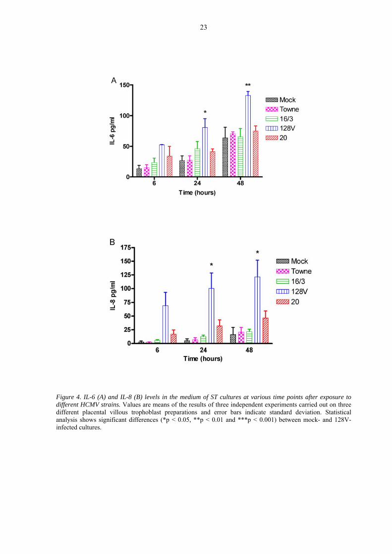
infected with any of the HCMV strains. The amount of IL-6 measured in the mock-infected
ST cultures was observed to increase in a time-dependent manner [Figure 4A]. Similar or
slightly increased amounts of IL-6 were found in most HCMV-infected ST cultures. One
strain (128V) induced the production of a significantly (p < 0.05) higher amount of IL-6 as
compared with that in the mock-infected cultures (Figure 4A). Analyses of the IL-8 levels in
the supernatants of the ST cultures revealed that, in contrast with the other strains, strain
128V was a potent IL-8 inducer. The second most potent IL-8-inducing strain was strain 20
(Figure 4B). The number of IE antigen-containing cells in ST cultures infected with different
HCMV strains seemed to be dependent on the amount of IL-8 produced (Table III).
Table III. Interrelationship between production of IL-8 and IE antigen in rensponse to infection with different
HCMV strains in ST cells
HCMV strain IL-8 No. of nuclei positive (pg/ml)a for IE antigen±SDb
- 3.1±1.5 -
Towne 28.2±7.6 9.0±3.0
16/3 21.5±8.1 12.0±2.0
128V 150.4±15.5 323±45.0
20 59.3±2.1 46.0±8.0
a Values are means ± SD of the results of three independent experiments. b Mean number ± SD of nuclei positive for IE (intermediate-early) antigen per microwell 48 h after infection of HCMV strains at an MOI of 0.1 from three independent experiments.
23
Figure 4. IL-6 (A) and IL-8 (B) levels in the medium of ST cultures at various time points after exposure to different HCMV strains. Values are means of the results of three independent experiments carried out on three different placental villous trophoblast preparations and error bars indicate standard deviation. Statistical analysis shows significant differences (*p < 0.05, **p < 0.01 and ***p < 0.001) between mock- and 128V-infected cultures.
24
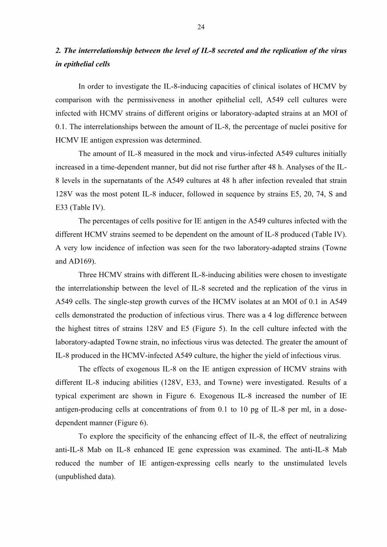
2. The interrelationship between the level of IL-8 secreted and the replication of the virus
in epithelial cells
In order to investigate the IL-8-inducing capacities of clinical isolates of HCMV by
comparison with the permissiveness in another epithelial cell, A549 cell cultures were
infected with HCMV strains of different origins or laboratory-adapted strains at an MOI of
0.1. The interrelationships between the amount of IL-8, the percentage of nuclei positive for
HCMV IE antigen expression was determined.
The amount of IL-8 measured in the mock and virus-infected A549 cultures initially
increased in a time-dependent manner, but did not rise further after 48 h. Analyses of the IL-
8 levels in the supernatants of the A549 cultures at 48 h after infection revealed that strain
128V was the most potent IL-8 inducer, followed in sequence by strains E5, 20, 74, S and
E33 (Table IV).
The percentages of cells positive for IE antigen in the A549 cultures infected with the
different HCMV strains seemed to be dependent on the amount of IL-8 produced (Table IV).
A very low incidence of infection was seen for the two laboratory-adapted strains (Towne
and AD169).
Three HCMV strains with different IL-8-inducing abilities were chosen to investigate
the interrelationship between the level of IL-8 secreted and the replication of the virus in
A549 cells. The single-step growth curves of the HCMV isolates at an MOI of 0.1 in A549
cells demonstrated the production of infectious virus. There was a 4 log difference between
the highest titres of strains 128V and E5 (Figure 5). In the cell culture infected with the
laboratory-adapted Towne strain, no infectious virus was detected. The greater the amount of
IL-8 produced in the HCMV-infected A549 culture, the higher the yield of infectious virus.
The effects of exogenous IL-8 on the IE antigen expression of HCMV strains with
different IL-8 inducing abilities (128V, E33, and Towne) were investigated. Results of a
typical experiment are shown in Figure 6. Exogenous IL-8 increased the number of IE
antigen-producing cells at concentrations of from 0.1 to 10 pg of IL-8 per ml, in a dose-
dependent manner (Figure 6).
To explore the specificity of the enhancing effect of IL-8, the effect of neutralizing
anti-IL-8 Mab on IL-8 enhanced IE gene expression was examined. The anti-IL-8 Mab
reduced the number of IE antigen-expressing cells nearly to the unstimulated levels
(unpublished data).

25
Table IV. Interrelationship between production of IL-8 and IE antigen in response to infection with different HCMV strains in A549 cells 1
HCMV strain2 IL-8 Nuclei positive (pg/ml) for IE3 antigen (%) - 108 -
128V 5009 3.5 ± 0.7
E33 385 1.4 ± 0.6
E5 764 1.8 ± 0.7
20 687 NT4
S 466 NT
74 508 NT
Towne 156 0.5 ± 0.1
AD169 329 0.7 ± 0.3
1 48 h after infection; 2 at an MOI of 0.1; 3 immediate-early antigen; 4 not tested
Figure 5. Replication of HCMV strains in A549 cells. F alysis of virus growth kinetics of HCMV strains of
different IL-8 inducing ability in A549 cultures, cells were infected at a MOI of 0.1. A549 culture lysates and
supernatants were pooled at varies times after infection d assayed for infectious virus. The virus titers were
determined by plaque assay. Results are representative of three experiments.
or an
an

26
Fi ure 6. Enhancement of IE gene expression of HCMV in A549 cells by IL-8. A549 cells were incubated with
th icated concentrations of IL-8 before and after infection with HCMV. Cells were infected at a MOI of
0. At 48 h after infection the IE antigen-positive cells were detected by using immunfluorescence assay. Data
are expressed as mean percentage ± of IE antigen-psitive cells per 30 microscope fields calculated.
g
e ind
1.
27
3. Distribution of the genotypes of the genes UL55, UL146 and UL139 among HCMV
strains, and the association between polymorphisms within the genes UL55, UL146 and
UL139 and the ability to induce IL-8 production
UL55 genotypes
gB genotyping was carried out on 26 samples. A region of high peptide variability in
the gene UL55 was amplified by nested PCR. Amplification products were analyzed by
electrophoresis and subjected to restriction analysis by using Hinf I and Rsa I. Four distinct
gB genotypes (gB1-gB4) could be identified though the different lengths of restriction
fragments (36-239 bp).
One sample (E24) contained 2 genotypes (gB1 and gB2). The overall distribution of the 27
genotypes was as follows: 19 gB1, 3 gB2, 4 gB3, and 1 gB4. In the isolates from 15
congenitally infected infants 11 gB1, 3 gB2 and 2 gB3 genotypes were detected (Table V).
UL146 and UL139 sequences
6 and UL139 genotypes in 36 samples were investigated by PCR and
es were amplified by PCR, using validated primer sets. In some
t
l. [7] was used for UL146 and for UL139.
s for which genotype data were obtained was 31, including
moth
ll fall into 14 genotypes defined previously [7] and are designated G1-G14.
The UL14
sequencing using primers in conserved regions. UL146 was amplified from 36 samples and
sequences were determined from 20, and UL139 was amplified from 36 samples and
sequences determined from 26. A total of 9 samples failed to yield products from either gene
(Table V).
The relevant gen
cases, nested PCR was used. Most products were sequenced directly, and some were cloned
into plasmids and then sequenced. For reference sequences, we included previously analyzed
strains Toledo, Towne, TB40/E, Davis and Merlin [7, 13]. The genotye system of Dolan e
a
The total number of strain
7 er-baby pairs.
UL146 genotypes
The UL146 coding sequences range in length in the interval 342 to 378 bp (114-126
codons). A
UL146 is hypervariable throughout its length and only a few amino acid residues are
completely conserved in all sequences.

28
Nine UL146 genotypes (G1-2, and G7-13) were apparent in our samples. Seven
different UL146 genotypes (G7-13) were detected in strains from congenital infections
L139 genotypes
coding sequences range in length from 372 to 444 bp (124-148 codons).
han in UL146, but that among genotypes is lower.
assigments still held.
The gB genotype (gB1) and the DNA sequence of genotypes UL146 (G11) and
infant did not change in the 17 months during which samples (16/3 and
is
mited
4 and E5) or UL139 (compare strain 128V with strains
(Table V).
U
The UL139
All fall into 8 genotypes and are designated G1-G8 [88]. The protein encoded by each
HCMV UL139 genotype contains a putative signal peptide sequence and a transmembrane
region. Variation is concentrated in the N-terminal portion of the protein. Variation within
genotypes tends to be higher in UL139 t
Seven UL139 genotypes (G1-7) were found in congenitally infected infants (Table
V). Isolate E24 is assigned to 2 genotypes (G4 and G7). After resequencing and analysis of 5
clones, these
UL139 (G2) in an
16/5) were obtained (Table V).
Polymorphisms within UL55, UL146 and UL139 genes and ability to induce IL-8
production
The number of strains whose ability to induce IL-8 production has been examined
li , and only one strain (128V) induces high levels (Table VI). However, there appears
to be no obvious correlation between the IL-8-inducing ability with the genotype of UL55
(compare strain 128V with strains 7
20 and E5). A correlation with UL146 remains possible, since no other strain has the same
genotype (G9) as strain 128V.

29
Table V. Genetic variations in the UL55, UL146 and UL139 genes of HCMV strains.
Straina Clinical b Age/sesource xc Clinical
detailsdgB
genotypee UL146 genotypef UL139 genotypef
16/5 U/P Infant C 1 G11 G2 128V U/P Infant C 3 G9 G4 E24 U/P Infant C 1,2 G9 G4, G7 16/3 U/P Infant C 1 G11 G2
E34AV U/P Mother C+ 1 G13 G2 5DV U/P Adult CH 1 G10 G4 E5 U/P Adult C+ 3 G1 G4
E33AV U Mother C+ 1 G7 G2 E33BV U/P Infant C 1 G7 G2 E36BV2 U Infant C ND G7 G4 E36AV1 U Mother C+ ND ND ND E41BV3 U Infant C 1 G12 G5 E41AV1 U Mother C+ 1 ND ND E42BV1 U Infant C 1 G8 G7 E42AV1 U Mother C+ 1 ND G1 E43BV1 U Infant C 2 G11 G4 E4 3AV1 U Mother C ND ND G1 E ND 62BV1 U Infant C G13 G4 E ND 62AV1 U Mother C+ ND G4 E4 1 5BV U Infant C 1 ND ND E ND 59BV1 U Infant C ND ND E28BV1 U Infant C ND ND G2
16/2 U Infant C 1 ND ND E3 V 4B/B U 5m/M C+ 1 ND ND E60BV1 U Infant C+ 1 ND ND E55BV1 U Infant C+ ND ND ND E63BV1 U 10 w/F C+ ND ND G1 E63AV1 U Mother C+ ND ND ND E49AV1 U Mother C+ 1 G2 ND E52BV1 U 14 m/M C+ 2 G10 G2
20 B/P Adult R 4 G1 G4 74 B/P Adult R 3 G12 G3 S U Infant C 1 G7 G2 B U Infant C 3 G7 G6 83 U Infant C 1 ND G2 89 U Infant C 1 ND G2
Davis B P/P Infant C 1 G5 G4 Toledo U/P Infant C 2 G1 G4 Towne U/P Infant C 1 G7 G5 M U/P ND erlin Infant C G2 G1 TB40/E T ND S/P Adult B G8 G4 AD169 B P/P Infant C+ 2 ND ND
a V b U, u P, , bloo S, throat swab; P, pass lture; s are in wee months . Sex ale; F, female; d C+, HCMV posi by PCR or IgM rum; C, con , chron emod t; R, renal transpl recipient; e marrow transp ient
e G re den gB1- L55 otypes are denoted G for UL146 an -G8 for UL139 ultiple gen s are s d by commas. ND, not termined.
irus strains; rine; B biopsy; B d; T aged in cell cu C Ageks (w) or (m) es: M, m tive in segenital; CHeno es a
ic h ialysis patieng U
ant B bon1 4
lant reciptyp oted B4 for f enG -G1 d G1. M otype eparate de

30
Table VI. R ionship n genoty and prod ion of IL-8 s and in A549 ce
Genotype IL-8 pg/ml
elat betwee pes uct in ST lls
Strain Clinica b
5c UL146d UL139e in STs in 9 cells
a l Source
UL5 A54
E 1 7 2 ND 5 33 U 38
S U 1 7 2 ND 6 46
16/ 1 11 2 21.5 ND 3 U
128V U 3 9 4 150.4 5009
20 B 4 1 4 59.3 687
74 B 3 12 3 ND 508
E5 U 3 1 4 ND 764
Towne U 1 7 5 28.5 156
avirus strain; bU, urine; B, blood; cGenotypes are denoted gB1-gB4 for UL55; dGenotypes are denoted G1-G14 for UL146; eGenotypes are denoted G1-G8 for UL139; ND, not determined

31
Discus
Congenital HCMV transm res ubst ng- urod l
morbidity in newborns, including m tal retard on and se rineural ring loss ( L)
[89] hile antivira herapies may limit the SNHL, therapeutic interv ns
have only a limited ability to reve other ologic in ries [90 e most e ive
strategies for the control of congenital HCMV y require the prevention of fetal infection
by preconceptual vaccination or anti-HCMV immunoglobulins [91, 92]. However, v es
and munoglobuli are not yet l nsed fo evention congeni CMV in on.
Continued study of the molecular pathophysiology of HCMV infection in cell types relevant
to nital transm ion is therefore warranted, in order to facilitate the develop of
1. Production of proinflammatory cytokines of ST cultures infected with clinical HCMV
isolates.
Numerous investigations have indicated that placental cells can be productively
infected by HCMV [42, 64-67]. Experiments have been performed in vitro by using
laboratory-adapted strains and a high MOI (10 or 1), which may not be entirely
representative of the behavior of wild strains. In the placenta, proinflammatory cytokines,
including IL-1, IL-6, IL-8 and TNF-α, and anti-inflammatory cytokines such as IL-4 and IL-
10, are produced by the trophoblasts [70-74]. Intrauterine infections are associated with the
expression of placental cytokines such as IL-1 and IL-8 [71]. IL-8 has been demonstrated to
upregulate the replication of laboratory-adapted HCMV strains in various cells, including the
ST [75-77]. The presence of this cytokine in the placental microenvironment may play a
regulatory role, potentially promoting the progression of latent HCMV infection in the
placenta into a fulminating infection that can be transmitted to the foetus.
We have found that HCMV strains induce the production of different amounts of IL-
8 in STs differentiated from first-trimester CTs, and the IE gene expression of the virus
increases with the amount of IL-8 produced. This observation indicates that IL-8 may be
involved in the materno-foetal transmission of HCMV. Our findings suggest that certain
HCMV strains induce a high level of IL-8 in the STs, which in turn enhances productive
HCMV expression in the placenta, while others replicate if the IL-8 is provided by co-
infection agents, i.e. human immunodeficiency virus (HIV), human herpesvirus-6 or
sion
ission ults in s antial lo term ne evelopmenta
en ati nso hea SNH
. W l t severity of entio
rse neur ju ]. Th ffect
ma
accin
im ns ice r pr of tal H fecti
conge iss ment
novel disease prevention strategies.

32
bacteria. The potential of various bacteria to stimulate cytokine production in highly purified
rimary trophoblast cultures varies. E. coli and B. fragilis exhibited the highest potencies to
ulate IL
potentially relevant functions. The key target
emokine similar to IL-8 [15].
2. The interrelationship between the level of IL-8 secreted and the replication of the virus
lial cells
the various
p
stim -8 expression [78].
The fact that the IL-6-mediated stimulation of trophoblast cells did not result in an
enhancement of IL-8 production [93] suggests that particular genotypes of the target HCMV
genes may be associated with IL-8 induction in physiologically relevant STs. In recent years,
it has become clear that many HCMV genes are strikingly variable in sequence between
strains, with at least 25 of the 165 genes present in wild-type virus strains being
hypervariable [4, 7]. The anticipated association between pathogenic properties and
genotypes is a subject of importance, but has yet to find conclusive experimental support.
We intend to focus on several hypervariable genes whose genotypic structures are well
characterized and which encode products with
is UL146, which encodes a potent CXC ch
in epithe
HCMV displays a broad host cell range and infects many different cell types [94].
Epithelial cells are major targets of permissive infection by HCMV and play a role in the
spread of the virus in infected tissues during acute infection. Little is known about the viral
factors that determine HCMV epithelial cell tropism [95]. Most analyses of HCMV
epithelial cell tropism have been performed with the use of laboratory-adapted strains and a
high MOI. Potential differences between HCMV strains were not taken into consideration.
A549 epithelial cells were infected with HCMV isolates of different origins with a
view to acquiring support for our earlier suggestion that interstrain differences in HCMV
epithelial cell tropism depend on the IL-8-inducing capacity and determine the outcome of
HCMV infection [96]. The data revealed that the IL-8-inducing capacities of
HCMV isolates differed in A549 cells too. The IE gene expression and the yield of
infectious virus were dependent on the amount of IL-8 produced in these cells.
IL-8 has been demonstrated to upregulate the replication of laboratory-adapted
HCMV strains in various cells [75-77]. Our results indicate that exogenous IL-8 increases
the number of IE antigen-producing cells, in a dose-dependent manner in A549 cells infected
with HCMV strains with different IL-8-inducing abilities. The presence of this cytokine in
the microenvironment may promote the progression of HCMV infection.

33
Our findings support our earlier suggestion that certain HCMV strains induce a high
level of IL-8 in epithelial cells, which in turn enhances productive HCMV infection in these
cells [95]. Other strains can replicate in epithelial cells if IL-8 is provided by coinfecting
We focused on hypervariable genes whose genotypic structures are well
6, which encodes a potent CXC chemokine similar to IL-8, and UL139,
hich maps about 5 kbp distant and potentially encodes a highly O-glycosylated membrane
emon
r many years
agents, i.e. viruses or bacteria. The potential of various bacteria to stimulate IL-8 production
in epithelial cells varies [78, 97].
Our results showed that HCMV replication in A549 cells is strain-dependent, in
support of the view that specific viral gene(s), including UL146, which encodes a potent
CXC chemokine similar to IL-8, are required for efficient replication in this cell type.
3. Distribution of the genotypes of the genes UL55, UL146 and UL139 among HCMV
strains, and the association between polymorphisms within the genes UL55, UL146 and
UL139 and the ability to induce IL-8 production
characterized and which encode products with potentially relevant functions. The primary
targets were UL14
w
protein. Genetic variability in the gB gene was also investigated. gB is an important target
for neutralizing antibodies and participates in cell-receptor interactions. With the same
approach, the genotypes of several paired HCMV samples from mothers and babies were
also determined.
The gB1, genotype is the most prevalent in congenital infections followed by
genotypes gB3, and gB2, as it observed in erlier studies in our laboratory [83, 98]. The
d strated predominance of gB genotype 1 could reflect the infection type of the study
population.
The gB genotype (gB1) and the DNA sequences of genotypes UL146 and UL139 did not
change in an infant in the period during which samples were obtained (16/3 and 16/5) as
observed in persistently infected renal transplant recipients [14]. Since the gene UL 146 is
variable, many studies have addressed the stability of UL146 upon isolation ove
[7, 12-14, 99].
As found in previous studies, mixed infections with different HCMV strains were
common [100]. In one sample (E24), a single UL146 genotype and multiple UL 55 (gB1 and
gB2) and UL139 genotypes (G4, G7) were detected, which indicates that this infant was

34
infected with two different HCMV isolates as seen in both congenital infections and immune
compromised patients[7, 8, 13].
This could be due to different strains happening to contain the same genotype at one locus
but not at the other, or to the limitations of amplifying sequences present as mixtures in
unequal proportions.
Evidence for a genetic linkage between the three loci was unconvincing, consistent
with a role for interstrain recombination during HCMV evolution [4]. The fact that the
genotyping of one gene therefore does not shed light on the genotypes of other genes
stifies the need to determine genotypes separately for each target locus.
frequence variation in the genes UL146
nd UL139. The small sample size we does not allow to draw a direct link between the
elial cells.
cation.
ju
The analysis to determine whether an association exists between the UL55, UL146
and UL139 genotypes and ability to induce IL-8 production in STs and A549 cells was
underpowered, because of the unexpectedly high
a
genotypes and the ability to induce IL-8 production. However, a correlation with UL146
remains possible.
The analysis of further strains is required, especially those able to induce high levels of IL-8
or those known to have a UL146 genotype G9. Additionally data for other loci (for which
data collection is ongoing) will broaden the analysis.
Continuation of the investigation will help us determine whether particular genotypes
of UL146 (and other genes) are associated with IL-8 induction in physiologically relevant
STs or other epith
A better understanding of the molecular basis of the interactions between HCMV
isolates and epithelial cells, including trophoblasts, may yield novel strategies with which to
prevent the progression of the virus. IL-8 may be an important new target for the control of
HCMV repli

35
Summary Forty per cent of women with primary HCMV infection during gestation transmit the
infection to their fetuses, which may result in abnormalities for the newborn, varying in
degree from mild to severe. Since placental HCMV infection has been detected both in the
on, clinical HCMV isolates and a low MOI were used.
oting the progression of HCMV infection in the placenta
f
between the level of IL-8 secreted and the replication of HCMV was
ined in another epithelial cell type, A459 cells.
The data revealed that the IL-8-inducing ability of the various HCMV isolates
gene expression and the yield of infectious virus were
ependent on the amount of IL-8 produced in these cells. Exogenous IL-8 increased the
umber of IE antigen-producing cells, in a dose-dependent manner, in A549 cells infected
ith HCMV strains with different IL-8-inducing abilities.
Our results showed, that HCMV replication in epithelial cells is strain-dependent,
suggesting that specific viral gene(s), including UL146, which encodes a potent CXC
chemokine similar to IL-8, are required for efficient replication in this cell type.
presence and in the absence of fetal infection, the placenta should be considered the most
important site of either protection of the fetus from HCMV infection or transmission, by
acting as a viral reservoir and allowing the infection to reach the fetal compartment.
The factors whereby HCMV in the placenta develops into a fulminating infection and
spreads to the fetus are not known.
This study concerned three main features:
1. The production of proinflammatory cytokines was investigated in ST cultures infected
with HCMV strains. The interrelationships between the cytokines produced in the ST
cultures and the number of nuclei of ST expressing the HCMV IE gene were examined. To
resemble a natural infecti
TNF-α and IL-1β were not detected in the supernatants of any ST cultures. Similar or
increased amounts of IL-6 were found in the HCMV-infected cultures. The IL-8-inducing
capacities of the HCMV strains differed in the ST cultures. The IE gene expression of the
virus provided was dependent on the amount of IL-8 produced in the STs.
Our observations indicate that IL-8 in the placental microenvironment may play a
regulatory role, potentially prom
into a ulminating infection that can be transmitted to the fetus. Certain HCMV strains
induce high amounts of IL-8, which in turn enhances HCMV replication in the placenta,
while others can replicate if the IL-8 is provided by a co-infecting agent.
2. The interrelationship
exam
differed in A549 cells too. The IE
d
n
w

36
3. Genotyping was carried out to elucidate whether there is any link between the ability of
CMV isolates to induce IL-8 production in STs and in another epithelial cell type, A549
elation between the IL-8-inducing ability and
ith UL146 remains possible. The analysis
H
cell. We focused on hypervariable genes (UL55, UL146 and UL139), whose genotype
structure has been well characterized, and which encode products with potentially relevant
functions.
The total number of strains for which genotype data were obtained was 31, including
7 mother-baby pairs. Four gB genotypes (gB1-gB4), 9 UL146 genotypes (G1-2, and G7-13),
and 7 UL139 genotypes (G1-7) were apparent in our samples. The number of strains whose
ability to induce IL-8 production in STs and A549 cells has been examined is limited.
However, there appears to be no obvious corr
the genotype of UL55 or UL139. A correlation w
of additional strains is required, especially those able to induce high levels of IL-8 or those
known to have the genotype G9 gene UL146.
A better understanding of the molecular basis of the interactions between HCMV
isolates and trophoblasts may yield novel strategies with which to prevent the vertical
transmission of the virus. IL-8 may be an important new target for the control of HCMV
replication.

37
Acknowledgements I would like to express my gratitude to all those who have given me the possibility to carry
out research work and prepare this thesis.
I express my deepest thanks to my supervisor, Professor Rozália Pusztai, for her invaluable
ientif
I also o
r granting me the possibility to spend 3 months in his laboratory and to study
ew molecular methods of HCMV research.
thank Mrs. Erzsébet Borzási, Mrs Erzsébet Dallos-Szilágyi, Mrs Ildikó Wellinger and Ms.
atalin Hegedűs for their advice on methodology and excellent technical assistance.
hanks are due to all of the contributors to the study.
This study was supported by grants from the Hungarian Scientific Research Fund (OTKA-
T26442) and the Higher Education Research and Development Fund (FKFP-113), a FEMS
Research Fellowship and a FEMS-ESCMID Joint Fellowship.
sc ic guidence and encouragement.
I am particularly grateful to Dr. Emőke Endreffy and Dr. Zoltán Maróti from professional
aspects; it was a great pleasure to work under their skillful guidance and with their tireless
support.
I would like to thank to Professor Yvette Mándi, Director of the Department of Medical
Microbiology and Immunobiology, for accepting me as a Ph.D. student.
I am grateful to Dr. Judit Deák for providing the testing routinely for bacterial infection of
the cytotrophoblasts.
we my thanks to Professor Attila Pál, Director of the Department of Gynecology and
Obstetrics, for providing the clinical specimens.
I would like to thank to Dr. Andrew Davison (Institute of Virology, University of Glasgow,
Scotland) fo
n
I
K
T

38
References 1. Britt, W., Stagno, S.: Cytomegalovirus. In Remington J.S., Klein, J.O., eds. Infectious
Cytomegalovirus: genetic
354, 332 - 346; 1980.
iology of cytomegalovirus infection in women and their infants. N Engl J Med
riation in cytomegalovirus
163
ssan-Walker, AF., Lee, L., Addison, C.,
diseases of the fetus and newborn infant. 6th ed. Philadelphia: W.B. Saunders 739-781;
2006.
2. Huang, ES., Huang, SM., Tegtmeier, GE., Alford, C.:
variation of viral genomes. Ann NY Acad Sci
3. Huang, ES., Alford, CA., Reynolds, DW., Stagno, S., Pass, RF.: Molecular
epidem
303, 958-962; 1980.
4. Pignatelli, S., Dal Monte, P., Rossini, G., Landini, M.: Genetic polymorphisms among
human cytomegalovirus (HCMV) wild-type strains. Rev Med Virol 14, 383-410; 2004.
5. Britt, WJ., Mach, M.: Human cytomegalovirus glycoproteins. Intervirology 39, 401-
412; 1996.
6. Chou, S., Dennison, KM.: Analysis of interstrain va
glycoprotein B sequences encoding neutralisation-related epitopes. J Infect Dis ,
1229-1234; 1991.
7. Dolan, A., Cunningham, C., Hector, RD., Ha
Dargan, DJ., McGeoch, DJ., Gatherer, D., Emery, VC., Griffiths, PD., Sinzger, C.,
McSharry, BP., Wilkinson, GW., Davison, AJ.: Genetic content of wild-type human
cytomegalovirus. J Gen Virol 85, 1301-1312; 2004.
8. Arav-Boger, R., Foster, CB., Zong, JC., Pass, RF.: Human Cytomegalovirus Encoded
-Chemokines Exhibit High Sequence Variability in Congenitally Infected Newborns. J
Infect Dis 193, 788-791; 2006.
9. Chee, MS., Bankier, AT., Beck, S. and 12 other authors: Analysis of the protein
coding content of the sequence of human cytomegalovirus strain AD169. Curr Top
Microbiol Immunol 154, 125–169; 1990.
10. Cha, TA., Tom, E., Kemble, GW., Duke, GM., Mocarski, ES., Spaete, RR.: Human
cytomegalovirus clinical isolates carry at least 19 genes not found in laboratory strains. J
Virol 70, 78–83; 1996.
11. Murphy, E., Yu, D., Grimwood, J., Schmutz, J., Dickson, M., Jarvis, MA., Hahn,
G., Nelson, JA., Myers, RM., Shenk, TE.: Coding potential of laboratory and clinical
strains of human cytomegalovirus. PNAS 100, 14976-14981; 2003.
39
12. Hassan-Walker, AF., Okwuadi, S., Lee, L., Griffiths, PD., Emery, VC.: Sequence
variability of the alpha-chemokine UL146 from clinical strains of human
13.
Analysis of the human cytomegalovirus genomic region
egalovirus Infection in the United States, 1988
cytomegalovirus. J Med Virol 74, 573-579; 2004.
Lurain, NS., Fox, AM., Lichy, HM., Bhorade, SM., Ware, CF., Huang, DD., Kwan,
SP., Garrity, ER., Chou, S.:
from UL146 through UL147A reveals sequence hypervariability, genotypic stability, and
overlapping transcripts. Virol J 3, 4; 2006.
14. Stanton, R., Westmoreland, D., Fox, JD., Davison, AJ., Wilkinson, GW.: Stability of
human cytomegalovirus genotypes in persistently infected renal transplant recipients. J
Med Virol 75, 42-46; 2005.
15. Penfold, ME., Dairaghi, DJ., Duke, GM., et al.: Cytomegalovirus encodes a potent α-
chemokine. Proc Natl Acad Sci USA 96, 9839–45; 1999.
16. Staras, SAS., Dollard, SC., Radford, KW., Flanders, WD., Pass, RF., and Cannon
MJ.: Seroprevalence of Cytom 1994.
17.
Arabia. J Infect Dis 152, 1351; 1985.
19. egalovirus, Epstein-Barr virus and
20. Stagno, S., Cloud, GA.:
Proc Natl Acad Sci USA 91, 2384-2389; 1994.
22. Isolation of cytomegalovirus
23.
mmune deficiency syndrome: Documentation
Clin Infect Dis 43, 1143–1151; 2006.
Ashraf, SJ., Paranse, CM., Arya, SC.: Cytomegalovirus antibodies of patients in the
Gizan area of Saudi
18. Wang, PS., Evans, AS.: Prevalence of antibodies to Epstein-Barr virus and
cytomegalovirus in sera from a group of children in the People’s Republic of China. J
Infect Dis 153, 150-152; 1986.
Pethő, E., Geiger, J.: Seroprevalence of cytom
toxoplasma in the normal population. Transfúzió 29, 3-7; 1996 (in Hungarian).
Working parents: the impact of day care and breast-feeding on
cytomegalovirus infections in offspring.
21. Pass, RF.: Cytomegalovirus, Field’s Virology, 4th Edition (Knipe, DM & Howley, PM,
eds), Lippincott Williams and Wilkins, Philadelphia, pp 2675-2705; 2001.
Hutto, C., Little, EA., Ricks, R., Lee, JD., Pass, RF.:
from toys and hands in a day care center. J Infect Dis. 154, 527–530; 1986.
Drew, WL., Sweet, ES., Miner, RC., Mocarski, ES.: Multiple infections by
cytomegalovirus in patients with acquired i
by Southern blot hybridization. J Infect Dis 150, 952-963; 1984.
40
24. Leach, CT., Detels, R., Hennessey, K., Zin, Z., Visscher, BR., Dudley, JP., Cherry,
JD.: A longitudinal study of cytomegalovirus infection in human immunodeficiency
virus type 1-seropositive homosexual men: Molecular epidemiology and association with
25.
27. Sule, O., Devi, R., Rice, P.: Cytomegalovirus infection in
28. V serology and CMV disease
29.
ent of cytomegalovirus and Epstein-Barr virus
30. ., Jabs, DA., Kern, ER. and
31. ngenital cytomegalovirus disease. Orvosi Hetilap 121,
32. Landini,
33. ., Jahn, G.:
35. ternal cytomegalovirus
infection and perinatal transmission. Clin Obstet Gynaecol 25, 563-576; 1982.
6. Griffiths, PD., Baboonian, C.: A prospective study of primary cytomegalovirus
infection during pregnancy: final report. Br J Obstet Gynaecol 91, 307-315; 1984.
disease progression. J Infect Dis 170, 293-298; 1994.
Chandler, SH., Handsfield, HH., McDougall, JK.: Isolation of multiple strains of
cytomegalovirus from women attending a clinic for sexually transmitted diseases. J
Infect Dis 155, 655-660; 1987.
26. Bale, JF., Petheram, SJ., Inara, ES., Murph, JR.: Cytomegalovirus reinfection in
young children. J Pediatr 128, 347-352; 1996.
Wreghitt, TG., Teare, EL.,
immunocompetent patients. Clin Infect Dis 37, 1603-1606; 2003.
Laing, RBS., Brettle, RP. and Leen, CLS.: Effect of CM
on AIDS morbidity and mortality. Infection 25, 255; 1997.
Razonable, RR., Paya, CV.: Herpesvirus infections in transplant recipients: current
challenges in the clinical managem
infections. Herpes 10, 60-65; 2003.
de Jong, MD., Galasso, GJ., Gazzard, B., Griffiths, PD
Spector, SA.: Summary of the II International Symposium on Cytomegalovirus.
Antiviral Res 39, 141-162; 1998.
Korányi, Gy., Krausz, J.: Co
1129-1134; 1980 (in Hungarian).
Lanari, M., Lazzarotto, T., Venturi, V., Papa, I., Gabrielli, L., Guerra, B.,
MP. and Faldella, G.: Neonatal cytomegalovirus blood load and risk of sequelae in
symptomatic and asymptomatic congenitally infected newborns. Pediatrics 117, 76–83;
2006.
Hamprecht, K., Maschmann, J., Vochem, M., Dietz, K., Speer, C
Epidemiology of transmission of cytomegalovirus from mother to preterm infant by
breastfeeding. The Lancet 357, 513-518; 2001.
34. Demmler, GJ.: Cytomegalovirus, Viral Diseases in Pregnancy. ed Gonik B, Springer-
Verlag, New York, Berlin, Heidelberg. p. 69-91; 1994.
Stagno, S., Pass, RF., Dworsky, ME., Alford, CA.Jr.: Ma
3

41
37. Nankervis, GA., Kumar, ML., Cox, FE., Gold, E.: A prospective study of maternal
cytomegalovirus infection and its effect on the fetus. Am J Obstet Gynecol 149, 435-440;
1984.
Alford, CA., Stagno, S., Pass, RF., Britt, 38. WJ.: Congenital and perinatal
39.
328; 1991.
on to maternal antibody status. N Engl J
41. ., Jahn, G.: Cell types
l 423, 249-256; 1993.
; 2000.
pe 1 variants even with cytomegalovirus coinfection
44. ., Tanaka, Y., Shimuzi, H., Yokota, S.: Ascending
45.
46. chke, K., Mendoza, GR., Bazeley, PL.: Placental and fetal manifestations of
47.
te. Arch Pathol Lab Med. 108, 403–406;
48. JA.: Surveillance for congenital
cytomegalovirus infection. Rev Infect Dis 174, 745-753; 1990.
Demmler, GJ.: Summary of a workshop on surveillance for congenital cytomegalovirus
disease. Rev Infect Dis 13, 315-
40. Fowler, KB., Stagno, S., Pass, RF., Britt, WJ., Boll, TJ., Alford, CA.: The outcome of
congenital cytomegalovirus infection in relati
Med 326, 663-667; 1992.
Sinzger, C., Müntefering, H., Löning, T., Stoss, H., Plachter, B
infected in human cytomegalovirus placentitis identified by immunohistochemical
double staining. Virchows Archiv A Pathol Anat Histopatho
42. Fischer, S., Genvacev, O., Maidji, E., Pereira, L.: Human cytomegalovirus infection
of placenta cytotrophoblast in vitro and in utero: implication for transmission and
pathogenesis. J Virol 74, 6808-6820
43. Kilani, RT., Chang, LJ., Garcia-Lloret, MI., Hemmings, D., Winkler-Lowen, B.,
and Guilbert, LJ.: Placental trophoblasts resist infection by multiple human
immunodeficiency virus (HIV) ty
but support HIV replication after provirus transfection. J Virol 71, 6359–6372; 1997.
Ohyama, M, Motegi, Y., Goto, A
placentofetal infection caused by cytomegalovirus. Br J Obstet Gyneacol 99, 770; 1992.
Hayes, K., Gibas, H.: Placental cytomegalovirus infection without fetal involvement
following primary infection in pregnancy. J Pediatr. 79, 401–405; 1971.
Benirs
cytomegalovirus infection. Virchows Arch B Cell Pathol. 16, 121–139; 1974.
Mostoufi-zadeh, M., Driscoll, SG., Biano, SA., Kundsin, RB.: Placental evidence of
cytomegalovirus infection of the fetus and neona
1984.
Istas, AS., Demmler, GJ., Dobbins, JG., Stewart,
cytomegalovirus disease: a report from the National Congenital Cytomegalovirus
Disease Registry. Clin Infect Dis. 20, 665–670; 1995.

42
49. Mühlemann, K., Miller, RK., Metlay, L., Menegus, MA.: Cytomegalovirus infection
of the human placenta: an immunocytochemical study. Hum Pathol. 23, 1234–1237;
1992.
50. Hemmings,, DG. and Guilbert, LJ.: Polarized Release of Human Cytomegalovirus
from Placental Trophoblasts J Virol 76, 6710–6717; 2002.
51. Pereira, L., Maidji, E., McDonagh, S., Tabata, T.: Insights into viral transmission at
the uterine–placental interface. Trends Microbiol 13, 164-174; 2005.
53. n, P.: Pathology of the human placenta. Springer, New York;
54.
1-440; 1999.
57.
irst
; 1967.
61. ann, P., Castellucci, M.: Extravillous trophoblast int he human placenta.
62.
63. r, S.: Human Cytomegalovirus
Transmission from the Uterus to the Placenta Correlates with the Presence of Pathogenic
Bacteria and Maternal Immunity. J Virol 77, 13301-13314; 2003.
52. Vicovac, L., Aplin, JD.: Epithelial-mesenchymal transition during trophoblast
differentiation. Acta Anat. 156, 202-216; 1996.
Bernischke, K., Kaufman
1995.
Yeh, IT., Kurman, RJ.: Functional and morphologic expressions of trophoblast. Lab
Invest 61, 1-4; 1989.
55. Zdravkovic, M., Aboagye-Mathiesen, G., Guimond, MJ., Hager, H., Ebbesen, P.,
Lala, PK.: Susceptibility of MHC class I expressing extravillous trophoblast cell lines to
killing by natural killer cells. Placenta 20, 43
56. Brosens, I., Robertson, WB., Dixon, HG.: Morphologic changes in uteroplacental
arteries during pregnancy. Am J Obstet Gynecol 132, 233; 1978.
Damsky, CH., Fitzgerald, ML., Fisher, SJ.: Distribution patterns of extracellular
matrix components and adhesion receptors are intricately modulated during f
trimester cytotrophoblast differentiation along the invasive pathway, in vivo. J Clin
Invest 89, 210-222; 1992.
58. Aplin, JD.: Implantation, trophoblast differentiation and haemochorial placentation:
mechanistic evidence in vivo and in vitro. J Cell Sci 99, 681-692; 1991.
59. Boyd, JD., Hamilton, WJ.: Development and structure of the human placenta from the
end of the 3rd month of gestation. J Obstet Gynecol Br Commonw 74, 161-226
60. Foidart, JM., Hustin, J., Dubois, M., Schaaps, JP.: The human placenta becomes
haemochorial at the 13th week of pregnancy. Int J Dev Biol 36, 451-453; 1992.
Kaufm
Trophoblast Research 10, 21-62; 1997.
Pijnenborg, R.: Trophoblast invasion. J Reprod Med 3, 53-73; 1994.
Pereira, L., Maidji, E., McDonagh, S., Fishe

43
64. Maidji, E., Percivalle, E., Gerna, G., Fisher, S., and Pereira, L.: Transmission of
human cytomegalovirus from infected uterine microvascular endothelial cells to
differentiating/invasive placental cytotrophoblasts. Virology 304, 53-69; 2002.
cells are permissive to the
66. ni, MP.: Complete
egalovirus Infection of Primary Villous Term and First Trimester Trophoblasts J
68. tson, SA., Wegmann, TG.: The trophoblast as an integral
69.
kine interleukin 10. J
71.
nces its production in intrauterine infection. Biol Reprod 47,
72. andwerger, S.:
.
o cells
74. Flanagan, BF., Hart, CA., Johnson, PM.:
nta, decidua and
65. Halwachs-Baumann, G., Wilders-Truschnig, M., Desoye, G., Hahn, T., Kiesel, L.,
Rieger, P., Jahm, G., Sinzger, C.: Human trophoblast
complete replicative cycle of human cytomegalovirus. J Virol 72, 7598-7602; 1998.
Gabrielli, L., Losi, L.,Varani, S., Lazarotto T., Eusebi, V., Landi
replication of human cytomegalovirus in explants of first trimester human placenta. J
Med Virol 64, 499-504; 2001.
67. Hemmings, DG., Kilani, R., Nykiforuk, C., Preiksaitis, J., Guilbert, LJ.: Permissive
Cytom
Virol 72, 4970–4979; 1998.
Guilbert, L., Rober
component of a macrophage-cytokine network. Immunol Cell Biol 71, 49-57; 1993.
Robertson, SA., Seamark, RF., Guilbert, LJ., Wegmann, TG.: The role of cytokines
in gestation. Crit Rev Immunol 14, 239-292; 1994
70. Roth, I., Corry, DB., Locksley, RM., Abrams, JS., Litton, MJ., Fisher, SJ.: Human
placental cytotrophoblasts produce the immunosuppressive cyto
Exp Med 184, 539-548; 1996.
Shimoya, K., Matsuzaki, N., Taniguchi, T., Kameda, T., Koyama, M.,. Neki, R.,
Saji, F., and Tanizawa, O.: Human placenta constitutively produces interleukin-8
during pregnancy and enha
220-226; 1992.
Stephanou, A., Myatt, L., Eis, AL., Sarlis, N., Jikihara, H., H
Ontogeny of the expression and regulation of interleukin-6 (IL-6) and IL-1 mRNAs by
human trophoblast cells during differentiation in vitro. J Endocrinol 147, 487-496; 1995
73. Knöfler, M., Kiss, H., Mösl, B., Egarter, C., Husslein, P.: Interleukin-1 stimulates
tumor necrosis factor-alpha (TNF-alpha) release from cytotrophoblastic BeW
independently of induction of the TNF-alpha mRNA. FEBS Lett 405, 213-218; 1997.
De Moraes-Pinto, MI., Vince, GS.,
Localization of IL-4 and IL-4 receptors in the human term place
amniochorionic membranes. Immunology 90, 87-94; 1997.

44
75. Murayama, T., Kuno, K., Jisaki, F., Obuchi, M., Sakamuro, D., Furukawa, T.,
Mukaida, N., Matsushima, K.: Enhancement of human cytomegalovirus replication in
human fibroblast cell line by interleukin-8. J Virol 68, 7582-7585; 1994.
76. Capobianchi, MR., Barresi, C., Borghi, P., Gessani, S., Fantuzzi, L., Ameglio, F.,
Belardelli, F., Papadia, S., Dianzani, F.: Human immunodeficiency virus type 1 gp120
stimulates cytomegalovirus replication in monocytes: possible role of endogenous
77.
óth, F.: Placental macrophage contact potentiates the
enic
79. , Bartfai, G., Dobozy, A., Pal, A.,
).
81. esen, G., Laugesen, J., Zdravkovic, M. and Ebbesen, P.: Isolation and
82.
83. an
84. e, 1998. Methods Mol Biol
132, 115-130; 2000.
interleukin-8. J Virol 71, 1591-1597; 1997.
Bácsi, A., Aranyosi, J., Beck, Z., Ebbesen, P., Andirkó, I., Szabó, I., Lampé, L.,
Kiss, J., Gergely, L., D. T
complete replicative cycle of human cytomegalovirus in syncytiotrophoblast cells: role
of interleukin-8 and transforming growth factor-β1. J Interferon and Cytokine Res 19,
1153-1160; 1999.
78. Griesinger, G., Saleh, L., Bauer, S., Husslein, P., Knöfler, M.: Production of pro- and
anti-inflammatory cytokines of human placental trophoblasts in response to pathog
bacteria. J Soc Gynecol Invest 8, 334-340; 2001.
Szony, BJ., Hegedus, K., Bata, Z., Kemeny, L.
Kovacs, L. and Pusztai, R.: Cytotrophoblats cells from human first-trimester placental
villi: isolation, characterization, cultivation and research applications. Orv Hetil 143,
2047-2054; 2002 (in Hungarian
80. Kliman, HJ., Nestler, JE., Sermasi, E., Sanger, JM. and Strauss, JF.: Purification,
characterisation and in vitro differentiation of cytotrophoblasts from human term
placentae. Endocrinology 118, 1567-1582; 1986.
Aboagye Mathi
characterization of human placental trophoblast subpopulations from first-trimester
chorionic villi. Clin Diagn Lab Immunol 3, 14-22; 1996.
Douglas, GC. and King, BF.: Isolation of pure villous cytotrophoblast from term
human placenta using immunomagnetic microspheres. J Immunol Methods 119, 259-
268; 1989.
Lukácsi, A., Taródi, B., Endreffy, E., Babinszki, Á., Pál, A. and Pusztai, R.: Hum
cytomegalovirus gB genotype 1 is dominant in Congenital Infections in South Hungary.
J Med Virol 65, 537-542; 2001.
Staden, R:, Beal, KF., Bonfield, JK.: The Staden packag

45
85. Thompson, JD., Higgins, DG., Gibson, TJ.: CLUSTAL W: improving the sensitivity
of progressive multiple sequencing alignment through sequence weighting, position-
specific gap penalties and weight matrix choice. Nucleic Acids Res 22, 4673-4680;
86.
87. ar, S.: MEGA4: Molecular Evolutionary
88.
or, A., Moss, PA., Pusztai, R., Rawlinson, WD., Schulz TF. and
89. Curr Opin Infect Dis 18, 241-5; 2005.
91. C.: Progress toward an elusive goal: current status of
92.
n. N Engl J Med 353, 1350-62; 2005.
tumour necrotic factor (TNF)-
94.
of human cytomegalovirus. Adv Virus Res 46, 195-261; 1996.
96. , A. and Pusztai, R.: Production of Proinflammatory
2007.
1994.
Katoh, K., Kuma, K., Toh, H., Miyata, T.: MAFFT version 5: improvement in
accuracy of multiple sequence alignment. Nucleic Acids Res 33, 511-518; 2005.
Tamura, K., Dudley, J., Nei, M., Kum
Genetics 500 Analysis (MEGA) software version 4.0. Mol Biol Evol 24, 1596-1599;
2007.
Bradley, AJ., Kovács, IJ., Gatherer, D., Dargan, DJ., Alkharsah, KR., Chan, PK.,
Carman, WF., Dedicoat, M., Emery, VC., Geddes, CC., Gerna, G., Ismaeil, BB.,
Kaye, S., McGreg
Davison, AJ.: Genotypic analysis of two hypervariable human cytomegalovirus genes J
Med Virol, 80, 1615-1623; 2008.
Griffiths, PD. and Walter, S.: Cytomegalovirus.
90. Schleiss, MR. and McVoy, MA.: Overview of congenitally and perinatally acquired
cytomegalovirus infections: recent advances in antiviral therapy. Expert Rev Anti Infect
Ther 2, 389-403; 2004.
Schleiss, MR. and Heineman, T
cytomegalovirus vaccines. Expert Rev Vaccines 4, 381-406; 2005.
Nigro, G., Adler, SP., La Torre, R., Best, AM.: Passive immunization during
pregnancy for congenital cytomegalovirus infectio
93. Shimoya, K., Moriyama, A., Matsuzaki, N., Ogata, I., Koyama, M., Azuma, C., Saji,
F. and Murata, Y.: Human placental cells show enhanced production of interleukin
(IL)-8 in response to lipopolysaccharide (LPS), IL-1 and
α, but not to IL-6. Mol Hum Reprod 5, 885-891; 1999.
Plachter, B., Sinzger, C. and Jahn, G.: Cell types involved in replication and
distribution
95. Sinzger, C., Digel, M., Jahn, G.: Cytomegalovirus cell tropism. Curr Top Microbiol
Immunol 325, 63-68; 2008.
Kovacs, IJ., Hegedus, K., Pal
Cytokines by Syncytiotrophoblasts Infected with Human Cytomegalovirus Isolates.
Placenta 28, 620-623;

46
97. Mikamo, H., Johri, AK., Paoletti, LC., Madoff, LC. and Onderdonk, AB.:
Adherence to, Invasion by, and Cytokine Production in Response to Serotype VIII
Group B Streptococci. Infect Immun 72, 4716-4722; 2004.
Nagy A98. ., Endreffy E., Streitman K., Pintér S., Pusztai R.: Incidence and outcome of
alpha and beta-chemokine receptors in congenital CMV disease. J Infect Dis 186,
100.
congenital cytomegalovirus infection in selected groups of preterm and full-term
neonates under intensive care. In Vivo 18, 819-23; 2004.
99. Arav-Boger, R., Willoughby, RE., Pass, RF., Zong, JC., Jang,WJ., Alcendor, D.,
Hayward, GS.: Polymorphisms of the cytomegalovirus (CMV)-encoded tumor necrosis
factor-
1057-64; 2002.
Puchhammer-Stockl, E. and Gorzer L.: Cytomegalovirus and Epstein-Barr virus
subtypes--the search for clinical significance. J Clin Virol 36, 239-48; 2006.