Contentspetit.lib.yamaguchi-u.ac.jp/.../20150603140511/DT14100341_FullText.pdf · Contents Contents...
47
Transcript of Contentspetit.lib.yamaguchi-u.ac.jp/.../20150603140511/DT14100341_FullText.pdf · Contents Contents...


Contents
Contents
Page No.
Publications 1
Abstract 2
General introduction 4
References 7
Chapter
Antitumor effects of high-temperature
Hyperthermia on a glioma rat model
Abstract 11
1. Introduction 11
2. Materials and Methods 13

Contents
3. Results 15
4. Discussion 17
References 19
Figures 24
Chapter
High temperature hyperthermia treatment
For canine superficial tumor A report of three cases
Abstract 29
1. Introduction 29
2. Case reports 30
3. Discussion 32

Contents
References 33
Figure legends 37
Conclusions 42
Acknowledgements 43

Publications
Publications
The contents of this thesis were published in the following journals.
Chapter :
Takagi H, Azuma K, Tsuka T, Imagawa T, Osaki T and Okamoto Y: Antitumor effects
of high temperature hyperthermia on a glinoma rat model. Oncol Lett 7: 1007-1010,
2014.
Chapter :
Takagi H, Azuma K, Osaki T, Azuma K, Itoh N, Nakazumi S, Taura Y and Okamoto Y:
High temperature hyperthermia treatment for canine superficial tumor: A report of
three cases. Oncol Lett 2014 Accepted).

Abstract
Abstract
High-temperature hyperthermia is an established treatment option for cancer.
The aim of the present study was to reveal the exact correlation between
high-temperature hyperthermia of 50~70 and the resulting antitumor effects, using
a glioma rat model. In the 60 and 70 high-temperature hyperthermia groups,
tumor growth rates were significantly suppressed compared with those in the
nontreatment group. In the 50 high-temperature hyperthermia group, tumor growth
rates were not suppressed with those in the nontreatment group. The numbers of
terminal dUTP nick-end labeling-positive cells in tumor tissue were significantly higher
in the 50 , 60 and 70 groups than those in the nontreatment group. The
Ki-67-positive areas were significantly decreased in the 70 group compared with
those in the nontreatment and 60 groups. Our data indicate that high temperature
hyperthermia at 60 and 70 suppresses tumor growth in a glioma rat model. In
particular, cell proliferation was significantly suppressed by high temperature
hyperthermia at 70 . We evaluated the effects of high temperature hyperthermia for
the treatment of spontaneous tumors in dog. In case 1, an 18-year-old female Papillon
presented with a right forelimb rhabdomyosarcoma. Case 2 was a 14-year-old male
Golden Retriever with a perianal gland adenocarcinoma surrounding the anus. High
temperature hyperthermia wasperformed for10 min at 65 with inhaled isoflurane.
Case 3 was a13-year-old male English Cocker Spaniel with a right external auditory
canal ceruminous adenocarcinoma. In case 1 the tumor disappeared 4 weeks after
high temperature hyperthermia therapy. In case 2, the tumor volume decreased by

Abstract
day 21. In case 3, high temperature hyperthermia was performed three times, and the
tumor disappeared after the third procedure. High temperature hyperthermia is a
simple procedure with no severe side effects. Consequently, this treatment modality is
expected to become a useful alternative therapy for superficial tumors in companion
animals.

General Introduction
General Introduction
The companion animal life span has lengthened with the advent of routine
vaccination, improved nutrition, improved environment, and advances in veterinary
medicine. As a result, the incidence of illnesses associated with aging has increased
in pet populations. In particular, cancer is a significant problem. As in human medicine,
there are three major treatments for cancer in veterinary medicine, surgery,
chemotherapy, and radiation therapy. However, it is difficult to treat all affected
patients with these therapies. Therefore, new treatments must be developed.
Hyperthermia has long been established as a treatment for cancer, particularly for
superficially located cancer [1]. Hyperthermia is used alone or as an adjunct to
radiotherapy or chemotherapy [2-5] and has been used to treat spontaneous tumors
in veterinary medicine [6-9]. Studies have focused on two common strategies,
conventional hyperthermia at mild temperature(42-45 ) [6, 7,8] and ablation therapy
at high temperatures(>70 ) [9]. To the best of knowledge, there are no previous
studies which aimed to determine the correlation between temperatures of 50-70
and antitumor effects.
In the chapter 1, The effects of high temperature hyperthermia at 50~70 were
evaluated in a glioma rat model by the resulting antitumor effects and
histopathological examination. As a result, in the 60 and 70 high temperature
hyperthermia groups, tumor growth rates were significantly suppressed compared
with those in the nontreatment group. In the 50 high temperature hyperthermia
group, tumor growth rates were not suppressed compared with those in the

General Introduction
nontreatment group. The numbers of terminal dUTP nick-end labeling-positive cells in
tumor tissue were significantly higher in the 50 60 and 70 groups than those
in the nontreatment group. This finding indicates that relatively low temperatures
induce apoptosis [10]. Our data also indicate that high temperature hyperthermia at
50~70 induces necrosis and apoptosis in a glioma rat model.
Ki-67 is a cell proliferation marker that is detected during all active phases of
the cell cycle, but is absent in resting cells [11]. Ki-67 expression increases during S
phase until mitosis, when its expression is maximal. Following cell division, cell in G1
phase exhibit decreased Ki-67 expression until they reenter the S phase when the
level of Ki-67 increases again [12].Ki-67 expression is useful for assessing the grade
of tumors. [13]. The Ki-67-posutive areas were significantly smaller in the 50 and
70 groups than in the nontreatment groups.
In the chapter 2, we evaluated the effects of high temperature hyperthermia for
the treatment of spontaneous tumors in dogs. In case 1, an 18-year-old female
Papillon presented with a right forelimd rhabdomyosarcoma. Case 2 was a
14-year-old male Golden Retriever with a perianal gland adenocarcinoma
surrounding the anus. High temperature hyperthermia was performed for 10 min at
65 with inhaled isoflurane. In case 1, the tumor disappeared 4 weeks after
hyperthermia therapy. In case 2, the tumor volume decreased by day 21. In case 3,
hyperthermia therapy was performed three times, and the tumor disappeared after the
third procedure.
The beneficial effects of high temperature hyperthermia in the treatment of
superficial cancer have not yet been reported in veterinary medicine. The high

General Introduction
temperature hyperthermia protocol we used was very simple and was only performed
on canine spontaneous tumors. In these three cases, the tumor volumes decreased
following high temperature hyperthermia therapy. Furthermore, no severe side effects
were observed in any of the cases.
In recent decades, several innovative minimally invasive cancer therapies have
been developed as alternatives to surgery. Ablation, which uses high temperature,
radio waves, or microwaves, is considered a potent alternative therapy [14].
High temperature (>46 ) can directly damage cells, resulting in severe protein
denaturation and DNA damage [15,16] and inducing irreversible changes that
ultimately result in cell death. Tumor cells express specific tumor-associated antigens.
In high temperature (>46 ) conditions, tumor cells swell and break into pieces, which
releases antigens; the large antigen load generates antitumor immunity. The high
temperature also cause severe protein denaturation, but this likely destroys the
immunogenicity of tumor cells [17-21]. When thermal ablation temperatures (>70 )
are achieved, there is a high risk of shock syndrome induced by the sudden and large
production of necrotic tumor material [22]. Therefore, the case for ablation therapy is
restricted in human medicine. In general, ablation therapy is performed to the tumor
within 3cm in diameter [23].
HTH therapy might indicate the sensitivity to the temperature of HTH vary by
tumor types. More extended study which performs HTH to various tumors is
necessary. The optimal therapeutic protocol including effective temperature, time, and
frequency must be established to expand HTH therapy for routine use in veterinary
oncology.

References
References
1. Soares PI, Ferreira IM, Igreja RA, Novo CM and Borges JP: Application of
hyperthermia for cancer treatment: recent patents review. Recent Pat Anticancer
Drug Discov 7: 64-73, 2012.
2. Wust P,Hildebrandt B, Sreenivasa G, Rau B, Gellermann J, Riess H, Felix R and
Schlag PM: Hyperthermia in combined treatment of cancer. Lancet Oncol 3:
487-497, 2002.
3. Falk MH and Issels RD:Hyperthermia in oncology. Int J Hyperthermia 17: 1-18,
2001.
4. Ross MI: Current status of hyperthermic limb perfusion for in-transit melanoma.
Int J Hyperthermia 24: 205-217, 2008.
5. Pennacchioli E, Fiore M and Gronchi A: Hyperthermia as an adjunctive treatment
for soft-tissue sarcoma. Expert Rev Anticancer Ther 9: 199-210, 2009.
6. Soares PI, Ferreira IM, Igreja RA, Novo CM and Borges JP: Application of
hyperthermia for cancer treatment: recent patents review. Recent Pat Anticancer
Drug Discov 7: 64-73, 2012.
7. Stojkovic R and Radacic M: Cell killing of melanoma B16 in vivo by hyperthermia

References
and cytotoxins. Int J Hyperthermia 18: 62-71, 2002.
8. Ito A, Fujioka M, Yoshida T, et al: 4-S-Cysteaminylphenol-loaded magnetite
cationic liposomes for combination therapy of hyperthermia with chemotherapy
against malignant melanoma. Cancer Sci 98:424-430, 2007.
9. Haen SP, Pereira PL, Salih HR, Rammensee HG and Gouttefangeas C: More
than just tumor destruction: immunomodulation by thermal ablation of cancer. Clin
Dev Immunol: 160250, 20011.
10. Moroz P, Jones SK and Gray BN: Magnetically mediated hyperthermia: current
status and future directions. Int J Hyperthermia 18: 267-284, 2002.
11. Brown DC and Gatter KC: Ki-67 protein: the immaculate deception?
Histopatholigy 40: 2-11, 2002.
12. Lopez F, belloc F,Lacombe F, Dumain P, Reiffers J, Bernard P and Boisseau MR
Modalities of synthesis of Ki67 antigen during the stimulation of lymphocytes.
Cytometry 12: 42-49, 1991.
13. Prayson RA: The utility of MIB-1/Ki-67 immunostaining in the evaluation of
central nervous system neoplasms. Adv Anat Pathol 12: 144-148, 2005.

References
14. Baisi A, De simone M,Raveglia F and Cioffi U: Thermal ablation in the treatment
of lung cancer:present and future. Eur J Cardiothorac Surg 43: 683-686, 2013.
15. Diederich CJ: Thermal ablation and high temperature thermal therapy: overview
of technology and clinical implementation. Int J Hyperthermia 21: 745-753, 2005.
16. Roti Roti JL: Cellular responses to hyperthermia (40-46 degrees C): cell killing
and molecular events. Int J Hyperthermia 24: 3-5, 2008.
17. Den Brok MH, Sutmuller RP, van der Voort R, Bennink EJ, Figdor CG, Ruers TJ
and Adema GJ: In situ tumor ablation creates an antigen source for the generation
of antitumor immunity. Cancer Res 64: 4024-4029, 2004.
18. Baronzio G, Gramaglia A and Fiorentini G: Hyperthermia and immunity. A brief
overview. In Vivo 20: 689-695, 2006.
19. Zerbini A, Pilli M, Penna A, et al: Radiofrequency thermal ablation of
hepatocellular carcinoma liver nodules can activate and enhance tumor-specific
T-cell responses. Cancer Res 66:1139-1146, 2006
20. Mukhopadhaya A, Mendecki J, Dong X, et al: Localized hyperthermia combined
with intratumoral dendritic cells induces systemic antitumor immunity. Cancer Res
67: 7798-7806, 2007
21. Zhang HG, Mehta K, Cohen P and Guha C: Hyperthermia on immune regulation:

References
-204, 2008
22. Moroz P, Jones SK and Gray BN: Magnetically mediated hyperthermia: current
status and future directions. Int J Hyperthermia 18: 267-284, 2002.
23. Wiggermann P, Puls R, Vasilj A, Sieron D, Schreyer AG, Jung EM, Wawrzynek W,
Stroszczynski C: Thermal ablation of unresectable liver tumors: factors associated
with partial ablation and theimpact on long-term survival. Med Sci Mont, 18:
CR88-92, 2012

Chapter
Chapter
Antitumor effects of high- temperature hyperthermia
on glioma rat model
Abstract. High-temperature hyperthermia (HTH) is an established treatment option
for cancer. The aim of the present study was to reveal the exact correlation between
HTH at temperatures of 50-70 and the resulting antitumor effects, using a glioma
rat model. In the 60 (T-60) and 70 (T-70) HTH groups. Tumor growth rates were
significantly suppressed compared with those in the nontreatment (NT) group. In the
50 (T-50) HTH group, tumor growth rates were not suppressed compared with
those in the NT group. The numbers of terminal dUTP nick-end labeling-positive cells
in tumor tissue were significantly higher in the T-50, T-60 and T-70 groups than those
in the NT group. The Ki-67-positive areas were significantly decreased in the T-70
group compared with those in the NT and T-60 groups. Our data indicate that HTH at
60 and 70 suppresses tumor growth in a glioma rat model. In particular, cell
proliferation was significantly suppressed by HTH at 70 . However, differences in
the mechanism of HTH at 60 and 70 were observed.
1. Introduction
Hyperthermia has long been established as a treatment option for cancer,
particularly for superficial types of cancer (1). Hyperthermia is used alone or as an

Chapter
adjunct to radiotherapy or chemotherapy (2-5). Traditional hyperthermia(41-43 , or
even lower) can synergistically enhance the therapeutic effects of radiotherapy by
inducing apoptosis. In hyperthermia with thermal ablation(>60 ), direct Killing of
tumor cells occurs by necrosis(6,7).
High temperature (>46 ) can directly induce cell damage, including severe protein
denaturation and DNA damage(8,9).These changes are irreversible and ultimately
result in cell death. Tumor cells express specific tumor-associated antigens. In
high-temperature (e.g.>46 ) conditions, tumor cells swell and break into pieces,
allowing antigen release. This creates a large antigen load for the generation of
antitumor immunity. Although high temperatures cause severe protein denaturation,
this is likely to destroy the immunogenicity of tumor cells (10-14). When thermal
ablation temperatures(>60 ) are achieved, there is a high risk of shock syndrome
induced by the sudden and large production of necrotic tumor material. However, a
relatively low temperature range (46-55 ) can increase the proportion of apoptotic
cells among the dead cells, which is likely to reduce the risk of shock syndrome (15).
To the best of our knowledge, there are no previous studies which aimed to determine
the correlation between temperatures of 50-70 and antitumor effects.
Glioma is the most common brain tumor. Surgery, chemotherapy and radiotherapy
form the basis of glioma treatment. However, the prognosis of patients with glioma is
poor (16). The development of alternative therapies for patients with glioma is
essential to improve their prognosis. Previously, it was indicated that hyperthermia
can prolong the survival time and rate of patients with glioma (17). Specific reports
have revealed the mechanisms of action of hyperthermia on glioma cell lines (18,19).

Chapter
However, mild-temperature hyperthermia was utilized in these studies.
The aim of the present study was to clarify the exact correlation between
high-temperature hyperthermia (HTH) at 50-70 and the resulting antitumor effects,
using a glioma rat model. The effects of HTH were evaluated in a glioma rat model by
histopathological examination.
2. Materials and methods
Treatment device. In this study, a tissue ablation device for veterinary medicine
(AMTC 200; AdMeTech Co., Ltd, Ehime, Japan) was used. This device can regulate
temperature between 50 and 70 .
Preparation of the glioma-bearing rat model. F344 rats (female; 31-37 weeks old)
were purchased from CLEA Japan (Osaka, Japan). The animals were maintained
under conventional conditions. The use of these animals and the procedures they
underwent were approved by the Animal Research Committee of Tottori University
(Tottori, Japan).
9L cells were maintained in E-MEM (Wako, Inc., Osaka, Japan) containing 10%
fetal bovine serum, 0,1 mg/ml neomycin, 0,05 mg/ml penicillin at 5% CO2 and 37
under a humidified atmosphere in an incubator. The rats were anesthetized via
inhalation of 3-5% isoflurane (Intervet, Inc., Tokyo, Japan). In total, 2,5x10 cells
(0,2 ml) were injected subcutaneously into the femoral regions of each rat. Rats with
tumor diameters exceeding 10 mm were used for the experiment.

Chapter
Study design. Rats (n=14) were randomized into four groups: Nontreatment (NT; n=3),
70 (T-70) HTH(N=3) 60 (T-60) HTH (n=4) and 50 (T-50) HTH (n=4) groups.
The tumor growth rates were calculated according to the tumor volumes (mm /day).
On day 0, HTH was performed for 10 min at 70, 60 or 50 . On days 0, 3, 6, 9 and 12,
the volumes of the tumor tissues were calculated by measuring the mediastinum,
transverse length and depth of each tumor. the tumor tissues were removed on day
12 and fixed in 10% buffered formalin.
Histological examination. Fixed samples were embedded in paraffin and sectioned in
a routine manner. The sections were subjected to hematoxylin-eosin (HE), Ki-67 and
terminal dUTP nick-end labeling (TUNEL) staining.
For Ki-67 staining, tissue sections(3 m) on glass slides were deparaffinized,
washed with ethanol and water, and soaked in phosphate-buffered saline (PBS). THE
sections were autoclaved with 0,01 M citrate buffer (pH 6,0) for 15 min (121 ). The
sections were then washed with PBS and incubated with rabbit polyclonal anti-Ki-67
antibody (1:50; E0468, Dako, Glostrup, Denmark) for 30 min at room temperature.
After washing with PBS, the sections were incubated with rat anti-IgG antibody(1:100;
sc-372; Vector Laboratories, Inc., Burlingame, CA, USA) for 30 min at room
temperature. The slides were washed with PBS and stained using the ABC method
(PK-4000, Vector Laboratories, Inc.) for 30 min.
For TUNEL staining, tissue sections (3 m) on glass slides were deparaffinized,
washed with ethanol and water, and soaked in diluted water, and soaked in diluted
water. TUNEL staining was performed using the In situ Apoptosis Detection Kit

Chapter
(Takara Bio, Inc., Shiga, Japan) according to the manufacturer
high-power field randomly selected and the positive cells were counted.
Image analysis of Ki-67 staining. Quantitative digital morphometric analysis of the
Ki-67-positive area was performed. Ten randomly-selected high-power fields
(magnification, x200) were photographed for each cross section using a digital
camera attached to an Olympus microscope system (Olympus Corporation, Tokyo,
Japan). The color wavelengths of the copied images were transformed into digital
readings using Lumina Vision software (Mitani Corporation, Tokyo, Japan), allowing
for quantification of the various color wavelengths with pixels as the unit of
measurement. The percentage of positive areas in the tumor tissues was calculated
by dividing the total pixel area of the positive areas by the total pixel area
corresponding to the entire tumor tissue in the field of view. The tumor tissues of three
rats were analyzed in each group. The mean scores for 30 fields were used to
determine the percentage of positive areas for each group.
Statistical analysis. Data are expressed as the mean standard error of the mean.
Statistical analyses were performed using one-way analysis of variance followed by
the Tukey-Kramer test or the Steel-Dwass test. P<0.05 was considered to indicate a
statistically significant difference.
3. Results

Chapter
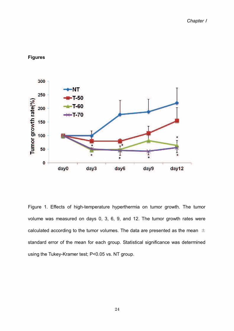
Tumor growth rates. The tumor growth rates of the various groups are shown in Fig.
1.
The tumor growth rates were significantly lower in the T-70 group than those in the NT
group on days 3, 6, 9 and 12 (P<0.05). The tumor growth rates were significantly
lower in the T-60 group than those in the NT group on days 3, 6 and 12. The tumor
growth rates were slightly, but insignificantly, lower in the T-50 group than those in the
NT group. The tumor growth rates were also lower in the T-70 and T-60 groups than
those in the T-50 group on days 3, 6, 9 and 12. The tumor growth rates in the T-60
and T-70 groups were similar on days 3, 6, and 12.
Histological evaluation. The Results of HE staining are shown in Fig. 2. In the NT
group, active cell proliferation was frequently observed. Cell proliferation was
markedly suppressed in the T-70 and T-60 groups compared with that in the NT group.
In addition, in the T-70 and T-60 groups, necrotic cells were widely observed. Cell
proliferation was slightly suppressed in the T-50 group compared with that in the NT
group, and only a few necrotic cells were observed.
TUNEL staining. The results of TUNEL staining are shown in Fig. 3A. The
TUNEL-positive cells are denoted by arrows. The numbers of TUNEL-positive cells
were significantly higher in The T-70 (106.1 14.2 cells/field), T-60 (131.4 12.4
cells/field) and T-50 groups (106.7 6.7 cells/field) than those in the NT group (47.1
9.5 cells/field) (P<0.01) (Fig. 3B).

Chapter
Ki-67 staining. The results of Ki-67 stains are shown in Fig. 4A. Ki-67-positive areas
are denoted by arrows. The Ki-67-positive area was significantly smaller in the T-70
group (1.7 0.1%/field) than that in the NT (3.9 0.4%/field) and T-60 groups (4.1
0.2%/field) P<0.01). The Ki-67-positive area was significantly smaller in the T-50
group (2.8 0.2%/field) than in the NT and T-60 groups (P<0.05, vs. NT; P0.01 vs.
T-60) (Fig. 4B).
4. Discussion
In the present study, the antitumor effects of HTH were evaluated using a glioma
rat model HTH at 60 and 70 significantly suppressed tumor growth. Previously,
specific reports indicated that HTH has potency as a treatment for melanoma in
experimental models (23, 24). Li et al (24) previously described that local HTH
(>50 ) inhibited tumor growth and stimulated a favorable antitumor immune
response in a malignant melanoma model. However, the authors did not investigate
the correlation between higher temperatures (>60 ) and antitumor effects.
Studies of hyperthermia have focused on two commonly applied strategies,
conventional hyperthermia at mild temperatures (42-45 ) (1, 20, 21) and ablation
therapy at high temperature (>70 ) (22). To the best of our knowledge, no study has
examined the difference in antitumor effects between mild (42-45 ) and high
temperatures (70 ) under the same conditions as performed in the present study.
Necrotic cells were more commonly observed in the T-60 and T-70 groups than in the
NT groups. Previous reports indicated that HTH directly induced cell damage and
necrosis (6, 7). In the T-50, T-60 and T-70 groups in the present study, the numbers of
TUNEL-positive cells in tumor tissues were significantly increased compared with

Chapter
those in the NT group. This finding indicates that relatively low temperatures induce
apoptosis (15). Our data also indicate that HTH at 50-70 induces necrosis and
apoptosis in a glioma rat model.
Ki-67 is a cell proliferation marker that is detected during all active phases of the
cell cycle, but is absent in resting cells (25). Ki-67 expression increases during S
phase until mitosis, when its expression is maximal. Following cell division, cells in G1
phase exhibit decreased Ki-67 expression until they reenter the S phase when the
level of Ki-67 increases again (26).Ki-67 expression is also useful for diagnosing and
assessing the grade of tumors in the central nervous system (27). The Ki-67-positive
areas were significantly smaller in the T-50 and T-70 groups than in the NT groups.
Our data also indicate that temperature exceeding 70 sufficiently suppress cell
proliferation. Following suppression of cell proliferation, apoptosis may be induced in
circumferential areas. The Ki-67-positive area was significantly smaller in the T-70
group than that in the T-60 group, although the tumor growth rates in these groups
were equally decreased. We cannot explain this difference, however one possible
explanation may be that there are differences in the percentages of apoptotic cells, as
the T-60 group had significantly more TUNEL-positive cells than the other groups.
HTH at 60 may suppress tumor growth by inducing apoptosis more significantly
than HTH at other temperatures. Further studies, including those of other molecules
associated with apoptosis, are required to clarify this point.
In conclusion, HTH at temperatures exceeding 60 suppressed tumor growth in
a glioma-bearing rat model. In addition, HTH at 50-70 induced necrosis and
apoptosis in a glioma rat model. Further studies is required to clarify the differences in

Chapter
the mechanisms of action for HTH at 60 and 70 .
References
1. Soares Pl, Ferreira IM, Igreja RA, Novo CM and Borges JP: Application of
hyperthermia for cancer treatment: recent patents review. Recent Pat
Anticancer Drug Discov 7:64-73, 2012.
2. Wust P, Hildebrandt B, Sreenivasa G. Rau B, Gellermann J, Riess h,Felix R and
Schlag PM: Hyperthermia in combined Treatment of cancer. Lancet Oncol 3:
487-497, 2002.
3. Falk MH and Issels. RD: Hyperthermia in oncology. Int J, Hyperthermia 17: 1-18,
2001.
4. Ross MI: Current status of hyperthermic limb perfusion for In-transit melanoma.
Int J Hyperthermia 24: 205-217, 2008.
5. Pennacchioli E, Fiore M and Gronchi A: Hyperthermia as an adjunctive
treatment for soft-tissue sarcoma. Expert Rev Anticancer Ther 9: 199-210, 2009
6. Harmon BV, Takano YS, Winterford CM and Gobe GC: The role of apoptosis in
the response of cells and tumors to mild hyperthermia. Int J Radiat Biol 59:
489-501, 1991

Chapter
7. Horsman MR: Tissue physiology and the response to heat. Int JHyperthermia
22: 197-203, 2006.
8. Diederich CJ: Thermal ablation and high-temperature thermal Therapy:
overview of technology and clinical implementation. Int J Hyperthermia
21:745-753, 2005.
9. Roti Roti JL: Cellular responses to hyperthermia (40-46 Degrees C): cell killing
and moleculer events. Int J hyperthermia 24: 3-15, 2008.
10. den Brok MH, Sutmuller RP, van der Voort R, Bennink EJ, Figdor CG, Ruers TJ
and Adema GJ: Insitu tumor ablation creates An antigen source for the
generation of antitumor immunity. Cancer Res 64: 4024-4029, 2004
11. Baronzio G, Gramaglia A and Fiorentini G: Hyperthermia and Immunity. A brief
overview. In Vivo 20: 689-695, 2006
12. Zerbini A, Pilli M, Penna A, Pelosi G,Schianchi C, Molinari A Schivazappa
S,Zibera C,Fagnoni FF, Ferrari C and Missale G: Radiofrequency thermal
ablation of hepatocellular carcinoma Liver nodules can activate and enhance
tumor-specific T-cell Responses. Cancer Res 66:1139-1146, 2006.
13. Mukhopadhaya A, Mendecki J, Dong X, Liu L, Kalnicki S. Garg M, Alfieri A and

Chapter
Guha C: Localized hyperthermia Combined with intratumoral dendritic cells
induces systemic Antitumor immunity. Cancer Res 67: 7798-7806, 2007.
14. Zhang HG, Mehta K, Cohen P and Guha C: Hyperthermia on Immune
191-204, 2008.
15. Moroz P, Jones SK and Gray BN: Magnetically mediated Hyperthermia: current
status and future directions. Int J Hyperthermia 18: 267-284, 2002.
16. Rees JH: Diagnosis and treatment in neuro-oncology: an oncological
perspective. Br J Radiol 84: S82-S89, 2011.
17. Fiorentini G, Giovanis P, Rossi S. Dentico P, Paola R, Trrisi G and Bernardeschi
P: A phase clinical study on relapsed malignant Gliomas treated with
electro-hyperthermia. In Vivo 20: 721-724, 2006.
18. Bidwell GL 3rd, Perkins E, Hughes J, Khan M, James JR and Raucher D:
Thermally targeted delivery of a c-Myc inhibitory Polypeptide inhibits tumor
progression and extends survival in a rat glioma model. PLoS One 8: e55104,
2013
19. Wang DC, Zhang Y, Chen HY, Li XL, Qin Li, Li YJ,Zhang HY and Wang
S:Hyperthermia promotes apoptosis and suppresses invasion In C6 rat glioma

Chapter
cells. Asian Pac J Cancer Prev 13:3239-3245, 2012.
20. Stojkovic R and Radacic M: Cell killing of melanoma B16 in vivo by
hyperthermia and cytotoxins. Int J Hyperthermia 18:62-71,2002.
21. Ito A , Fujioka M , Yoshida T, et al: 4-S-Cysteaminylphenol-loaded magnetic
cationic liposomes for combination therapy of hyperthermia with chemotherapy
against malignant melanoma. Cancer Sci 98: 424-430, 2007
22. Haen SP, Pereira PL, Salih HR, Rammensee HG and Gouttefangeas C: More
than just tumor destruction: immunomodulation by thermal ablation of cancer.
LClin Dev Immunol: 16025o, 2011.
23. Xia QS, Liu X, Xu B, Zhao TD, Li HY, Chen ZH, Xiang Q, Geng CY, Pan L, Hu
RL, et al: Feasibility study of high-temperature thermoseed inductive
hyperthermia in melanoma treatment. Oncol Rep 25:953-962, 2011.
24. Li DY, Tang YP, Zhao LY, Geng CY and Tang JT: Antitumor effect and immune
response induced by local hyperthermia in B16 murine melanoma: Effect of
thermal dose. Oncol Lett 4:711-718, 2012.
25. Brown DC and Gatter KC: Ki-67 protein: the immaculate deception?
Histopathology 40:2-11, 2002

Chapter
26. Lopez F, Belloc F, Lacombe F, Dumain P, Reiffers J, Bernard P and Boisseau
MR: Modalities of synthesis of Ki-67 antigen during the stimulation of
lymphocytes. Cytometry 12: 42-49, 1991.
27. Prayson RA: The utility of MIB-1/Ki-67 immunostaining in the evaluation of
central nervous system neoplasms. Adv Anat Pathol 12: 144-148, 2005.
Chapter
Figures
Figure 1. Effects of high-temperature hyperthermia on tumor growth. The tumor
volume was measured on days 0, 3, 6, 9, and 12. The tumor growth rates were
calculated according to the tumor volumes. The data are presented as the mean
standard error of the mean for each group. Statistical significance was determined
using the Tukey-Kramer test; P<0.05 vs. NT group.

Chapter
(a) (b)
(c) (d)
Figure 2. Effects of high-temperature hyperthermia on histological changes.
The tumor tissue sections were stained with hematoxylin and eosin (bar, 200 m).
Data are presented for one rat each from the (a) nontreatment ,(b) 50, (c) 60 and (d)
70 high-temperature hyperthermia groups.

Chapter
A (a) (b)
(c) (d)
B
Figure 3. Effects of high-temperature hyperthermia on the number of TUNEL-positive

Chapter
cells in the tumor tissue. A The tumor tissue sections were stained with TUNEL (bar,
100 m). Data are presented for one rat each for the (a) NT (b) T-50 (c) T-60 (d) T-70
groups. (B) The numbers of TUNEL-positive cells were calculated. The data are
presented as the mean standard error of the mean for each group. Statistical
significance was determined according to the Steel-Dwass test; **P<0.01. TUNEL,
terminal dUTP nick-end labeling; NT, nontreatment; T-50, 50 high-temperature
hyperthermia; T-60, 60 high-temperature hyperthermia; T-70, 70
high-temperature hyperthermia.
A (a) ( b)
(c) (d)

Chapter
B
Figure 4. Effects of high-temperature hyperthermia on the size of the Ki-67-positive
areas in the tumor tissue (bar, 100 m). (A) Tumor tissue sections were
immunohistochemically stained with Ki-67. Data are presented for one rat each for
the (a) NT (b) T-50, (c) T-60 and (d) T-70 groups. (B) Proportions of Ki-67-positive
areas were calculated. The data are presented as the mean standard error of
the mean for each group. Statistical significance was determined according to the
Steel-Dwass test; *P<0.05; **P<0.01. NT, nontreatment; T-50, 50
high-temperature hyperthermia; T-60, 60 high-temperature hyperthermia; T-70,
70 high-temperature hyperthermia.

Chapter
Chapter
High temperature hyperthermia treatment for canine superficial tumor: A report
of three cases
Abstract
High temperature hyperthermia (HTH) has been demonstrated to suppress
tumor growth in a tumor-bearing rat model. In the present study, we evaluated the
effects of high temperature hyperthermia for the treatment of spontaneous tumors in
dogs. In case 1, an 18-year-old female Papillon presented with a right forelimb
rhabdomyosarcoma. Case 2 was a 14-year-old male Golden Retriever with a perianal
gland adenocarcinoma surrounding the anus. HTH was performed for 10 min at 65
with inhaled isoflurane. In case 1, the tumor disappeared 4 weeks after HTH therapy.
In case 2, HTH was performed three times, and the tumor disappeared the third
procedure. In case 3 the tumor volume decreased by day 21.HTH is a simple
procedure with no severe side effects. Consequently, this treatment modality is
expected to become a useful alternative therapy for superficial tumors in companion
animals.
1. Introduction
The companion animal life span has lengthened with the advent of routine
vaccination, improved environment, and advances in veterinary medicine. As a result,
the incidence of illnesses associated with aging has increased in pet populations. In

Chapter
particular, cancer is a significant problem. As in human medicine, there are three
major treatments for cancer in veterinary medicine, surgery, chemotherapy, and
radiation therapy. However, it is difficult to treat all affected patients with these
therapies. Therefore, new treatments must be developed.
Hyperthermia has long been established as a treatment for cancer, particularly
for superficially located cancer [1]. Hyperthermia is used alone or as an adjunct to
radiotherapy or chemotherapy [2-5] and has been used to treat spontaneous tumors
in veterinary medicine [6-9] Studies have focused on two common strategies,
conventional hyperthermia at mild temperatures (42-45 ) [1,10, 11] and ablation
therapy at high temperatures (>70 ) [12]. We previously demonstrated that high
temperature hyperthermia (HTH) ranging 60-70 suppressed glioma tumor growth
and induced necrosis and apoptosis in a rat model [13]. In the present study, we
evaluated the efficacy of HTH in the treatment of spontaneous tumor in dogs.
2. Case reports
Case 1: An 18-year-old female Papillon (3.2kg) presented for evaluation of a
right forelimb tumor (Figure 1A). Surgical removal of the tumor was performed twice
previously, but the tumor recurred, A biopsy and histopathological analysis revealed
that the tumor was a rhabdomyosarcoma. On initial exam, the caudal right forelimb
was covered by the tumor, and the patient was lame in the affected limb. We
explained the risk of recurrence and the treatment options to the owners, including
surgery, radiation therapy, and chemotherapy. Complete surgical excision was too
difficult because the tumor border was unclear. We recommended HTH experimental

Chapter
consent. A tissue ablation device for veterinary medicine (AMTC 200; AdMeTech Co.,
Ltd, Ehime, Japan) was used to administer the HTH treatment. On day 0, HTH was
performed with no anesthesia and sedation. Three needle of the device were inserted
into tumor tissue at 6-mm intervals, and HTH was performed for 10 min at 45-65 .
On day 21, the tumor volume decreased from that on day 0, and the lameness
improved (Figure 1B). After 4 weeks of HTH, the tumor disappeared.
Case 2: A 14-year-old male Golden Retriever (32.7 kg) presented for evaluation
of a tumor surrounding the anus (Figure 2a). Biopsy and histopathological analysis
identified the tumor as perianal gland adenocarcinoma. We explained the risk of
recurrence and the treatment options to the owners, including surgery, radiation
therapy, and chemotherapy. Complete surgical excision was too difficult because the
tumor border was unclear. We recommended HTH experimental therapy, and the pet
HTH was performed under general anesthesia administered by inhalation of
isoflurane. Five needles of the device were inserted into the tumor at 1-cm intervals,
and HTH was performed for 10min at 65 (Figure 2B) and then repeated one
additional time. On day 21,the tumor volume was decreased from that on day 0
(Figure 2C). HTH was repeated using the same protocol, but the dog died 1week later
due to senility.
Case 3: A 13-year-old male English Cocker Spaniel (12.3 kg) presented for
evaluation of a tumor in the right external auditory canal (Figure 3A). We performed a
right total ear canal ablation, and subsequent histopathological analysis revealed a
ceruminous adenocarcinoma. Several months after intervention, the tumor recurred

Chapter
at the surgical site. We explained the risk of recurrence and the treatment options to
the owner, including surgery, radiation therapy, and chemotherapy. In particular,
surgery presented risks of vestibular disorder and facial paralysis complications. We
recommended HTH experimental therapy, and the pet was enrolled in a clinical trial,
with the owner s signed informed consent. On day 0, HTH was performed under
general anesthesia maintained with inhaled isoflurane. Five needles of the device
were inserted into the tumor, and HTH was performed for 10 min at 65 (Figure 3B).
On day 22, the tumor volume was decreased from that on day 0. On day 28, the HTH
was repeated using the same protocol. On day 78, the tumor volume had decreased
further, and a third HTH procedure was performed. On day 133, the tumor had
disappeared and did not recur.
3. Discussion
The beneficial effects of HTH in the treatment of superficial cancer have not yet
been reported in veterinary medicine. The HTH protocol we used was very simple
and was only performed on canine spontaneous tumors, In these three cases, the
tumor volumes decreased following HTH therapy. Furthermore, no severe side effects
were observed in any of the cases.
In recent decades, several innovative minimally invasive cancer therapies
have been developed as alternatives to surgery. Ablation, which uses high
temperature, radio waves, or microwaves, is considered a potent alternative therapy
[14].
High temperature (>46 ) can directly damage cells, resulting in severe protein
denaturation and DNA damage [15, 16] and inducing irreversible changes that

Chapter
ultimately result in cell death. Tumor cells express specific tumor-associated antigens.
In high temperature (>46 ) conditions, tumor cells swell and break into pieces, which
releases antigens; the large antigen load generates antitumor immunity. The high
temperatures also cause severe protein denaturation, but this likely destroys the
immunogenicity of tumor cells [17-21]. When thermal ablation temperature (>70 )
are achieved, there is a high risk of shock syndrome induced by the sudden and large
production of necrotic tumor material [22]. Therefore, the case for ablation therapy is
restricted in human medicine. In general, ablation therapy is performed to the tumor
within 3cm in diameter [23]. In the present cases, tumor sizes were over 3 cm in
diameter although we did not measure exactly. In a previous study, we reported that
HTH administered at 50-70 induces necrosis in arat glioma model [13]. However,
HTH at 50 did not have adequate suppressive effects compared to treatment at 60
and 70 . In case 1, however, the adequate suppressive effect was showed by HTH
at 45-65 . This result might indicate the sensitivity to the temperature of HTH vary by
tumor types. More extended study which performs HTH to various is necessary. The
optimal therapeutic protocol including effective temperature, time, and frequency
must be established to expand HTH therapy for routine use in veterinary oncology.
In conclusion, HTH is a simple therapeutic option with no severe side effects.
This treatment modality is expected to become a useful alternative therapy for
superficial tumors in companion animals.
References
1. Soares Pl, Ferreira IM, Igreja RA, Novo CM and Borges JP: Application of
hyperthermia for cancer treatmrnt: recent patents review. Recent Pat Anticancer

Chapter
Drug 64-73, 2012.
2. Wust P, Hidebrandt B, Sreenivasa G, et ai: Hyperthermia in combined treatment of
cancer. Lancet Oncol 3: 487-497, 2002.
3. Falk MH and Issels RD: Hyperthermia in oncology. Int J Hyperthermia 17: 1-18,
2001.
4. Ross MI: Current status of Hyperthermic limb perfusion for in-transit melanoma.
Int J Hyperthermia 24: 205-217, 2008.
5. Pennacchioli E, Fiore M and Gronchi A. Hyperthermia as an adjunctive treatment
for soft-tissue sarcoma. Expert Rev Anticancer Ther 9: 199-210, 2009.
6. Brewer WG Jr and Turrel JM: Radiotherapy and hyperthermia in the treatment of
fibrosarcomas in the dog. J Am Vet Med Assoc 181: 146-150, 1982.
7. Page RL and Thrall DE: Clinical indications and applications of radiotherapy and
hyperthermia in veterinary oncology. Vet Clin North Am Small Anim Pract 20:
1075-1092, 1990.
8. Gillette EL: Hyperthermia effects in animals with spontaneous tumors. Natl Cancer
Inst Monogr 61: 361-364, 1982.
9. Grier RL, Brewer WG Jr and Theilen GH: Hyperthermic treatment of superficial
tumors in cats and dogs. J Am Vet Med Assoc 177: 227-233, 1980.
10. Stojkovic R and Radacic M: Cell killing of melanoma B16 in vivo by hyperthermia
and cytotoxins. Int J Hyperthermia 18:62-71, 2002.
11. Ito A, Fujioka M, Yoshida T, et al: 4-S-Cysteaminylphenol-loaded magnetite
cationic liposomes for combination therapy of hyperthermia with chemotherapy
against malignant melanoma. Cancer Sci 98: 424-430, 2007

Chapter
12. Haen SP, Pereira PL, Salih HR, Rammensee HG and Gouttefangeas C: More
than just tumor destruction: immunomodulation by thermal ablation of cancer. Clin
Dev Immunot: 160250, 2011.
13. Takagi H, Azuma K, Tsuka T, Imagawa T, Osaki T and Okamoto Y:Antitumor
Effects of High-temperature hyperthermia on a Glioma-bearing Rat Model
According to Temperature. Oncol Lett 2013 Accepted.
14. Baisi A, De Simone M, Raveglia F and Cioffi U: Thermal ablation in the treatment
of lund cancer: present and future. Eur J Cardiothorac Surg 43: 683-686, 2013.
15. Diederich CJ: Thermal ablation and high temperature thermal therapy: overview
of technology and clinical implementation. Int J Hyperthermia 21: 745-753, 2005.
16. Roti Roti JL: Cellular responses to hyperthermia (40-46 degrees C): cell killing and
molecular events. Int J Hyperthermia 24: 3-15, 2008.
17. Den Brok MH, Sutmuller RP, van der Voort R, Bennink EJ, Figdor CG, Ruers TJ
and Adema GJ: In situ tumor ablation creates an antigen source for the generation
of antitumor immunity. Cancer Res 64: 4042-4029., 2004.
18. Baronzio G, Gramaglia A and Florentini G: Hyperthermia and immunity. A brief
overview. In Vivo 20: 689-695,2006.
19. Zerbini A, Pilli M, Penna A et al: Radiofrequency thermal ablation of hepatocellular
carcinoma liver nodules can activate and enhance tumor-specific T-cell responses.
Cancer Res66: 1138-1146, 2006.
20. Mukhopadhaya A, Mendecki J, Dong X, et al: Localized hyperthermia combined
with intratumoral dendritic cells induces systermic antitumor immunity. Cancer
Res 67: 7798-7806, 2007.

Chapter
21. Zhang HG, Mehta K, Cohen P and Guha C: Hyperthermia on immune regulation:
a temperature`s story. Cancer Lett 271: 191-204, 2008.
22. Moroz P, Jones SK and Gray BN: Magnetically mediated hyperthermia: current
status and future directions. Int J Hyperthermia 18: 267-284, 2002
23. Wggermann P, Puls R, Vasilj A, Sieron D. Schreyer AG, Jung EM, Wawrzynek W,
Stroszczynski C:Thermal ablation of unresectable liver tumors: factors associated
with partial ablation and the impact on long-term survival. Med Sci Mont. 18:
CR88-92, 2012.
Chapter
Figure legends
Figure 1. Gross appearance of case 1. (A) A tumor with right forelimb
(rhabdomyosarcoma). (B) On day 21, the tumor volume was decreased compared
with that on day 0.
Figure 2. Gross tumor appearance in case 2. (A) The tumor surrounding the anus,
later diagnosed as perianal gland adenocarcinoma. (B) HTH was performed with
inhalation of isoflurane. Five needles of the ablation device were inserted into the
tumor, and HTH was performed for 10 min at 65 . (C) the tumor volume was
decreased on day 21.
Figure 3. Gross tumor appearance in case 3. (A) The tumor, diagnosed as a
ceruminous adenocarcinoma, recurred in the right external auditory canal after a total
ear canal ablation. (B) HTH was performed with inhalation of isoflurane. Fiveneedles
of the ablation device were inserted into the tumor, and HTH was performed for 10
min at 65 . (C) The tumor volume was decreased, and HTH was reported using the
same protocol on day 28. (D) The gross appearance of the affected ear on day 133
reveals that the tumor disappeared

Chapter
(A)
(B)
Figure 1.

Chapter
(A)
(B)
(C)
Figure 2

Chapter
(A)
(B)
(C)

Chapter
(D)
Figure 3

Conclusions
Conclusions
High temperature hyperthermia at temperatures exceeding 60 suppressed
tumor growth in a glioma-bearing rat model. In addition, high temperature
hyperthermia at 50-70 induced necrosis and apoptosis in a glioma rat model.
Further studies is required to clarify the differences in the mechanisms of action for
high temperature hyperthermia at 60 and 70 .
High temperature hyperthermia is a simple therapeutic option with no severe side
effects. This treatment modality is expected to become a useful alternative therapy for
superficial tumors in companion animals.

Acknowledgements
Acknowledgements
I wish to pay sincere acknowledgement to Professor Yasuho Taura, D.V.M.,
Ph. D., Laboratory of Veterinary Surgery, Department of Veterinary Clinical Medicine,
Joint Faculty of Veterinary Medicine, Yamaguchi University, for his sincere
supervising and encouragement during the present investigation.
I also wish to thank Professor Yoshiharu Okamoto, D.V.M., Ph.D., Laboratory of
Veterinary Surgery, Department of Veterinary Medicine, Faculty of Agriculture, Tottori
University, Associate Professor Kenji Tani, D.V.M., Ph.D., Laboratory of Veterinary
Surgery, Department of Veterinary Clinical Medicine, Joint Faculty of Veterinary
Medicine, Yamaguchi University, for their helpful suggestion as co-supervisor. I am
grateful to Professor Munekazu Nakaichi, D.V.M., Ph.D., Laboratory of Veterinary
Radiology, Department of Veterinary Clinical Medicine, Joint Faculty of Veterinary
Medicine, Yamaguchi University, and Associate Professor Kazuhito Itamoto, D.V.M.,
Ph.D., Laboratory of Companion Animal Surgery, Animal Medical Center, Joint
Faculty of Veterinary Medicine, Yamaguchi University.
Finaly, I wish to express special thanks to my family to my wife Hitomi for her
hearty support and encouragement.