
. Chapter 6: A Tour of the Cell Cell theory Cell theory Cell organization and homeostasis Cell...
114
. Chapter 6: A Tour of the Cell Cell theory Cell organization and homeostasis Studying cells – microscopy and fractionation Eukaryotic vs. prokaryotic cells Compartments in eukaryotic cells (cell regions, organelles) Cytoskeleton Outside the cell
-
Upload
natalie-henbest -
Category
Documents
-
view
217 -
download
2
Transcript of . Chapter 6: A Tour of the Cell Cell theory Cell theory Cell organization and homeostasis Cell...
.
Chapter 6: A Tour of the Cell Cell theory
Cell organization and homeostasis
Studying cells – microscopy and fractionation
Eukaryotic vs. prokaryotic cells
Compartments in eukaryotic cells (cell regions, organelles)
Cytoskeleton
Outside the cell
.
Chapter 6: A Tour of the Cell Cell theory
Cell organization and homeostasis
Studying cells – microscopy and fractionation
Eukaryotic vs. prokaryotic cells
Compartments in eukaryotic cells (cell regions, organelles)
Cytoskeleton
Outside the cell
.
• What are the main tenets of cell theory?
• What are the major lines of evidence that all presently living cells have a common origin?
.
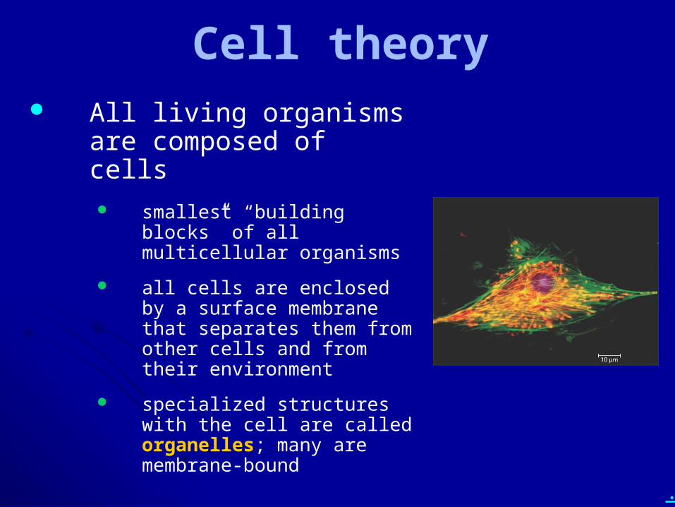
Cell theory All living organisms are
composed of cells smallest “building blocks” of all
multicellular organisms
all cells are enclosed by a surface membrane that separates them from other cells and from their environment
specialized structures with the cell are called organelles; many are membrane-bound
.
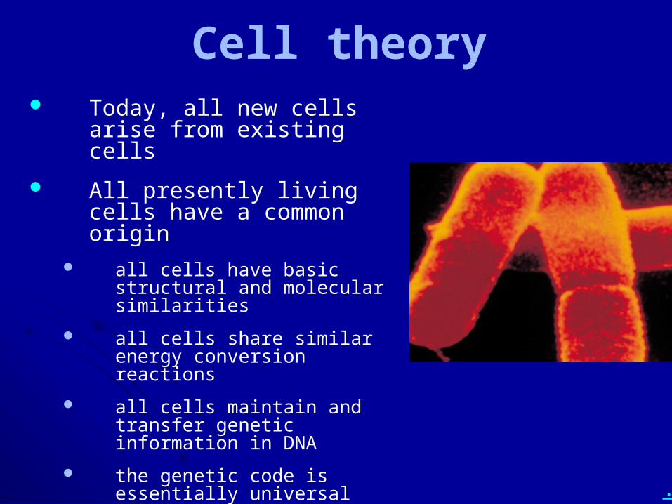
Cell theory Today, all new cells arise from
existing cells
All presently living cells have a common origin
all cells have basic structural and molecular similarities
all cells share similar energy conversion reactions
all cells maintain and transfer genetic information in DNA
the genetic code is essentially universal
.
• What are the main tenets of cell theory?
• What are the major lines of evidence that all presently living cells have a common origin?
.
Chapter 6: A Tour of the Cell Cell theory
Cell organization and homeostasis
Studying cells – microscopy and fractionation
Eukaryotic vs. prokaryotic cells
Compartments in eukaryotic cells (cell regions, organelles)
Cytoskeleton
Outside the cell
.
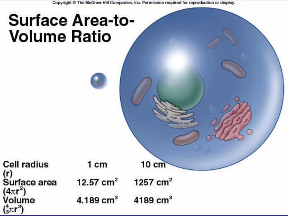
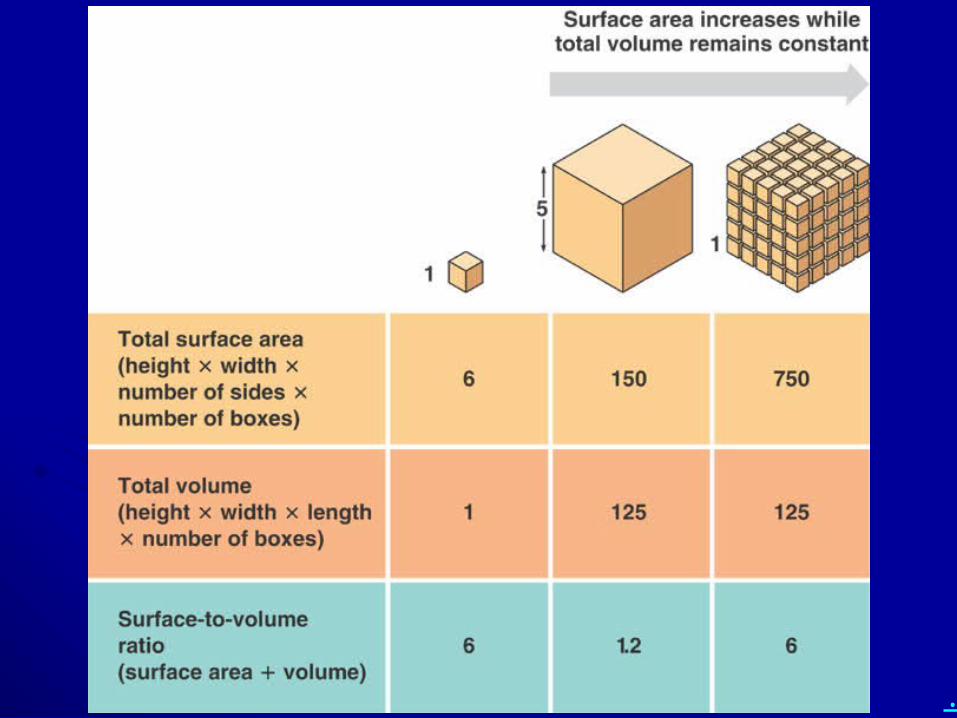
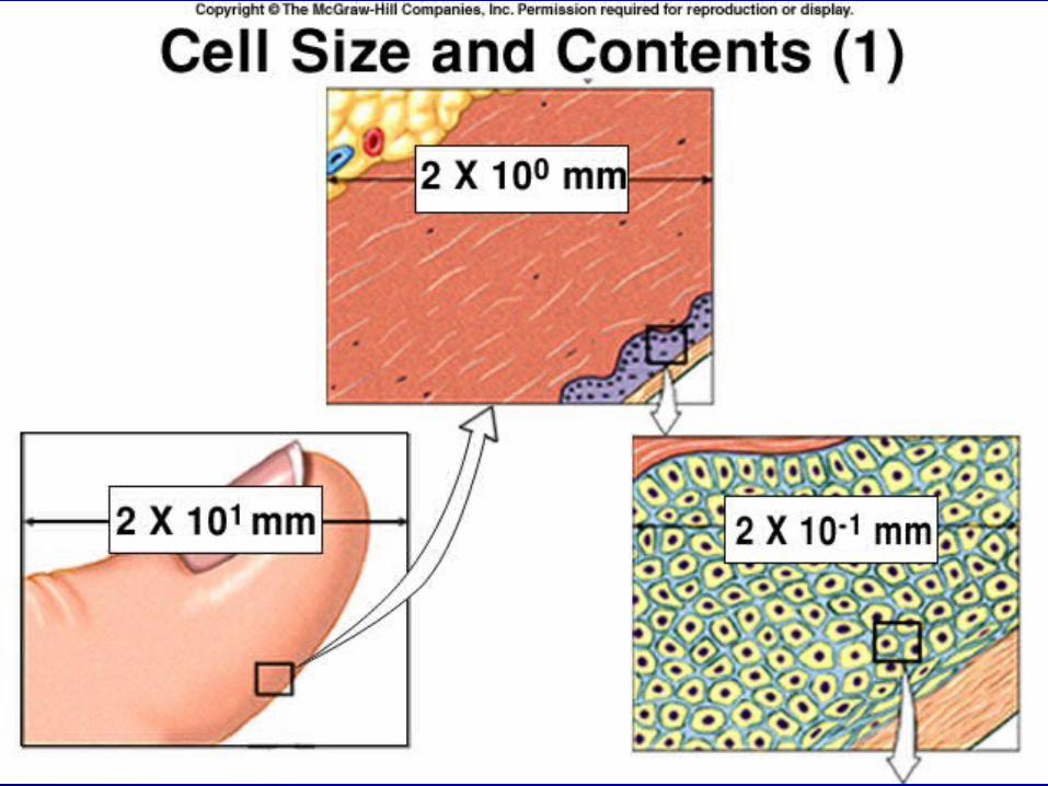
• What is surface area to volume ratio, and why is it an important consideration for cells?
• What (usually) happens to surface area to volume ratio as cells grow larger?
.
Cell organization and homeostasis
Plasma membrane surrounds cells and separates their contents from the external environment
Cells are heterogeneous mixtures, with specialized regions and structures (such as organelles)
.
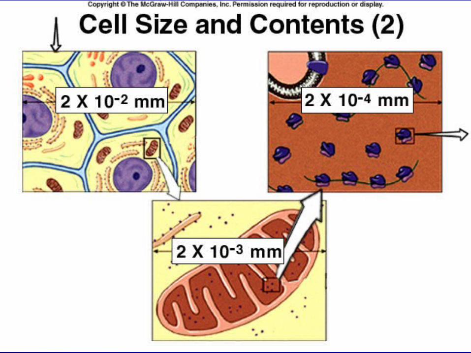
Cell organization and homeostasis
Cell size is limited
surface area to volume ratio puts a limit on cell size
food and/or other materials must get into the cell
waste products must be removed from the cell
cells need a high surface area to volume ratio
BUT volume increases faster than surface area as cells grow larger…so cells usually must divide
.
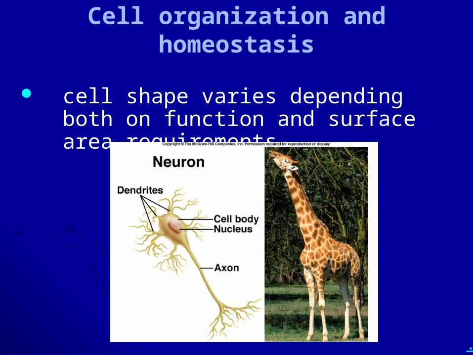
Cell organization and homeostasis
cell shape varies depending both on function and surface area requirements
.
• What is surface area to volume ratio, and why is it an important consideration for cells?
• What (usually) happens to surface area to volume ratio as cells grow larger?
.
Chapter 6: A Tour of the Cell Cell theory
Cell organization and homeostasis
Studying cells – microscopy and fractionation
Eukaryotic vs. prokaryotic cells
Compartments in eukaryotic cells (cell regions, organelles)
Cytoskeleton
Outside the cell
.
• Compare and contrast:– LM and EM– SEM and TEM
• Include the terms resolution and magnification in your discussions.
.
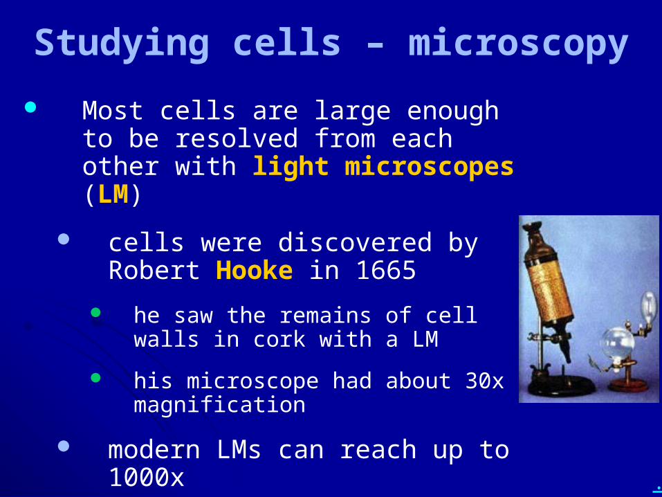
Studying cells – microscopy
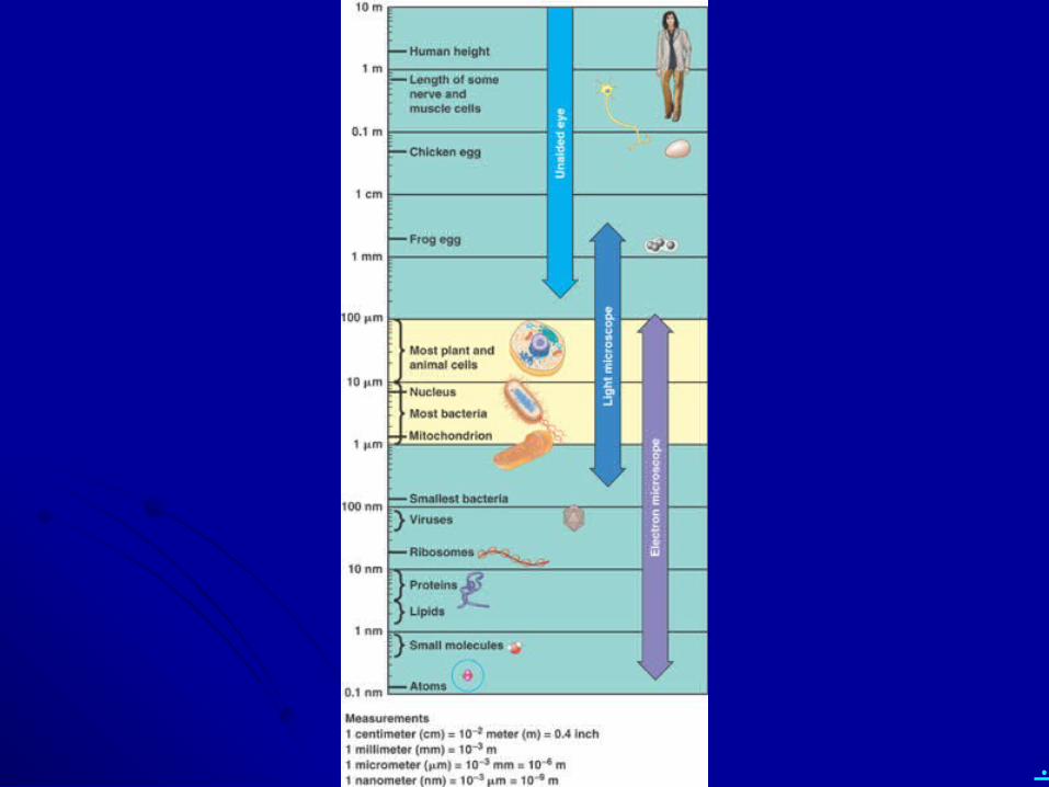
Most cells are large enough to be resolved from each other with light microscopes (LM)
cells were discovered by Robert Hooke in 1665
he saw the remains of cell walls in cork with a LM
his microscope had about 30x magnification
modern LMs can reach up to 1000x
.
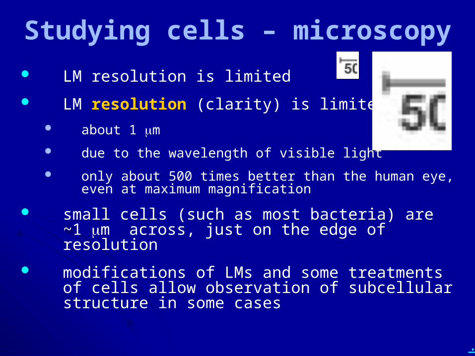
Studying cells – microscopy
LM resolution is limited
LM resolution (clarity) is limited about 1 mm
due to the wavelength of visible light
only about 500 times better than the human eye, even at maximum magnification
small cells (such as most bacteria) are ~1 mm across, just on the edge of resolution
modifications of LMs and some treatments of cells allow observation of subcellular structure in some cases
.
Studying cells – microscopy
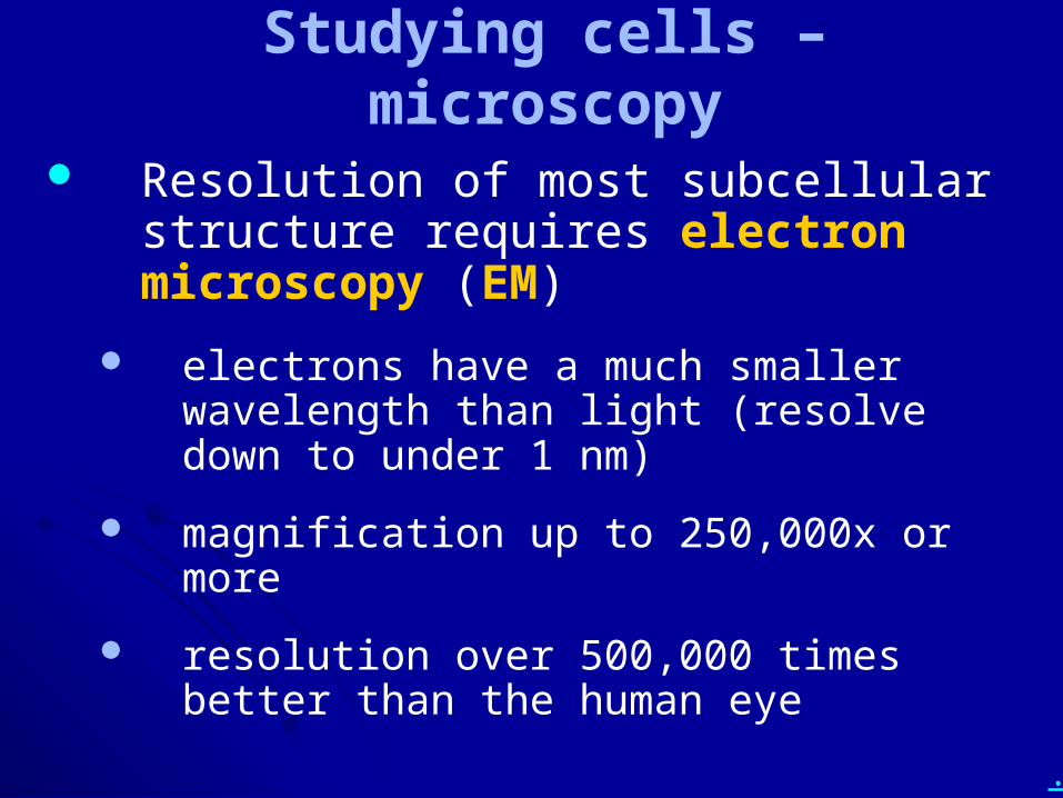
Resolution of most subcellular structure requires electron microscopy (EM)
electrons have a much smaller wavelength than light (resolve down to under 1 nm)
magnification up to 250,000x or more
resolution over 500,000 times better than the human eye
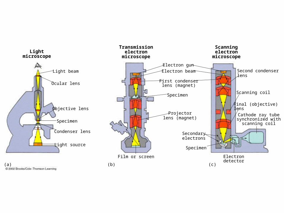
Lightmicroscope
Transmissionelectron
microscope
Scanningelectron
microscope
Light beam
Ocular lens
Objective lens
Specimen
Condenser lens
Light source
Electron gun
Electron beam
First condenserlens (magnet)
Specimen
Projectorlens (magnet)
Secondaryelectrons
Film or screen
Specimen
Second condenserlens
Scanning coil
Final (objective)lens
Cathode ray tubesynchronized with
scanning coil
Electrondetector
(a) (b) (c)
.
Studying cells – microscopy
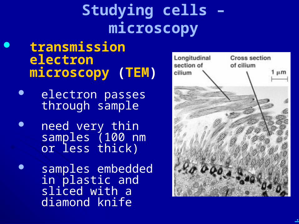
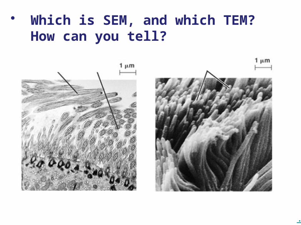
transmission electron microscopy (TEM)
electron passes through sample
need very thin samples (100 nm or less thick)
samples embedded in plastic and sliced with a diamond knife
.
Studying cells – microscopy
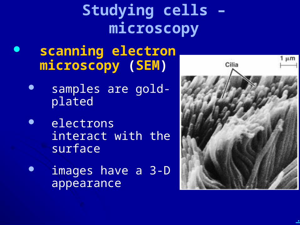
scanning electron microscopy (SEM)
samples are gold-plated
electrons interact with the surface
images have a 3-D appearance
.
• Compare and contrast:– LM and EM– SEM and TEM
• Include the terms resolution and magnification in your discussions.
.
• Which is SEM, and which TEM? How can you tell?
.
• Describe cell fractionation. Why is it done, and how is it done? Include the terms lyse, centrifugation, pellet, and supernatant in your discussion.
.
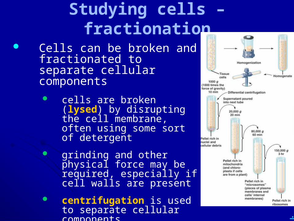
Studying cells – fractionation
Cells can be broken and fractionated to separate cellular components cells are broken (lysed) by
disrupting the cell membrane, often using some sort of detergent
grinding and other physical force may be required, especially if cell walls are present
centrifugation is used to separate cellular components
.
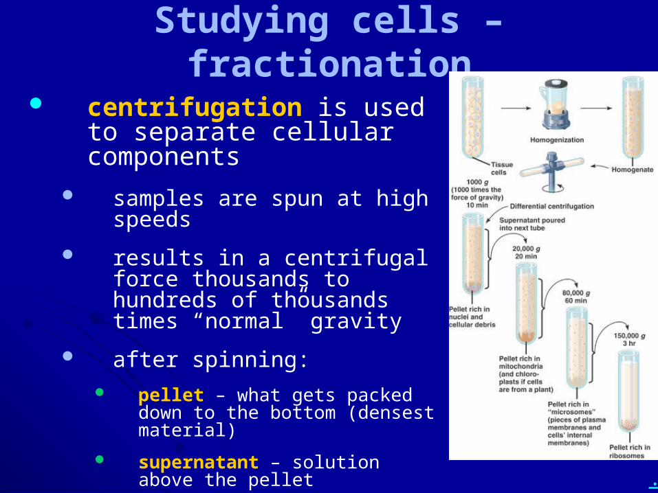
Studying cells – fractionation
centrifugation is used to separate cellular components
samples are spun at high speeds
results in a centrifugal force thousands to hundreds of thousands times “normal” gravity
after spinning: pellet – what gets packed down to
the bottom (densest material)
supernatant – solution above the pellet
.
Studying cells – fractionation
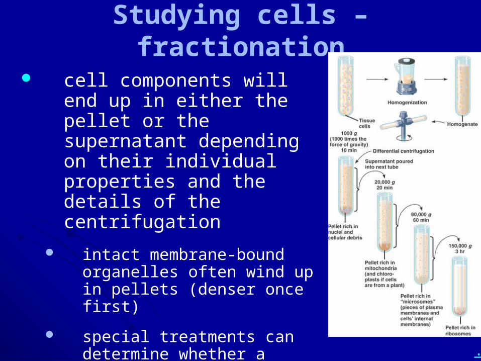
cell components will end up in either the pellet or the supernatant depending on their individual properties and the details of the centrifugation
intact membrane-bound organelles often wind up in pellets (denser once first)
special treatments can determine whether a component ends up in the pellet or supernatant
.
Studying cells – fractionation
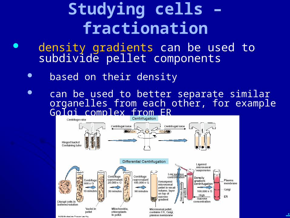
density gradients can be used to subdivide pellet components
based on their density
can be used to better separate similar organelles from each other, for example Golgi complex from ER
.
• Describe cell fractionation. Why is it done, and how is it done? Include the terms lyse, centrifugation, pellet, and supernatant in your discussion.
.
Chapter 6: A Tour of the Cell Cell theory
Cell organization and homeostasis
Studying cells – microscopy and fractionation
Eukaryotic vs. prokaryotic cells
Compartments in eukaryotic cells (cell regions, organelles)
Cytoskeleton
Outside the cell
.
• How do prokaryotic cells and eukaryotic cells differ from each other in typical size and general organization?
• Describe cytoplasm, cytosol, nucleoplasm, and the general role of membranes in cells.
.
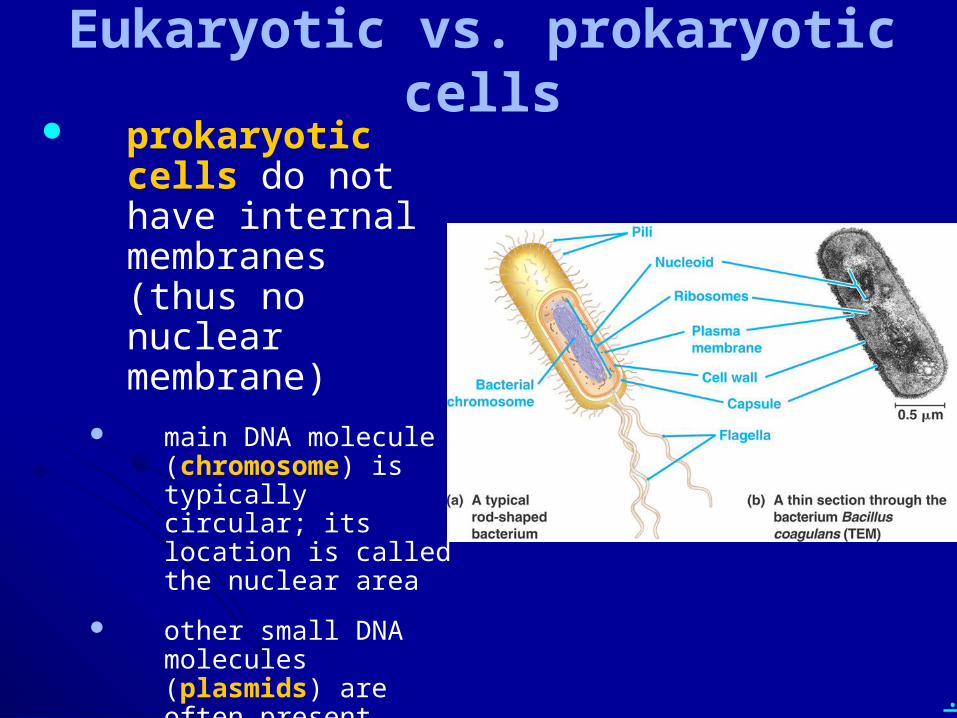
Eukaryotic vs. prokaryotic cells prokaryotic cells
do not have internal membranes (thus no nuclear membrane)
main DNA molecule (chromosome) is typically circular; its location is called the nuclear area
other small DNA molecules (plasmids) are often present, found throughout the cell
.
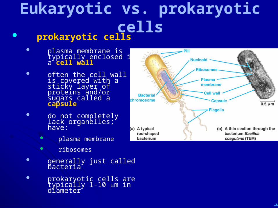
Eukaryotic vs. prokaryotic cells prokaryotic cells
plasma membrane is typically enclosed in a cell wall
often the cell wall is covered with a sticky layer of proteins and/or sugars called a capsule
do not completely lack organelles; have:
plasma membrane
ribosomes
generally just called bacteria
prokaryotic cells are typically 1-10 mm in diameter
.
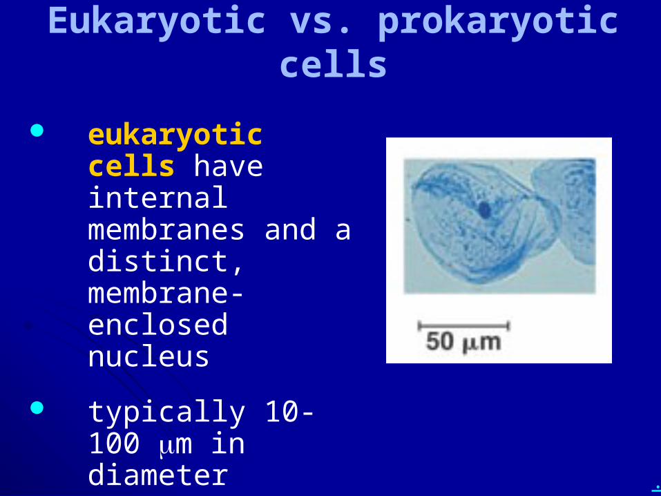
Eukaryotic vs. prokaryotic cells
eukaryotic cells have internal membranes and a distinct, membrane-enclosed nucleus
typically 10-100 mm in diameter
.
• How do prokaryotic cells and eukaryotic cells differ from each other in typical size and general organization?
• Describe cytoplasm, cytosol, nucleoplasm, and the general role of membranes in cells.
.
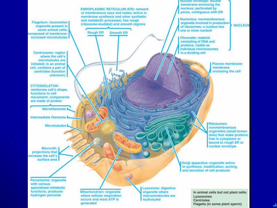
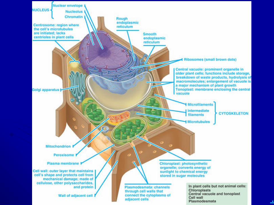
• List as many organelles as you can think of. Describe their structures and key functions.
• Draw and label a typical animal cell and a typical plant cell, including organelles.
.
• List as many organelles as you can think of. Describe their structures and key functions.
• Draw and label a typical animal cell and a typical plant cell, including organelles.
.
Chapter 6: A Tour of the Cell Cell theory
Cell organization and homeostasis
Studying cells – microscopy and fractionation
Eukaryotic vs. prokaryotic cells
Compartments in eukaryotic cells (cell regions, organelles)
Cytoskeleton
Outside the cell
.
How do proteins get outside of a cell?
How do proteins get into a cell membrane?
How does a cell digest its food?
How does a cell commit suicide?
Why would a cell commit suicide?
.
• Describe the nuclear envelope, nuclear pores, chromatin, chromosomes, and nucleoli in terms of structures and key functions.
• Name something that you KNOW must get out of the nucleus for cells to function.
.
Compartments in eukaryotic cells
two general regions inside the cell: cytoplasm and nucleoplasm
cytoplasm – everything outside the nucleus and within the plasma membrane
contains fluid cytosol and organelles
nucleoplasm – everything within the nuclear membrane
.
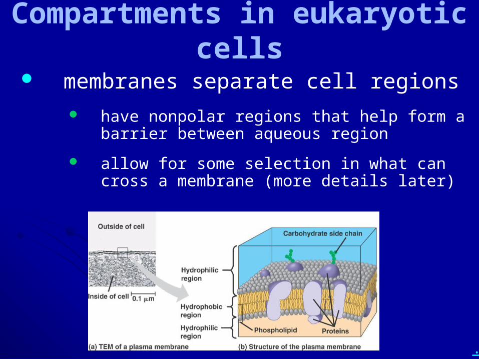
Compartments in eukaryotic cells
membranes separate cell regions
have nonpolar regions that help form a barrier between aqueous region
allow for some selection in what can cross a membrane (more details later)
.
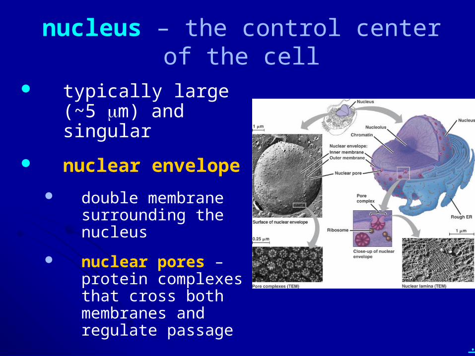
nucleus – the control center of the cell
typically large (~5 mm) and singular
nuclear envelope
double membrane surrounding the nucleus
nuclear pores – protein complexes that cross both membranes and regulate passage
.
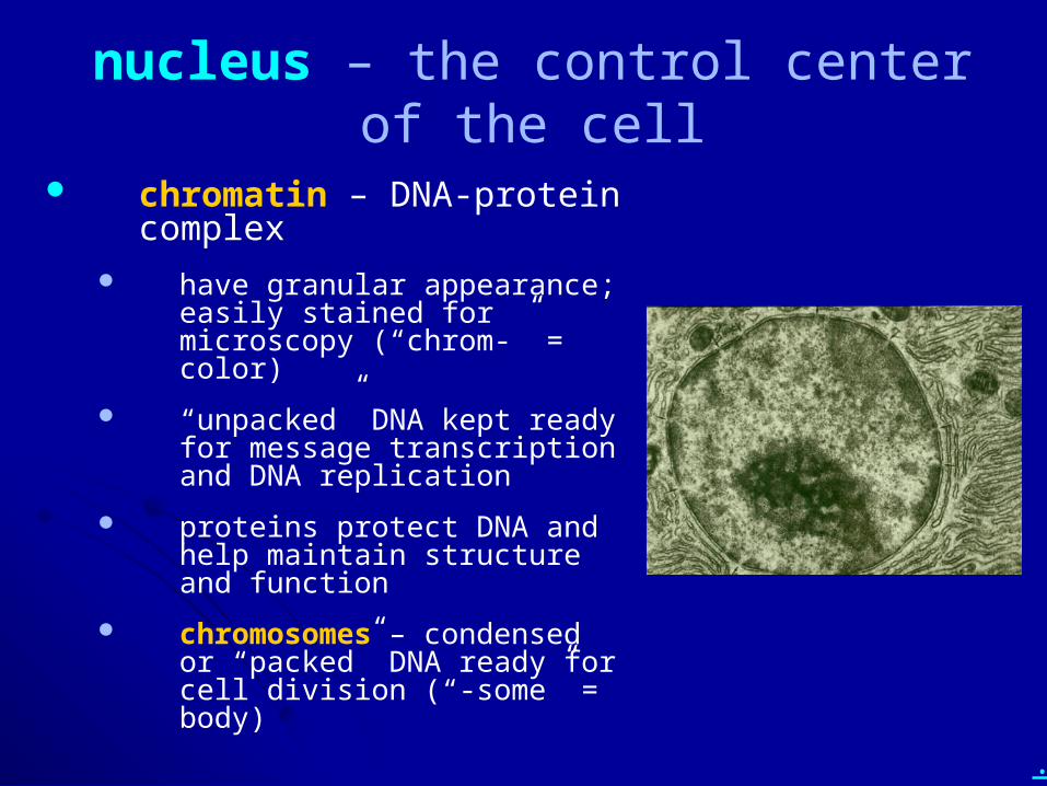
nucleus – the control center of the cell
chromatin – DNA-protein complex
have granular appearance; easily stained for microscopy (“chrom-” = color)
“unpacked” DNA kept ready for message transcription and DNA replication
proteins protect DNA and help maintain structure and function
chromosomes – condensed or “packed” DNA ready for cell division (“-some” = body)
.
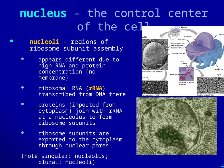
nucleus – the control center of the cell
nucleoli – regions of ribosome subunit assembly
appears different due to high RNA and protein concentration (no membrane)
ribosomal RNA (rRNA) transcribed from DNA there
proteins (imported from cytoplasm) join with rRNA at a nucleolus to form ribosome subunits
ribosome subunits are exported to the cytoplasm through nuclear pores
(note singular: nucleolus; plural: nucleoli)
.
• Describe the nuclear envelope, nuclear pores, chromatin, chromosomes, and nucleoli in terms of structures and key functions.
• Name something that you KNOW must get out of the nucleus for cells to function.
.
• Describe the structure and function of ribosomes.
.
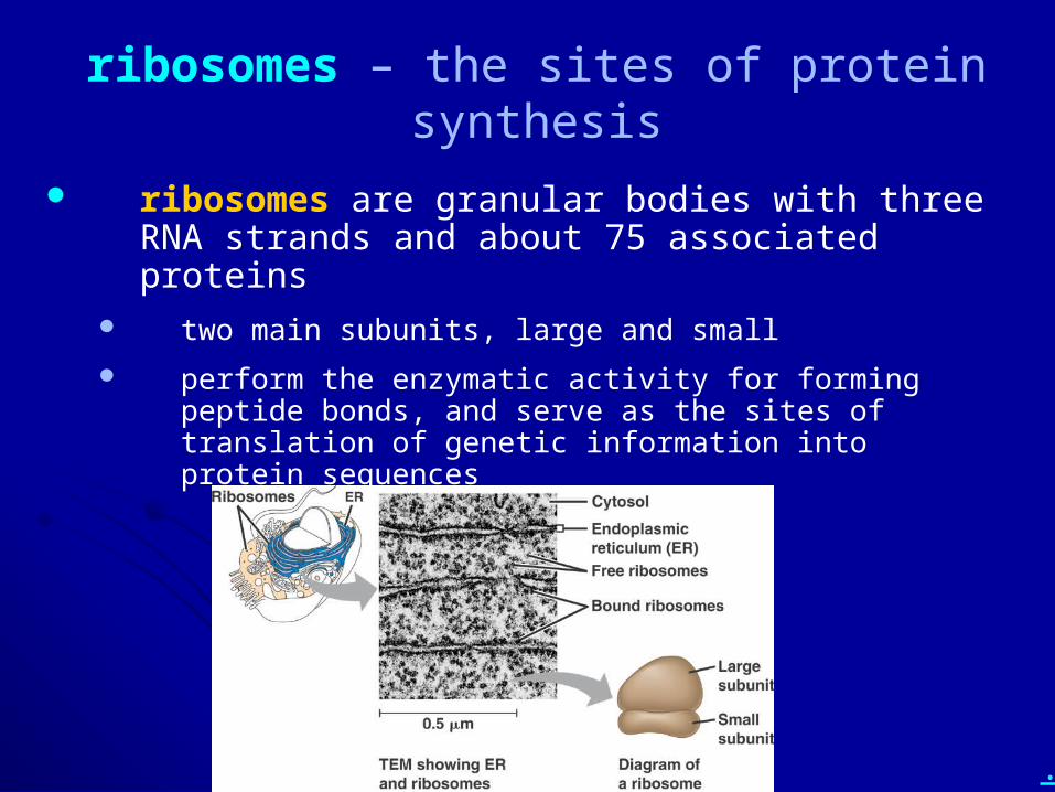
ribosomes – the sites of protein synthesis
ribosomes are granular bodies with three RNA strands and about 75 associated proteins
two main subunits, large and small
perform the enzymatic activity for forming peptide bonds, and serve as the sites of translation of genetic information into protein sequences
.
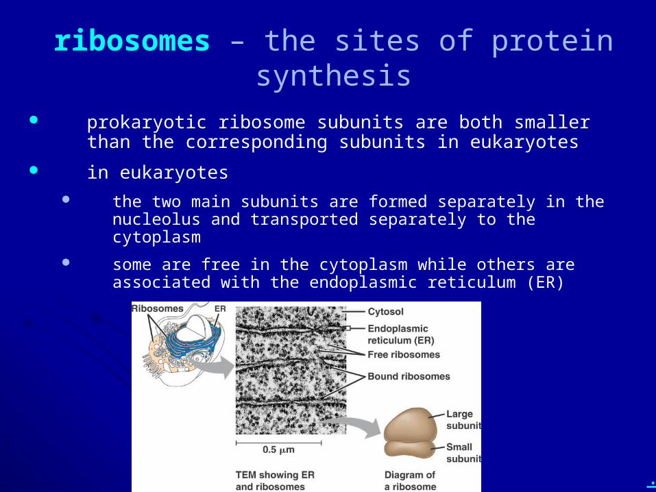
ribosomes – the sites of protein synthesis
prokaryotic ribosome subunits are both smaller than the corresponding subunits in eukaryotes
in eukaryotes the two main subunits are formed separately in the nucleolus and
transported separately to the cytoplasm
some are free in the cytoplasm while others are associated with the endoplasmic reticulum (ER)
.
• Describe the structure and function of ribosomes.
.
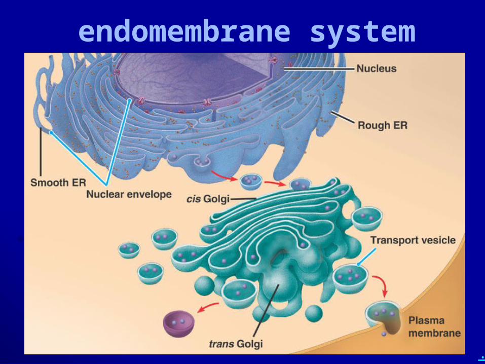
• What is the endomembrane system (include organelle components)?
.
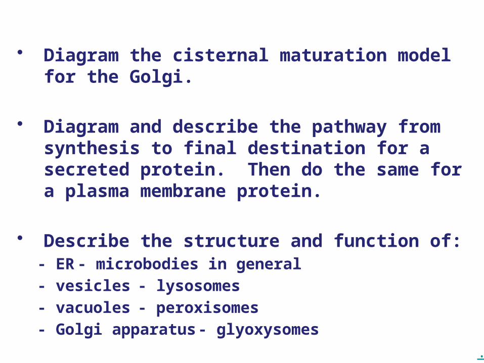
• Diagram the cisternal maturation model for the Golgi.
• Diagram and describe the pathway from synthesis to final destination for a secreted protein. Then do the same for a plasma membrane protein.
• Describe the structure and function of: - ER - microbodies in general
- vesicles - lysosomes
- vacuoles - peroxisomes
- Golgi apparatus - glyoxysomes
.
endomembrane system
endomembrane system – a set of membranous organelles that interact with each other via vesicles
includes ER, Golgi apparatus, vacuoles, lysosomes, microbodies, and in some definitions the nuclear membrane and the plasma membrane
.
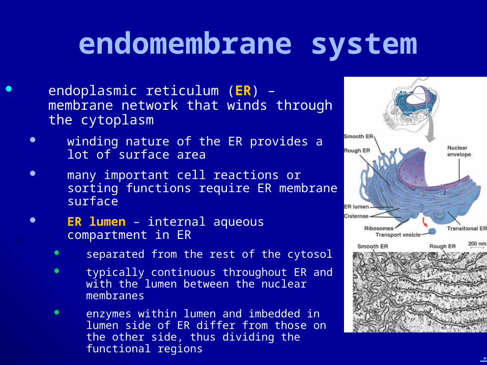
endomembrane system endoplasmic reticulum (ER) – membrane
network that winds through the cytoplasm winding nature of the ER provides a lot of surface
area
many important cell reactions or sorting functions require ER membrane surface
ER lumen – internal aqueous compartment in ER separated from the rest of the cytosol
typically continuous throughout ER and with the lumen between the nuclear membranes
enzymes within lumen and imbedded in lumen side of ER differ from those on the other side, thus dividing the functional regions
.
endomembrane system
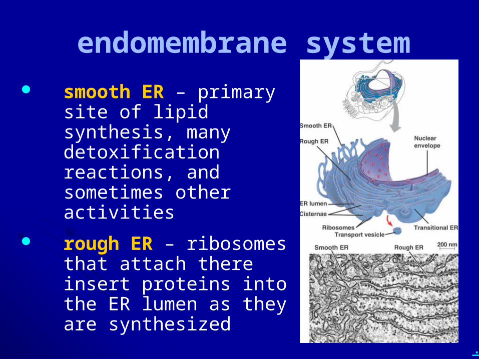
smooth ER – primary site of lipid synthesis, many detoxification reactions, and sometimes other activities
rough ER – ribosomes that attach there insert proteins into the ER lumen as they are synthesized
.
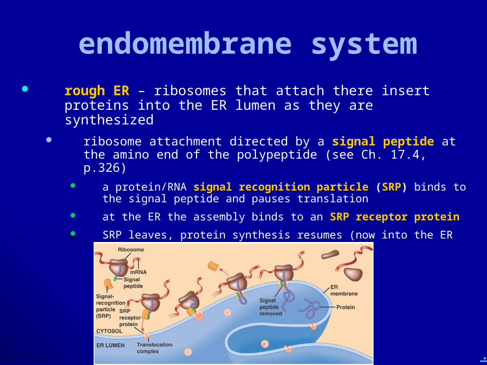
endomembrane system rough ER – ribosomes that attach there insert proteins into the ER
lumen as they are synthesized ribosome attachment directed by a signal peptide at the amino end of
the polypeptide (see Ch. 17.4, p.326) a protein/RNA signal recognition particle (SRP) binds to the signal
peptide and pauses translation
at the ER the assembly binds to an SRP receptor protein
SRP leaves, protein synthesis resumes (now into the ER lumen), and the signal peptide is cut off
.
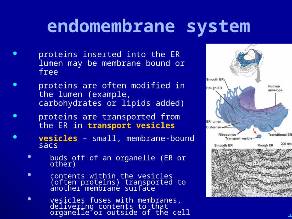
endomembrane system proteins inserted into the ER lumen may be
membrane bound or free
proteins are often modified in the lumen (example, carbohydrates or lipids added)
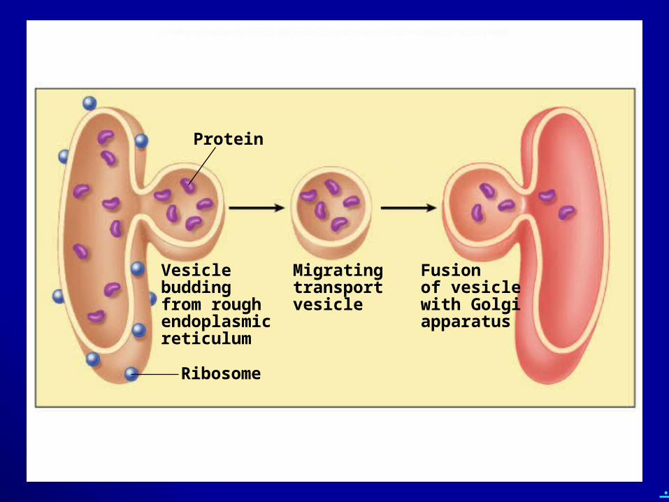
proteins are transported from the ER in transport vesicles
vesicles – small, membrane-bound sacs buds off of an organelle (ER or other) contents within the vesicles (often proteins)
transported to another membrane surface vesicles fuses with membranes, delivering
contents to that organelle or outside of the cell
.
Fig. 5.16d (TEArt)
Vesiclebuddingfrom roughendoplasmicreticulum
Fusionof vesiclewith Golgiapparatus
Migratingtransportvesicle
Protein
Ribosome
.
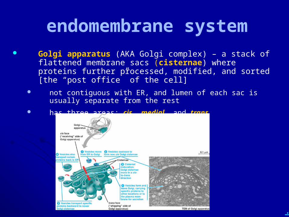
endomembrane system Golgi apparatus (AKA Golgi complex) – a stack of flattened
membrane sacs (cisternae) where proteins further processed, modified, and sorted [the “post office” of the cell]
not contiguous with ER, and lumen of each sac is usually separate from the rest
has three areas: cis, medial, and trans
.
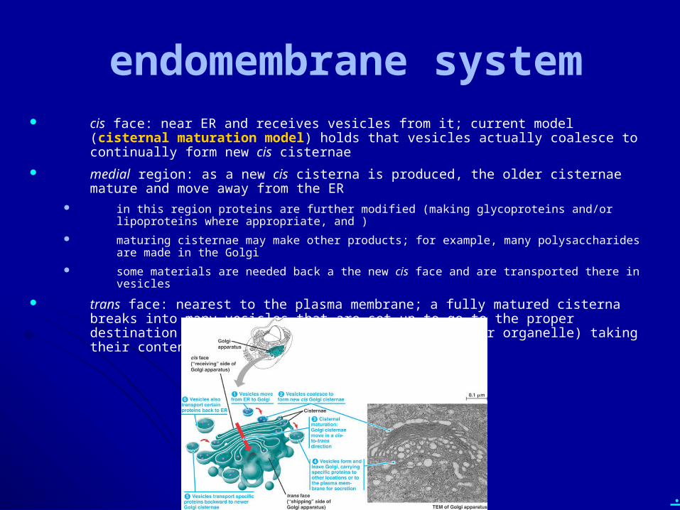
endomembrane system cis face: near ER and receives vesicles from it; current model (cisternal maturation model)
holds that vesicles actually coalesce to continually form new cis cisternae
medial region: as a new cis cisterna is produced, the older cisternae mature and move away from the ER
in this region proteins are further modified (making glycoproteins and/or lipoproteins where appropriate, and )
maturing cisternae may make other products; for example, many polysaccharides are made in the Golgi
some materials are needed back a the new cis face and are transported there in vesicles
trans face: nearest to the plasma membrane; a fully matured cisterna breaks into many vesicles that are set up to go to the proper destination (such as the plasma membrane or another organelle) taking their contents with them
.
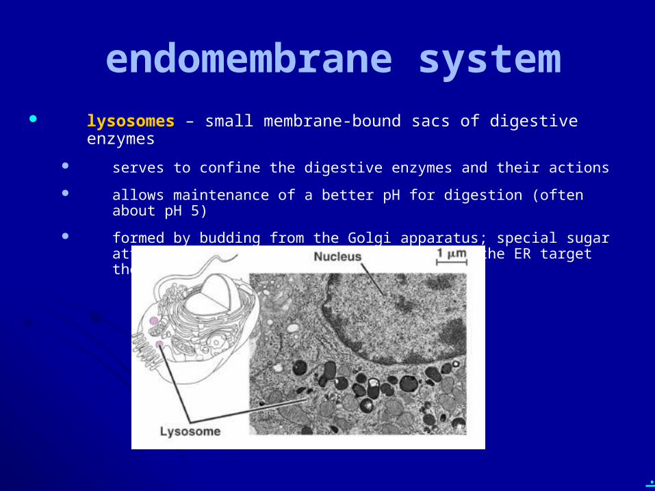
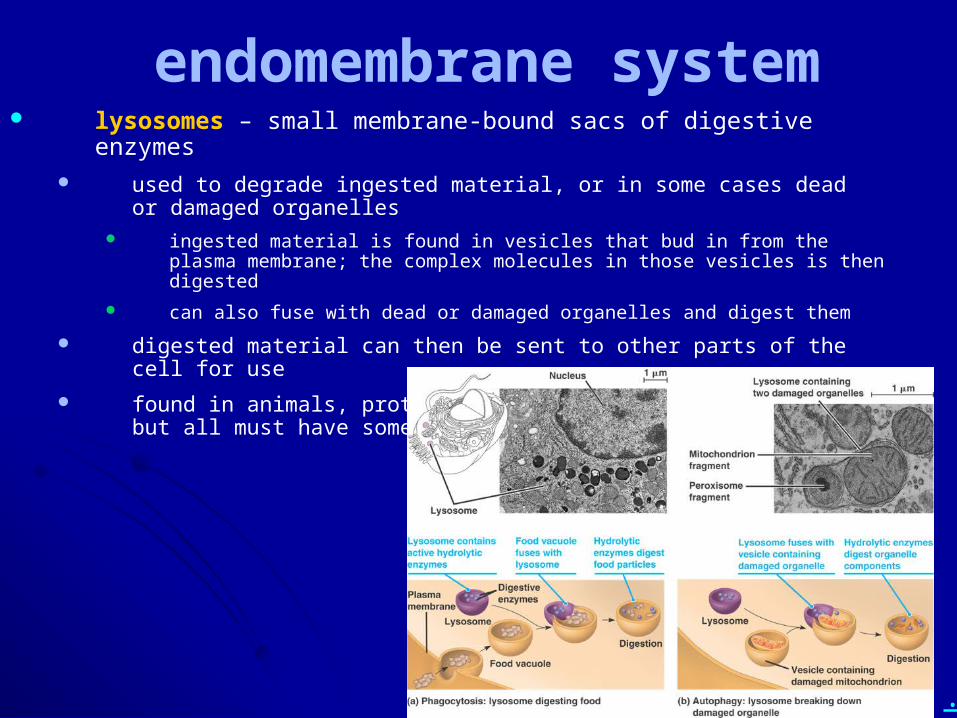
endomembrane system lysosomes – small membrane-bound sacs of digestive enzymes
serves to confine the digestive enzymes and their actions
allows maintenance of a better pH for digestion (often about pH 5)
formed by budding from the Golgi apparatus; special sugar attachments to hydrolytic enzymes made in the ER target them to the lysosome
.
endomembrane system lysosomes – small membrane-bound sacs of digestive enzymes
used to degrade ingested material, or in some cases dead or damaged organelles
ingested material is found in vesicles that bud in from the plasma membrane; the complex molecules in those vesicles is then digested
can also fuse with dead or damaged organelles and digest them
digested material can then be sent to other parts of the cell for use
found in animals, protozoa; debatable in other eukaryotes, but all must have something like a lysosome
.
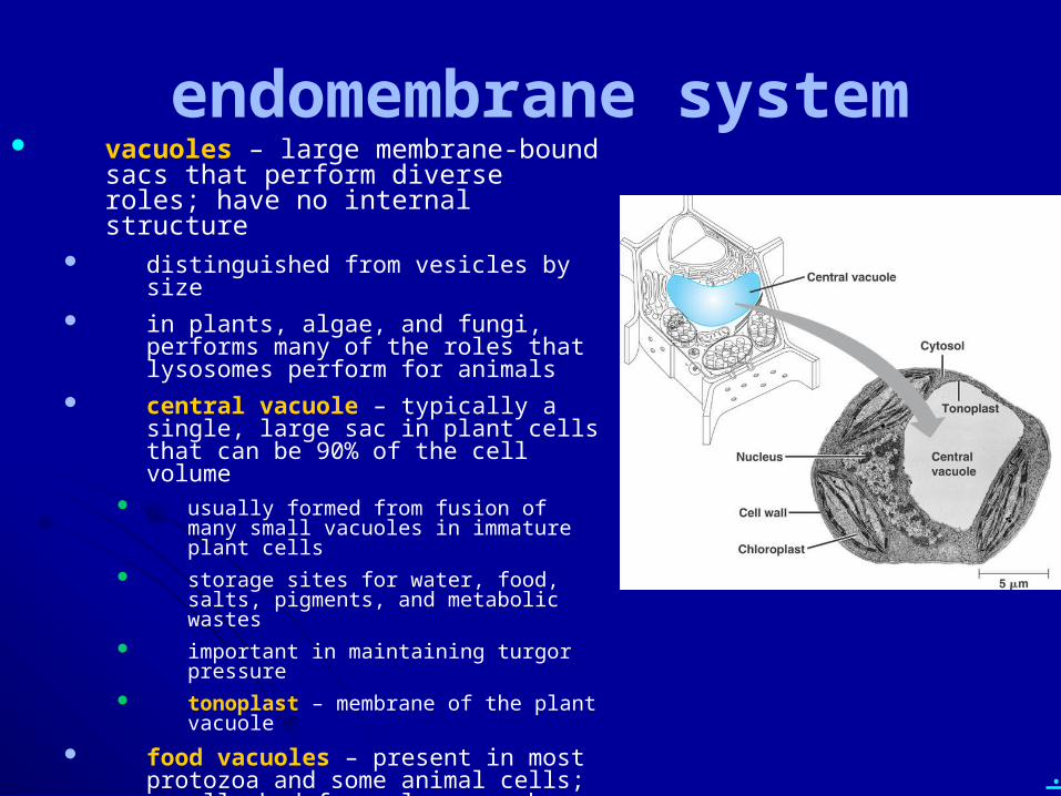
endomembrane system vacuoles – large membrane-bound sacs
that perform diverse roles; have no internal structure
distinguished from vesicles by size in plants, algae, and fungi, performs many
of the roles that lysosomes perform for animals
central vacuole – typically a single, large sac in plant cells that can be 90% of the cell volume
usually formed from fusion of many small vacuoles in immature plant cells
storage sites for water, food, salts, pigments, and metabolic wastes
important in maintaining turgor pressure tonoplast – membrane of the plant vacuole
food vacuoles – present in most protozoa and some animal cells; usually bud from plasma membrane and fuse with lysosomes for digestion
contractile vacuoles – used by many protozoa for removing excess water
.
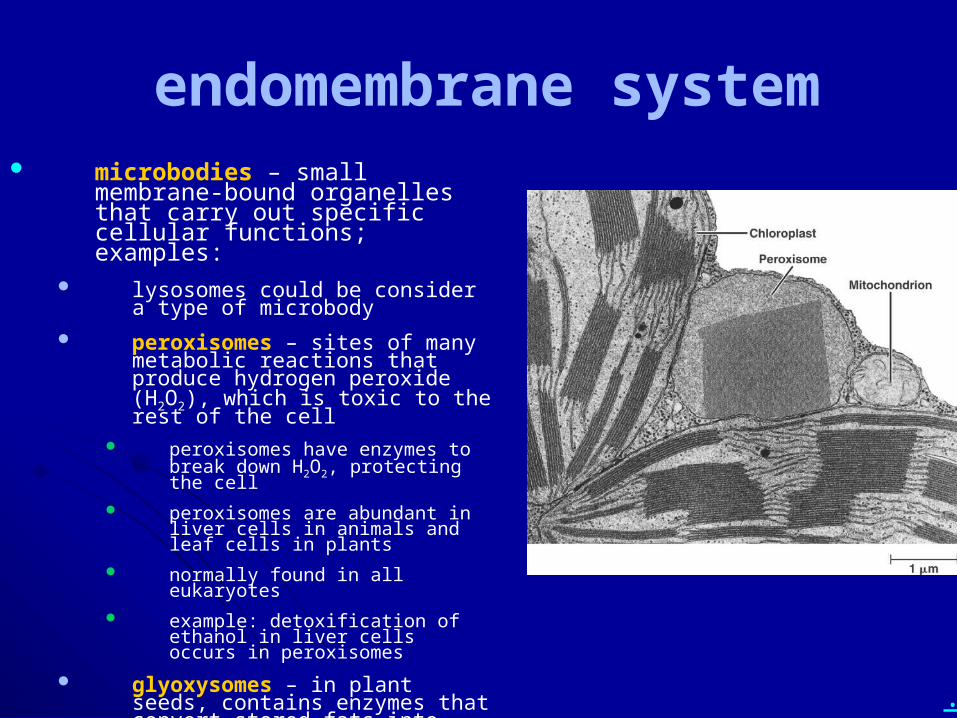
endomembrane system microbodies – small membrane-
bound organelles that carry out specific cellular functions; examples:
lysosomes could be consider a type of microbody
peroxisomes – sites of many metabolic reactions that produce hydrogen peroxide (H2O2), which is toxic to the rest of the cell
peroxisomes have enzymes to break down H2O2, protecting the cell
peroxisomes are abundant in liver cells in animals and leaf cells in plants
normally found in all eukaryotes example: detoxification of ethanol in
liver cells occurs in peroxisomes
glyoxysomes – in plant seeds, contains enzymes that convert stored fats into sugar
.
• What is the endomembrane system (include organelle components)?
.
• Diagram the cisternal maturation model for the Golgi.
• Diagram and describe the pathway from synthesis to final destination for a secreted protein. Then do the same for a plasma membrane protein.
• Describe the structure and function of: - ER - microbodies in general
- vesicles - lysosomes
- vacuoles - peroxisomes
- Golgi apparatus - glyoxysomes
.
Energy Converting Organelles
energy obtained from the environment is typically chemical energy (in food) or light energy
mitochondria are the organelles where chemical energy is placed in a more useful molecule
chloroplasts are plastids where light energy is captured during photosynthesis
.
• Draw a mitochondrion in cross-section and describe its structure and functions.
.
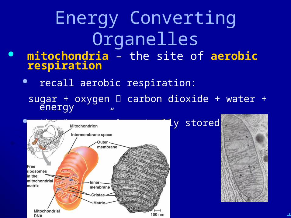
Energy Converting Organelles mitochondria – the site of aerobic respiration
recall aerobic respiration:
sugar + oxygen carbon dioxide + water + energy the “energy” is actually stored in ATP
.
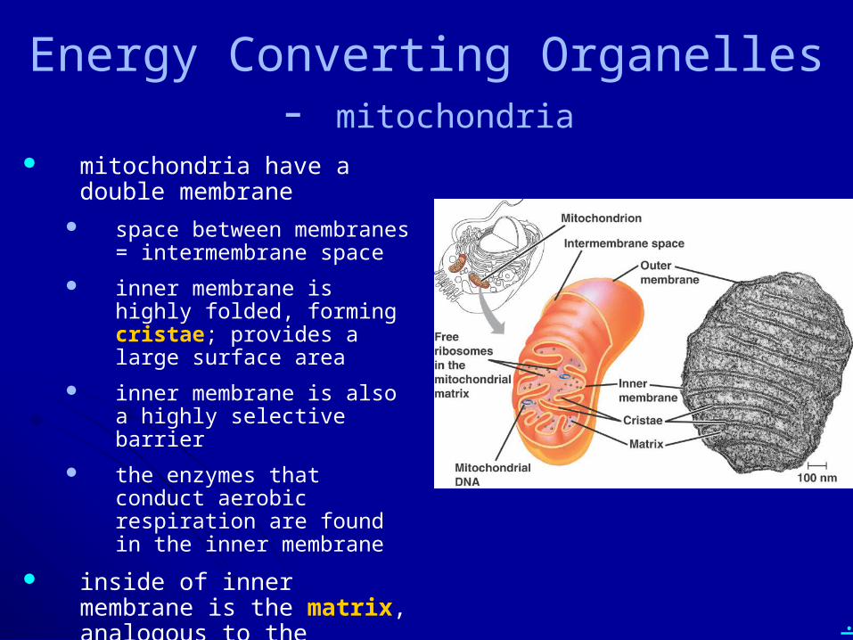
Energy Converting Organelles - mitochondria
mitochondria have a double membrane
space between membranes = intermembrane space
inner membrane is highly folded, forming cristae; provides a large surface area
inner membrane is also a highly selective barrier
the enzymes that conduct aerobic respiration are found in the inner membrane
inside of inner membrane is the matrix, analogous to the cytoplasm of a cell
.
Energy Converting Organelles – mitochondria
mitochondria have their own DNA, and are inherited from the mother only in humans
mitochondria have their own division process, similar to cell division; each cell typically has many mitochondria, which can only arise from mitochondrial division
some cells require more mitochondria than others
mitochondria can leak electrons into the cell, allowing toxic free radicals to form
mitochondria play a role in initiating apoptosis (programmed cell death)
.
• Draw a mitochondrion in cross-section and describe its structure and functions.
.
• Draw a chloroplast in cross-section and describe its structure and functions.
.
Energy Converting Organelles
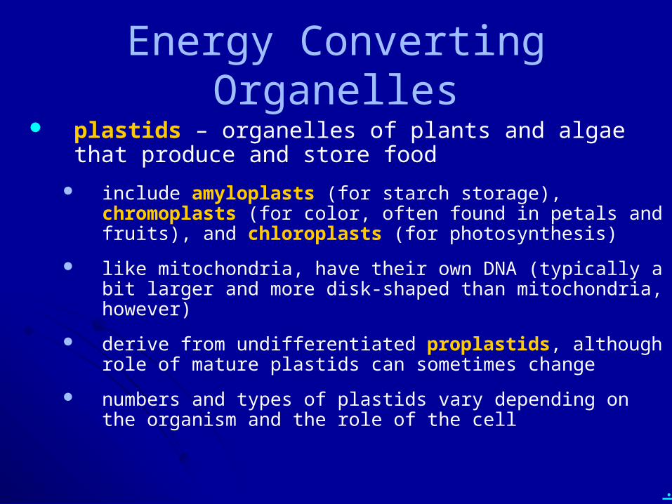
plastids – organelles of plants and algae that produce and store food
include amyloplasts (for starch storage), chromoplasts (for color, often found in petals and fruits), and chloroplasts (for photosynthesis)
like mitochondria, have their own DNA (typically a bit larger and more disk-shaped than mitochondria, however)
derive from undifferentiated proplastids, although role of mature plastids can sometimes change
numbers and types of plastids vary depending on the organism and the role of the cell
.
Energy Converting Organelles – plastids (chloroplasts)
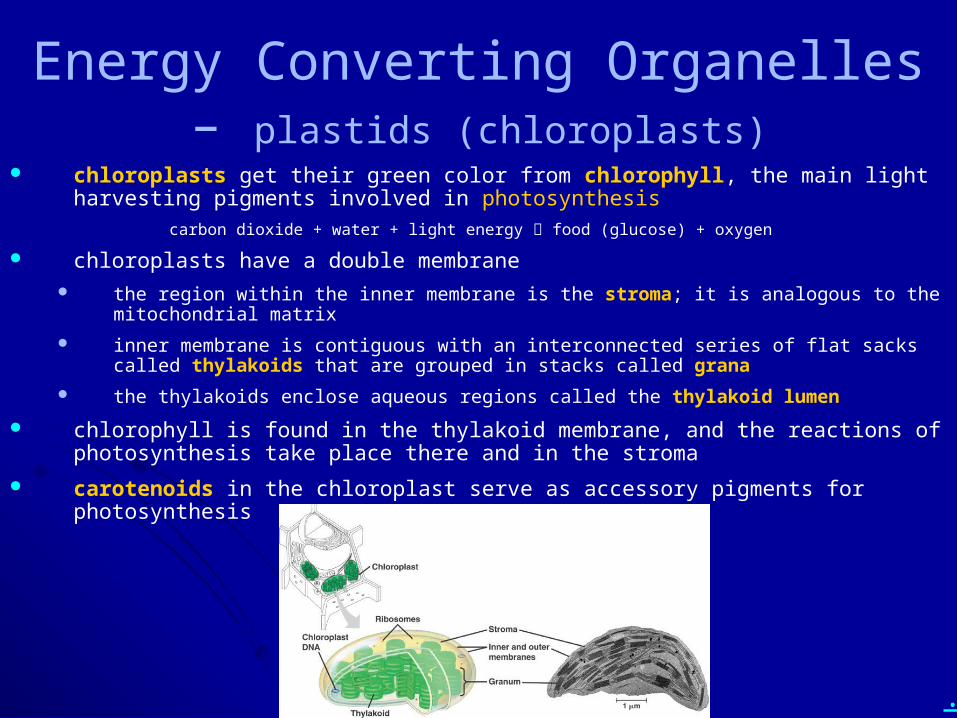
chloroplasts get their green color from chlorophyll, the main light harvesting pigments involved in photosynthesis
carbon dioxide + water + light energy food (glucose) + oxygen
chloroplasts have a double membrane the region within the inner membrane is the stroma; it is analogous to the mitochondrial matrix
inner membrane is contiguous with an interconnected series of flat sacks called thylakoids that are grouped in stacks called grana
the thylakoids enclose aqueous regions called the thylakoid lumen
chlorophyll is found in the thylakoid membrane, and the reactions of photosynthesis take place there and in the stroma
carotenoids in the chloroplast serve as accessory pigments for photosynthesis
.
• Draw a chloroplast in cross-section and describe its structure and functions.
.
• Describe the endosymbiont theory. Include evidence for it, including predictions that have proven true.
.
Energy Converting Organellesendosymbiont theory
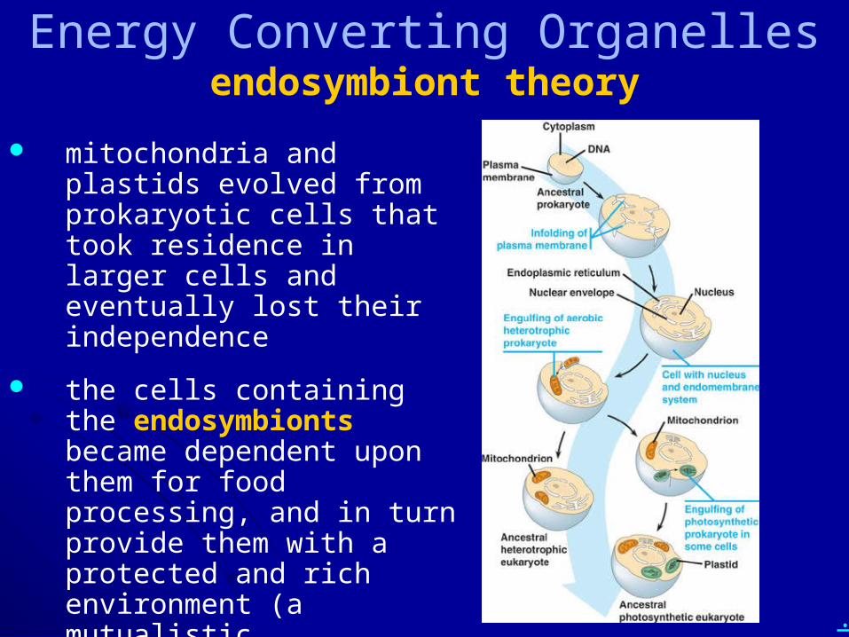
mitochondria and plastids evolved from prokaryotic cells that took residence in larger cells and eventually lost their independence
the cells containing the endosymbionts became dependent upon them for food processing, and in turn provide them with a protected and rich environment (a mutualistic relationship)
.
Energy Converting Organellesendosymbiont theory
supporting evidence
the size scale is right - mitochondria and plastids are on the high end of the size of typical bacteria
endosymbionts also have their own DNA and their own “cell” division; in many ways they act like bacterial cells
the DNA sequence and arrangement (circular chromosomes)of endosymbionts is closer to that of bacteria than to that found in the eukaryotic nucleus
endosymbionts have their own ribosomes, which are much like bacterial ribosomes
there are other known, more modern endosymbiotic relationships: algae in corals, bacteria within protozoans in termite guts
.
Energy Converting Organellesendosymbiont theory
some genes appear to have been shuttled out of the endosymbionts to the nucleus
many of the proteins used by endosymbionts are actually encoded by nuclear genes and translated in the cytoplasm (or on rough ER) and transported to the endosymbionts
DNA sequencing of endosymbionts is being used to trace the evolutionary history of the endosymbionts
appears that endosymbiosis began about 1.5 to 2 billion years ago (around when the first eukaryotic cells appeared)
mitochondria appear to have a monophyletic origin (one initial endosymbiotic event, giving rise to all mitochondria in eukaryotic cells today)
plastids appear to have a polyphyletic origin (more than one initial endosymbiotic event giving rise to different plastid lines present today in algae and plants)
.
• Describe the endosymbiont theory. Include evidence for it, including predictions that have proven true.
.
Chapter 6: A Tour of the Cell Cell theory
Cell organization and homeostasis
Studying cells – microscopy and fractionation
Eukaryotic vs. prokaryotic cells
Compartments in eukaryotic cells (cell regions, organelles)
Cytoskeleton
Outside the cell
.
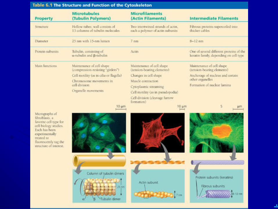
• What are the functions of the cytoskeleton?
• What are the three main types of cytoskeleton? Describe the structure and function(s) of each type.
.
• Describe the structure and function(s) of:
– motor proteins– MTOCs– centrosomes– centrioles– cilia and flagella
.
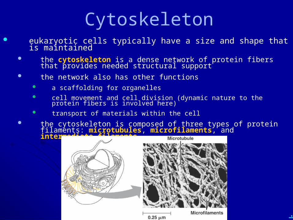
Cytoskeleton eukaryotic cells typically have a size and shape that is maintained
the cytoskeleton is a dense network of protein fibers that provides needed structural support
the network also has other functions a scaffolding for organelles cell movement and cell division (dynamic nature to the protein fibers is involved
here) transport of materials within the cell
the cytoskeleton is composed of three types of protein filaments: microtubules, microfilaments, and intermediate filaments
.
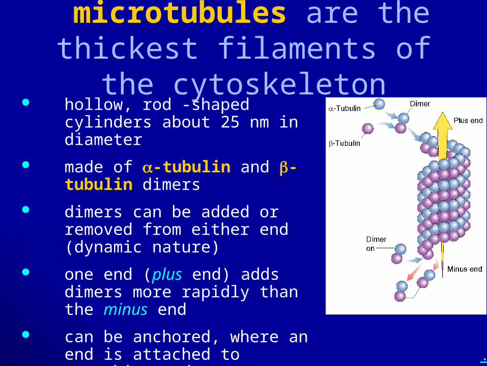
microtubules are the thickest filaments of the cytoskeleton
hollow, rod -shaped cylinders about 25 nm in diameter
made of a-tubulin and b-tubulin dimers
dimers can be added or removed from either end (dynamic nature)
one end (plus end) adds dimers more rapidly than the minus end
can be anchored, where an end is attached to something and can no longer add or lose dimers
.
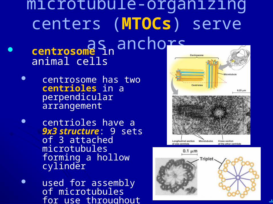
microtubule-organizing centers (MTOCs) serve as anchors
centrosome in animal cells
centrosome has two centrioles in a perpendicular arrangement
centrioles have a 9x3 structure: 9 sets of 3 attached microtubules forming a hollow cylinder
used for assembly of microtubules for use throughout the cell
.
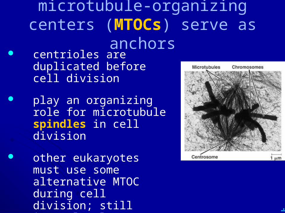
microtubule-organizing centers (MTOCs) serve as anchors
centrioles are duplicated before cell division
play an organizing role for microtubule spindles in cell division
other eukaryotes must use some alternative MTOC during cell division; still incompletely described
.
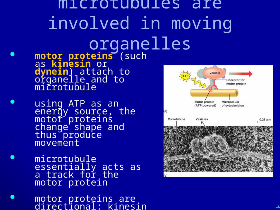
microtubules are involved in moving organelles
motor proteins (such as kinesin or dynein) attach to organelle and to microtubule
using ATP as an energy source, the motor proteins change shape and thus produce movement
microtubule essentially acts as a track for the motor protein
motor proteins are directional; kinesin moves toward the plus end, dynein away from it
.
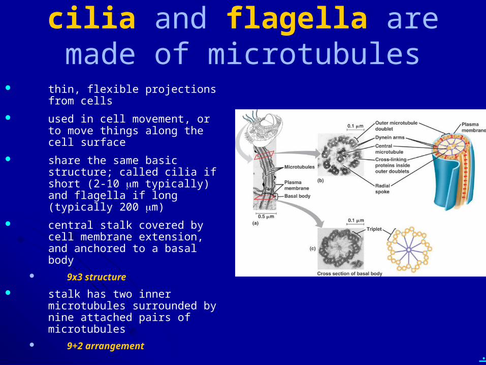
cilia and flagella are made of microtubules
thin, flexible projections from cells
used in cell movement, or to move things along the cell surface
share the same basic structure; called cilia if short (2-10 mm typically) and flagella if long (typically 200 mm)
central stalk covered by cell membrane extension, and anchored to a basal body
9x3 structure
stalk has two inner microtubules surrounded by nine attached pairs of microtubules
9+2 arrangement
.
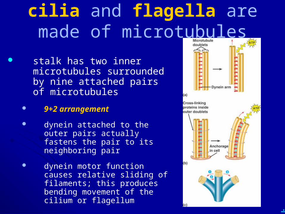
cilia and flagella are made of microtubules
stalk has two inner microtubules surrounded by nine attached pairs of microtubules
9+2 arrangement
dynein attached to the outer pairs actually fastens the pair to its neighboring pair
dynein motor function causes relative sliding of filaments; this produces bending movement of the cilium or flagellum
.
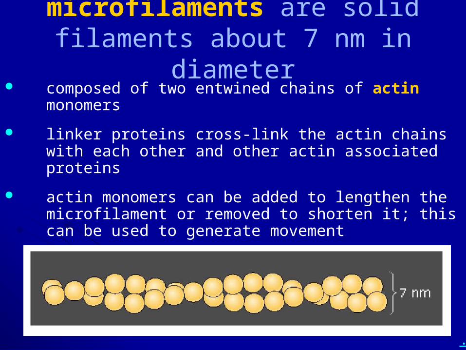
microfilaments are solid filaments about 7 nm in diameter
composed of two entwined chains of actin monomers
linker proteins cross-link the actin chains with each other and other actin associated proteins
actin monomers can be added to lengthen the microfilament or removed to shorten it; this can be used to generate movement
.
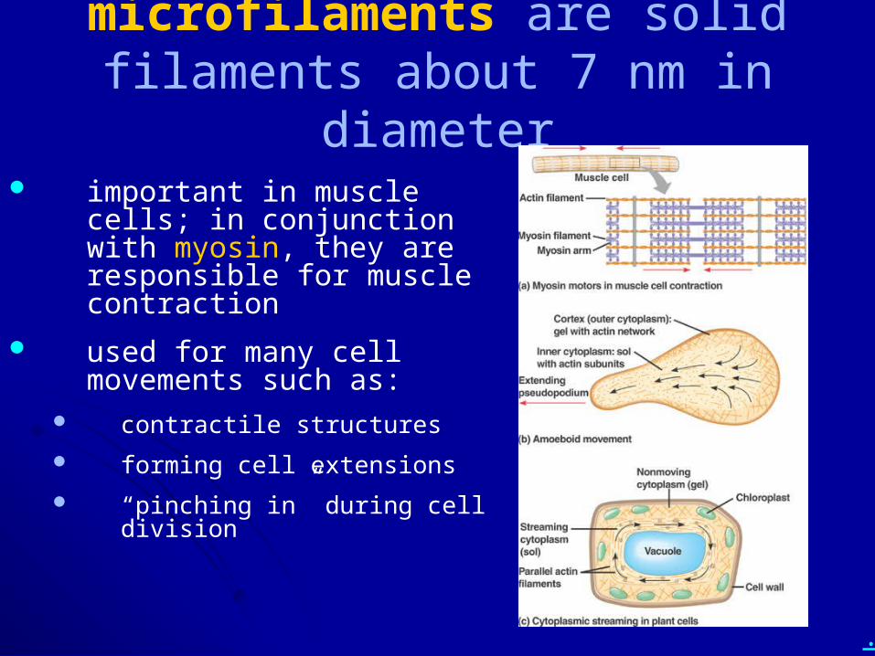
microfilaments are solid filaments about 7 nm in diameter
important in muscle cells; in conjunction with myosin, they are responsible for muscle contraction
used for many cell movements such as:
contractile structures
forming cell extensions
“pinching in” during cell division
.
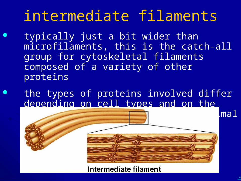
intermediate filaments typically just a bit wider than microfilaments, this is
the catch-all group for cytoskeletal filaments composed of a variety of other proteins
the types of proteins involved differ depending on cell types and on the organism; apparently limited to animal cells and protozoans
.
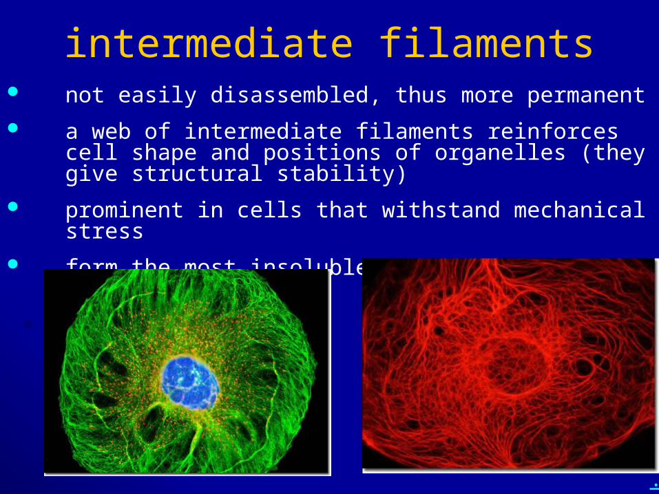
intermediate filaments not easily disassembled, thus more permanent
a web of intermediate filaments reinforces cell shape and positions of organelles (they give structural stability)
prominent in cells that withstand mechanical stress
form the most insoluble part of the cell
.
• What are the functions of the cytoskeleton?
• What are the three main types of cytoskeleton? Describe the structure and function(s) of each type.
.
• Describe the structure and function(s) of:
– motor proteins– MTOCs– centrosomes– centrioles– cilia and flagella
.
Chapter 6: A Tour of the Cell Cell theory
Cell organization and homeostasis
Studying cells – microscopy and fractionation
Eukaryotic vs. prokaryotic cells
Compartments in eukaryotic cells (cell regions, organelles)
Cytoskeleton
Outside the cell
.
• Describe the outer part and outside interface of a:
– typical prokaryotic cell– typical plant cell– typical fungal cell– typical animal cell
• Diagram and describe the animal cell glycocalyx and ECM interaction (include collagen, fibronectin, and integrin).
.
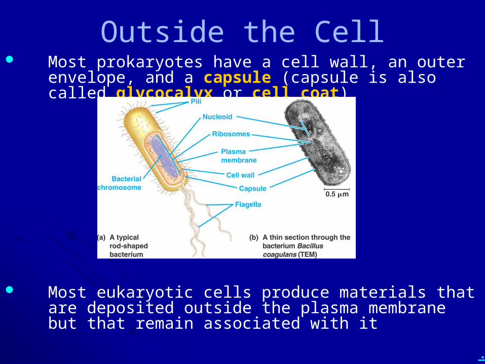
Outside the Cell Most prokaryotes have a cell wall, an outer envelope, and a
capsule (capsule is also called glycocalyx or cell coat)
Most eukaryotic cells produce materials that are deposited outside the plasma membrane but that remain associated with it
.
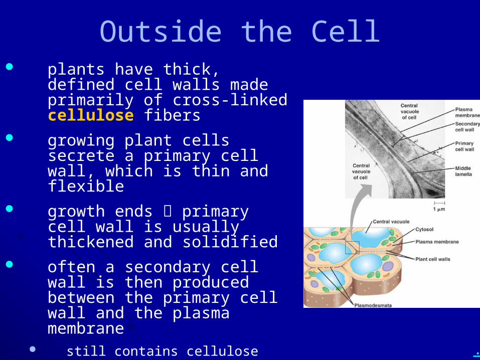
Outside the Cell plants have thick, defined cell
walls made primarily of cross-linked cellulose fibers
growing plant cells secrete a primary cell wall, which is thin and flexible
growth ends primary cell wall is usually thickened and solidified
often a secondary cell wall is then produced between the primary cell wall and the plasma membrane
still contains cellulose typically has more strengthening
material (for example, lignin in wood)
.
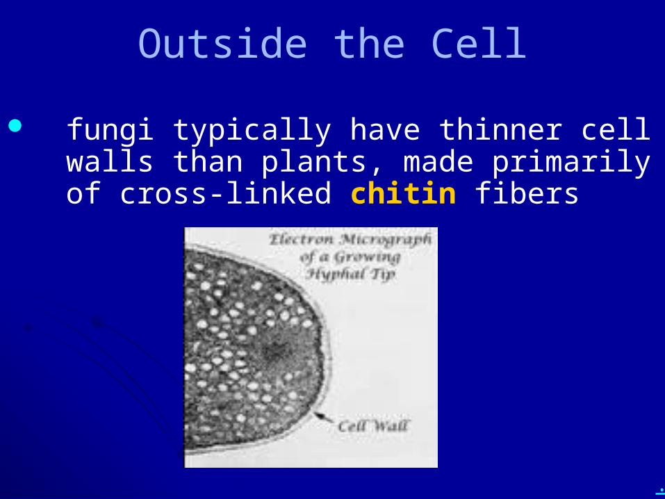
Outside the Cell
fungi typically have thinner cell walls than plants, made primarily of cross-linked chitin fibers
.
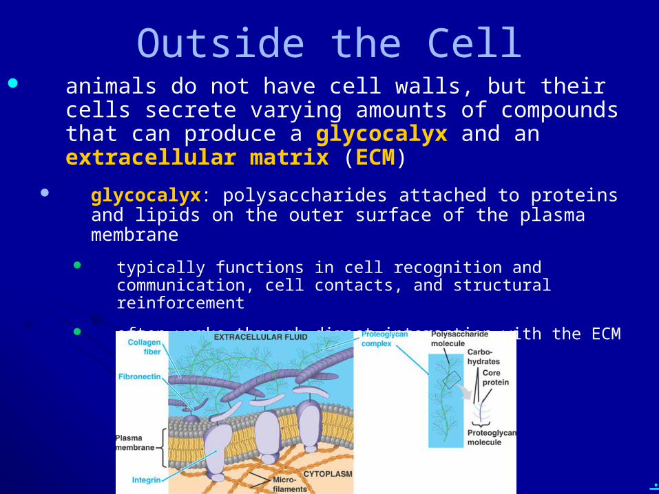
Outside the Cell animals do not have cell walls, but their cells secrete
varying amounts of compounds that can produce a glycocalyx and an extracellular matrix (ECM)
glycocalyx: polysaccharides attached to proteins and lipids on the outer surface of the plasma membrane
typically functions in cell recognition and communication, cell contacts, and structural reinforcement
often works through direct interaction with the ECM
.
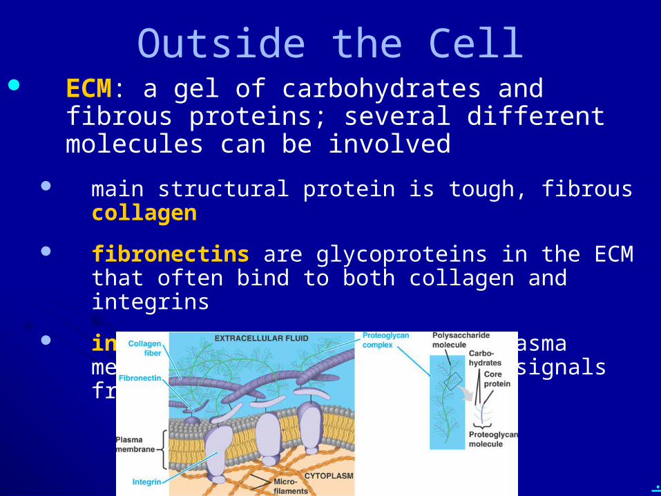
Outside the Cell ECM: a gel of carbohydrates and fibrous proteins;
several different molecules can be involved
main structural protein is tough, fibrous collagen
fibronectins are glycoproteins in the ECM that often bind to both collagen and integrins
integrins are proteins in the plasma membrane that typically receive signals from the ECM
.
• Describe the outer part and outside interface of a:
– typical prokaryotic cell– typical plant cell– typical fungal cell– typical animal cell
• Diagram and describe the animal cell glycocalyx and ECM interaction (include collagen, fibronectin, and integrin).