(+)-4R-limonene synthase from C. sinensis - Brandeis University
62
Kinetic characterization, crystallization, and photosynthetic expression of (+)-4R-limonene synthase from C. sinensis A Master’s Thesis Presented to The Faculty of the Graduate School of Arts and Sciences Brandeis University Department of Biochemistry Daniel Oprian, Advisor In Partial Fulfillment of the Requirements for Master’s Degree by Sonya Entova May, 2013
Transcript of (+)-4R-limonene synthase from C. sinensis - Brandeis University

Kinetic characterization, crystallization, and photosynthetic expression of (+)-4R-limonene synthase from C. sinensis
A Master’s Thesis
Presented to
The Faculty of the Graduate School of Arts and Sciences Brandeis University
Department of Biochemistry Daniel Oprian, Advisor
In Partial Fulfillment of the Requirements for
Master’s Degree
by Sonya Entova
May, 2013

Copyright by
Sonya Entova
© 2013

iii
ACKNOWLEDGEMENTS
I would like to express my most sincere gratitude to the many people without whose help
this thesis would not have been possible. Above all, I want to thank Dr. Daniel Oprian for
the privilege of working in his lab. I thank him for his patience and insight as a mentor, as
well as for challenging me to think critically and creatively, plan accordingly, and
persevere, no matter how daunting the task at hand. It is only by virtue of his unwavering
guidance and encouragement that I now feel prepared to continue my journey as a
scientist.
In addition, I would like to thank all the members of the Oprian lab, both past and
present, and my fellow classmates in the biochemistry department. Learning from them
has been a true honor, and I would not be the scientist, or person, that I am today without
their advice, support, and companionship.
I would like to thank my parents from the very bottom of my heart for always
demanding only the best from me, and for teaching me to do the same. I could never
have made it here were it not for their constant love and encouragement. And finally, I
would like to thank my close friends and family, whose endless support has made this
thesis a reality. To list all their names here would be impossible, but I trust that they
recognize who they are, and how much their support has meant to me.
iv
ABSTRACT
Kinetic Characterization, Crystallization, and Photosynthetic Expression
of (+)-4R-Limonene Synthase from C. sinensis
A Thesis Presented to the Department of Biochemistry
Graduate School of Arts and Sciences Brandeis University
Waltham, Massachusetts
By Sonya Entova
The terpenoid family represents one of the most abundant and diverse classes of organic
molecules found in nature. Because limonene synthase catalyses the simplest of all
monoterpene (C10) cyclization reactions, it is often used as a model for monoterpene
biosynthesis. The study described here was done with the enzyme (+)-(4R)-limonene
synthase, which was cloned previously from the flavedo of Citrus sinensis. In this thesis,
we discuss the kinetic characterization of this enzyme, in which we observe kinetic
properties very different from those previously found for the same enzyme cloned from
Citrus limon. We also describe a crystal structure of this enzyme in apoprotein form, and
the soaking of such crystals in a substrate-containing solution, where we observe time-
dependent changes in electron density for three coordinating Mn2+ atoms in the enzyme
active site. We hope that these results constitute key steps in identifying the structural
components of the enzyme mechanism. Finally, we relate the initial stages of an effort to
produce (+)-limonene, a potential biofuel, in the cyanobacterium Synechocystis by
engineering our (+)-(4R)-limonene synthase gene into the Synechocystis genome.

v
TABLE OF CONTENTS
ABSTRACT iv TABLE OF CONTENTS v LIST OF TABLES vi LIST OF FIGURES vii INTRODUCTION 1 Terpenoids 1 Monoterpenes 3 Limonene Synthase 5 Synechocystis 8 CHAPTER 1: LIMONENE SYNTHASE EXPRESSION AND PURIFICATION OPTIMIZATION 11 Materials and Methods 11 Results 13 CHAPTER 2: KINETIC CHARACTERIZATION 16 Materials and Methods 16 Results 18 CHAPTER 3: STRUCTURE DETERMINATION BY CRYSTALLIZATION 22 Materials and Methods 22 Results 24 CHAPTER 4: LIMONENE PRODUCTION IN SYNECHOCYSTIS 29 Materials and Methods 29 Results 36 DISCUSSION 41 Kinetic Characterization 41 Crystal Structure Determination 42 Limonene Production in Synechocystis 45 Future Directions 47 APPENDIX: SUPPLEMENTARY FIGURES 51 REFERENCES 53

vi
LIST OF TABLES
1 Enzyme activity of protein expressed at 20°C and 37°C 18
2 Data collection and refinement statistics 24
3 Primers used in plasmid construction 31

vii
LIST OF FIGURES
1 IPP and DMAPP production (simplified) 2
2 Monoterpene, sesquiterpene, and diterpene synthesis 4
3 Limonene enantiomers 6
4 Limonene synthase elution profile 13
5 Optimization of expression and purification 14
6 Product analysis by GC-MS 17
7 Limonene synthase kinetic analysis 20
8 Apoprotein crystal structure 25
9 Manganese atoms bound to the active site 26
10 Positioning of the N-terminal tail and the active site loops 28
11 Generating the KSusds plasmid 32
12 Insertion of GPPS’ and LS into KSusds 34
13 Gibson (isothermal) assembly 35
14 Restriction digest of KSusds 36
15 The pUC19a vector 38
16 The pBluescript vectors 39
17 Relative plasmid and fragment lengths for isothermal assembly 40
A1 BG11 mineral media composition 51
A2 Upstream and downstream psbA2 flanking sequences 52
A3 GPPS silent mutation 52

1
INTRODUCTION
Terpenoids
The terpenoid (or isoprenoid) family represents one of the most abundant and diverse
families of organic compounds found in nature, comprising over 55,000 members, many
of which play important biological roles in plant defense, attraction of pollinators, and
plant signaling. Many terpenoids have been implicated in a variety of industrial and
agricultural roles as flavorings, perfumes, solvents, pesticides, and the like. More
recently, a variety of pharmaceutical applications have also been discovered for
terpenoids, including as anti-inflammatory, [1] anti-tumor and anti-metastatic [2] agents.
The extensive commercial importance of terpenoids, together with the relative difficulties
often associated with extracting large amounts of them from their native sources, has
spurred an effort to apply methods of genetic and metabolic engineering to producing
these compounds recombinantly in a variety of alternate hosts [3, 4].
Despite the broad diversity of the terpenoid family, all terpenes are composed of
multiples of one common C5 precursor, isoprene, linked together in a “head-to-tail”
manner. The first step in the synthesis of any terpene is the production of two isomeric
activated forms of isoprene: isopentenyl pyrophosphate (IPP) and dimethylallyl
pyrophosphate (DMAPP). Because these two isomeric subunits are products of both the
mevalonic acid pathway and the mevalonic acid-independent pathway [5], they are amply
available in virtually all living species (Figure 1).

2
FIG . 1 IPP and DMAPP production (simplified) – Isoprene, the subunit from which all terpenoids are synthesized, is found in two isomeric activated forms: isopentyl pyrophosphate (IPP) and dimethylallyl pyrophosphate (DMAPP) (A). IPP and DMAPP are products of both the mevalonic acid pathway (B) and the mevalonic acid-indpendent pathway (C).
One IPP unit and one DMAPP unit can be joined together to form the C10 terpene
precursor, geranyl pyrophosphate (GPP). The 1-4 addition of an additional IPP unit to
GPP gives the C15 terpene precursor, farnesyl pyrophosphate (FPP), and the subsequent
addition of a third IPP addition to FPP gives the C20 terpene precursor, geranylgeranyl
pyrophosphate (GGPP) [6] (Figure 2). The cyclization of GPP and subsequent redox
modification yields the wide variety of C10 terpenes known as monoterpenes. Similarly,
the cyclization and modification of FPP yields a variety of C15 terpenes, which are known
as sesquiterpenes, and the cyclization and modification of GGPP yields a variety of C20
terpenes, called diterpenes. The focus of this study will be on the enzyme (+)-4R-
limonene synthase, which produces the monoterpene, limonene.

3
Monoterpenes
The conversion of GPP into monoterpenes occurs through a series of carbocation
intermediates, beginning with the removal of the pyrophosphate group to form the linalyl
cation. Non-cyclic monoterpenes such as linalool can be formed from this cation, but the
formation of cyclic monoterpenes requires a subsequent isomerization of the linalyl
cation via a (+)-3S-linalyl pyrophosphate intermediate, generating a conformation that
sterically favors cyclization to the α-terpinyl cation [5]. The α-terpinyl intermediate can
then be transformed into various cyclic monoterpenes, the simplest of which is limonene.
These basic monoterpene skeletons can then be modified further via a series of oxidation
reactions to yield the great diversity of cyclic monoterpenes observed in nature (Figure
2).
A number of monoterpene synthases from various plant sources have been studied
and characterized in either their native or recombinant form [6-9]. As a rule, these
enzymes have a molecular mass of 50- to 100-kDa in their native form, and are found as
either monomers or homodimers. They are generally soluble, but are often targeted to the
plant’s plastid membrane when expressed in vivo. Several studies have confirmed the
presence a highly conserved RR motif in monoterpene synthases located approximately
50-70 residues from the N-terminus. This arginine pair is believed to stabilize the
negatively-charged diphosphate that is hydrolyzed from the GPP substrate [10]. It is

4
FIG. 2 Monoterpene, sesquiterpene, and diterpene synthesis – The three terpenoid precursors geranyl pyrophosphate (GPP), farnesyl pyrophosphate (FPP), and geranylgeranyl pyrophosphate (GGPP) are generated by the addition of IPP and DMAPP subunits. GPP then undergoes a cyclization reaction to form the monoterpene limonene, which can be modified to form other monoterpenes. FPP undergoes a similar process to generate various sesquiterpenes, and GGPP undergoes a similar process to generate various diterpenes. Adapted from Dewick [5].
believed that the sequence upstream of this motif corresponds to an N-terminal transit
domain which targets the monoterpene to its final destination within the plastid, and it
has been shown that enzymes truncated at a position just prior to this conserved motif
take on their mature, native conformation and are fully functional when expressed
5
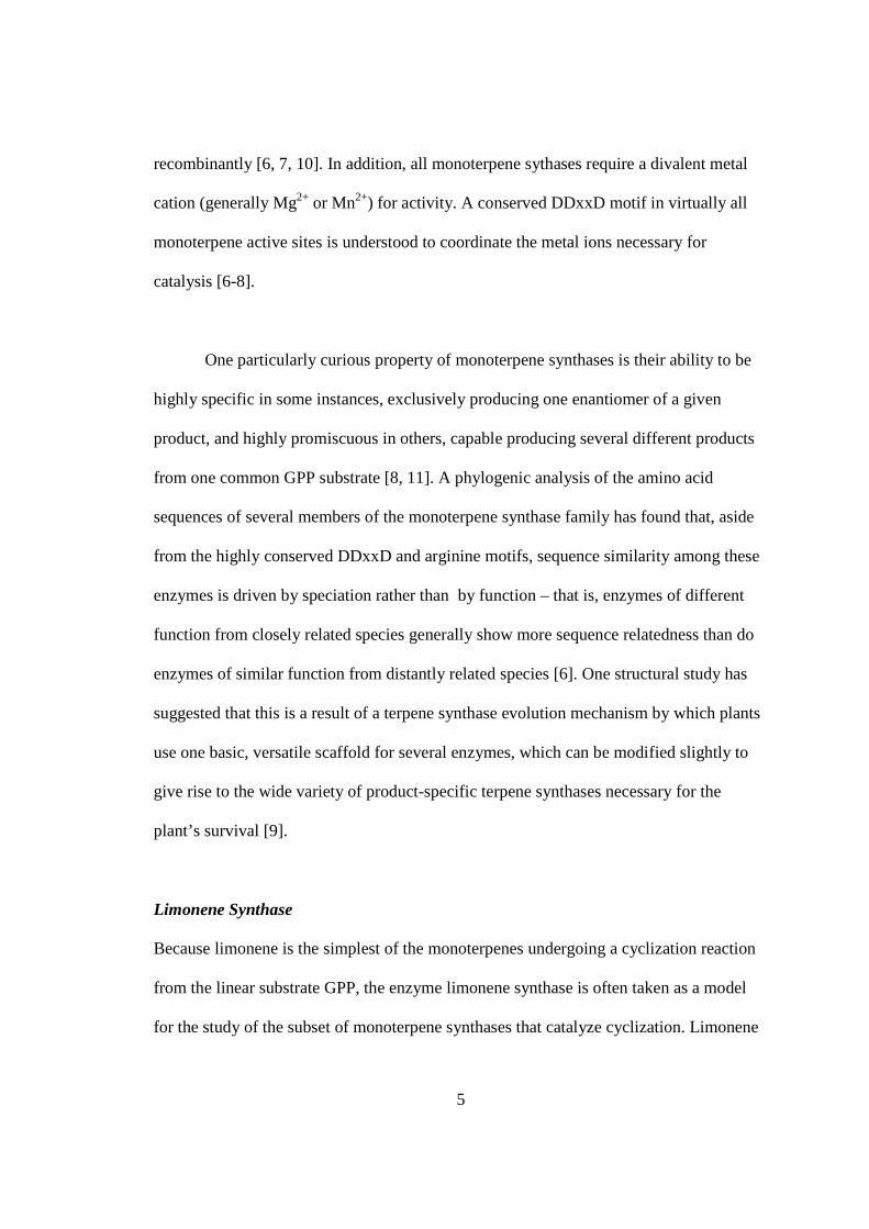
recombinantly [6, 7, 10]. In addition, all monoterpene sythases require a divalent metal
cation (generally Mg2+ or Mn2+) for activity. A conserved DDxxD motif in virtually all
monoterpene active sites is understood to coordinate the metal ions necessary for
catalysis [6-8].
One particularly curious property of monoterpene synthases is their ability to be
highly specific in some instances, exclusively producing one enantiomer of a given
product, and highly promiscuous in others, capable producing several different products
from one common GPP substrate [8, 11]. A phylogenic analysis of the amino acid
sequences of several members of the monoterpene synthase family has found that, aside
from the highly conserved DDxxD and arginine motifs, sequence similarity among these
enzymes is driven by speciation rather than by function – that is, enzymes of different
function from closely related species generally show more sequence relatedness than do
enzymes of similar function from distantly related species [6]. One structural study has
suggested that this is a result of a terpene synthase evolution mechanism by which plants
use one basic, versatile scaffold for several enzymes, which can be modified slightly to
give rise to the wide variety of product-specific terpene synthases necessary for the
plant’s survival [9].
Limonene Synthase
Because limonene is the simplest of the monoterpenes undergoing a cyclization reaction
from the linear substrate GPP, the enzyme limonene synthase is often taken as a model
for the study of the subset of monoterpene synthases that catalyze cyclization. Limonene

6
has a single chiral center located at C4 and is thus found in two different enantiomeric
forms: (+)-4R-limonene, commonly found in citrus fruit [12], and (-)-4S-limonene, found
in a wide variety of plants, including mint, pine, sage, and caraway [5] (Figure 3).
FIG. 3 Limonene enantiomers – The monoterpene limonene exists in two enantiomeric forms: (+)-4R-limonene, commonly found in citrus fruit, and (-)-4S-limonene, found in a wide variety of plants, including mint, pine, sage, and caraway.
While some plants, such as orange and mint, are known to contain a single limonene
enantiomer almost exclusively, others have been found to contain both enatiomers in
various ratios [7]. The production of either enantiomer is thought to be dictated by the
orientation in which the GPP substrate initially enters the active site, indicating that more
promiscuous enzymes must be able to bind both right- and the left-handed conformations
of GPP, while more specific enzymes must select for a single conformation [13, 14].
Much work has already been done to characterize limonene synthase from a wide
variety of sources. Studies have shown that both (+)- and (-)-limonene synthase share the
N-terminal transit peptide and the RR and DDxxD motifs conserved in other
monoterpene synthases [7, 10, 11]. The kinetics of limonene synthase from a variety of
native and recombinant sources including (-)-limonene synthase from Mentha x piperita
(peppermint) and Mentha spicata (spearmint) [15] and (+)-limonene synthase from C.
limon (lemon) have also been reported [11]. It has been found that both (+)- and (-)-
limonene synthase prefer the Mn2+ cation over the Mg2+ cation in vitro [14, 16].

7
A crystal structure of (-)-4S-limonene from Mentha spicata liganded to a
substrate analog and Mn2+ has been previously determined. This cocrystallization
technique yielded crystals of the I4 space group that diffracted to 2.7-Å resolution [14].
The structure of (-)-limonene synthase was found to be similar to those previously
determined for (+)-boranyl pyrophosphate synthase from Salvia [17] and 5-epi-
aristolochene synthase from tobacco [18], in that it is composed of two helical domains –
the C-terminal domain, which contains the active site, and an N-terminal domain, which
has been observed in several other terpenoid synthases but whose function remains
undetermined [19]. Like previous structures, (-)-limonene synthase was found to have an
N-terminal tail that folds over the C-terminal active site, forming a cap to shield the
carbocation intermediates in the active site from solvent during catalysis. Significant rms
deviations for all Cα in the (-)-limonene synthase structure indicated that the enzyme is
generally flexible, particularly around the active site [14]. This agrees well with a
reaction mechanism in which the enzyme must switch between open and closed
conformations upon substrate binding and product release. Three Mn2+ metal ions,
coordinated by the DDxxD motif, are easily observable in this structure.
This thesis will focus on a recombinant (+)-4R-limonene synthase, which has
previously been cloned from Citrus sinensis, the navel orange [16]. The kinetics of (+)-
4R-limonene synthase from Citrus limon (lemon) have been previously characterized,
showing a Km of 0.7µM and an activity profile consistent with substrate inhibition [11].
Initial activity assays of our (+)-limonene synthase from C. sinensis indicated that the Km

8
of this enzyme is significantly higher than that of its counterpart from C. limon [16]. In
this study we present a more complete analysis of the kinetics of (+)-limonene synthase
from C. sinensis that confirms these early findings. In fact, the Km determined for our
enzyme is 130µM, more than 100-fold greater than the published Km for enzyme from C.
limon, and shows no indication of substrate inhibition.
As of the time of this publication, no structures have been published of (+)-4R-
limonene synthase. The optimization of expression and purification of this enzyme from
Escherichia coli has allowed us to obtain enzyme of high enough purity to crystallize the
(+)-limonene apoprotein and perform initial soaking experiments with Mn2+ and GPP
substrate, in an attempt to observe bound reaction intermediates in the enzyme active site.
These results, when compared with the known structure for (-)-4S-limonene synthase
described above, represent a significant step towards pinpointing the particular structural
elements involved in the enzyme’s reaction mechanism.
Synechocystis
Synechocystis sp. PCC 6803 is a photoautotrophic freshwater cyanobacterium. The
physiology and genetics of this organism have been well-documented [20]. It is known
that the Synechocystis genome consists of a single, 3.6 Mbp chromosome in addition to
several plasmids of various sizes [21], and that it is able to spontaneously take up foreign
DNA into its own genome by double homologous recombination with very high
efficiency [22, 23], making it a popular candidate for the recombinant expression of
various proteins.

9
Synechocystis utilizes photosynthetic oxygen-generating machinery very similar
to that of plants. It has been shown that the genes that code for photosystem II in
Synechcystis, psbA2 and psbA3, are redundant, meaning that Synechocystis can continue
to function normally in the absence of either one of these two genes [24]. One study has
been able to successfully replace psbA2 with the isoprene synthase gene from Pueraria
montana (kudzu) [25], allowing isoprene synthase to be expressed under the psbA2
promoter. This resulted in isoprene being produced photosynthetically by the
Synechocystis [26]. This finding is significant because, as a combustible hydrocarbon,
isoprene produced this way is a model for the photosynthetic production of related
terpene biofuels. In fact, the manipulation of various microorganisms to produce bulk
amounts of biofuels is currently an active field of study [27, 28]. While this method has
not been used yet to produce more than one recombinant protein at a time in
Synechocystis, a different study, which was able to heterologously express several
different [NiFe] hydrogenases polycistronically in the related cyanobacterium
Synechococcus elongatus [29], suggests that polycistronic expression in Synechocystis
may be possible as well, allowing for the photosynthetic production of more complex
hydrocarbons.
In this study we complete the initial stages necessary to engineer a pathway in
Synechocystis which would produce (+)-limonene photosynthetically. Because limonene
contains more energy per molecule and has a much higher boiling point than does
isoperene (176 °C vs. 34 °C), it has more tangible applications as a biofuel. However, the

10
heterologous production of limonene is also more complex than that of isoprene, as it
must occur in two steps: the production of GPP first from IPP and DMAPP subunits, and
the cyclization of GPP into (+)-limonene second. Accordingly, this pathway requires two
enzymes to be functionally expressed in Synechocystis: GPP pyrophosphate synthase for
the production of GPP, and (+)-limonene synthase for the cyclization into the final (+)-
limonene product. In this thesis, we describe the first steps taken to insert the GPP
synthase and (+)-limonene synthase genes into the Synechocystis genome, following a
protocol modeled closely after that of Lindberg et al. for the production of isoprene in
the same organism [26].

11
CHAPTER 1:
L IMONENE SYNTHASE EXPRESSION AND PURIFICATION OPTIMIZATION
MATERIALS AND METHODS
Expression
The gene used to express (+)-4R-limonene synthase has been isolated in the lab
previously from C. sinensis (orange), truncated to remove the plastidial targeting
sequence, and tagged with an N-terminal 6•His tag. The truncated and tagged gene had
been cloned into the pET28a vector (Novagen) and expressed in E. coli strain BL21-
CodonPlus-RIL (Agilent Technologies), which is abundant in additional copies of
tRNAs found rarely in E. coli, and thereby facilitates the expression of arginine-,
isoleucine- or leucine rich proteins such as (+)-limonene synthase [16]. The pET28a
vector confers kanamycin resistance, while the CodonPlus-RIL strain confers
chloramphenicol resistance; thus, selection was performed by plating on 25 µg/mL
kanamycin/34 µg/mL chloramphenicol LB plates. Starter cultures were grown overnight
in LB supplemented with 25 µg/mL kanamycin and 50 µg/mL chloramphenicol, at 37°C
and shaking at 220 rpm, and used to inoculate 1L cultures of unsupplemented LB the next
morning.

12
Cultures were grown at 37°C to an OD600 of 0.6, then induced with 1 mM IPTG
and incubated at 37°C for 2 hours to allow for expression of the protein. For expression at
20°C, cultures were grown at 37°C to an OD600 of 0.6, then allowed to equilibrate at 20°C
for 30 minutes before induction with 1 mM IPTG, and finally incubated at 20°C for
approximately 20 hours. To test for protein stability over the course of the 20-hour
expression, culture samples were taken at induction and at points 3 hours, 8 hours, and 20
hours post-induction. Following expression at either temperature, cells were harvested by
centrifugation at 4°C and stored in pellet form at -80°C.
Purification
Frozen cell pellets were thawed and resuspended in 50 mL breaking buffer (50mM Tris,
100 mM NaCl, 20 mM imidazole, 10 µg/ml lysozyme, 10 µg/ml DNase I, pH 7.5)
supplemented with a protease inhibitor cocktail (La Roche) per the manufacturer’s
instructions. The cell suspension were sonicated with five 20-second pulses at
approximately 45 watts separated by 40-second rest periods, and then centrifuged at
16,000g at 4°C for 45 minutes. Supernatant was then filtered and loaded onto a 5-mL Ni-
Sepharose column (GE). After washing with wash buffer (20mM Tris, 100mM NaCl,
40mM imidazole, pH 7.5), limonene synthase was eluted using a linear 40-500mM
imidazole gradient (also in 20mM Tris,100mM NaCl, pH 7.5) over 12 column volumes
and stored short-term at 4°C.
13
RESULTS
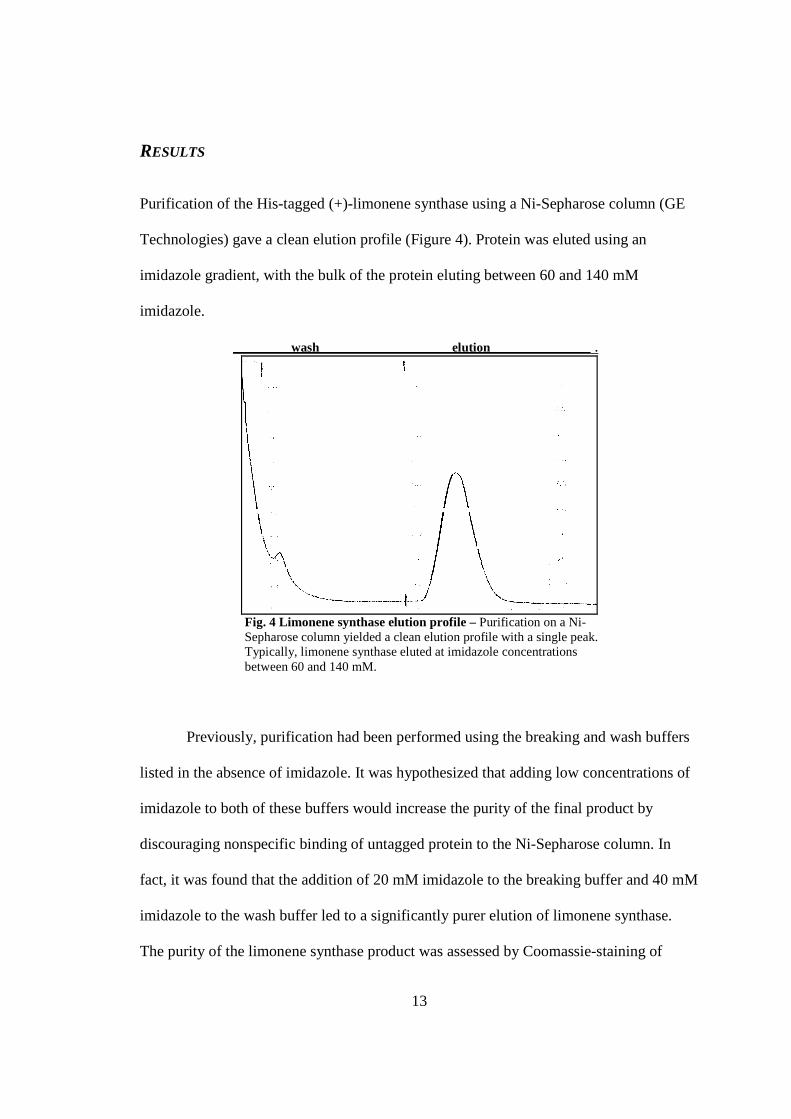
Purification of the His-tagged (+)-limonene synthase using a Ni-Sepharose column (GE
Technologies) gave a clean elution profile (Figure 4). Protein was eluted using an
imidazole gradient, with the bulk of the protein eluting between 60 and 140 mM
imidazole.
wash elution .
Fig. 4 Limonene synthase elution profile – Purification on a Ni-Sepharose column yielded a clean elution profile with a single peak. Typically, limonene synthase eluted at imidazole concentrations between 60 and 140 mM.
Previously, purification had been performed using the breaking and wash buffers
listed in the absence of imidazole. It was hypothesized that adding low concentrations of
imidazole to both of these buffers would increase the purity of the final product by
discouraging nonspecific binding of untagged protein to the Ni-Sepharose column. In
fact, it was found that the addition of 20 mM imidazole to the breaking buffer and 40 mM
imidazole to the wash buffer led to a significantly purer elution of limonene synthase.
The purity of the limonene synthase product was assessed by Coomassie-staining of

14
purified fractions on SDS-PAGE gels, which showed that the addition of imidazole lead
to a reduction of contaminants in these fractions (see Figure 5, A and B).
Figure 5 – Optimization of expression and purification– Protein bands corresponding to limonene synthase in eluted fractions are shown outlined in rectangles. Purification with the addition of imidazole to the breaking and wash buffers (B) yielded a significantly purer product than purification using imidazole-free buffers (A), as indicated by the presence of fewer contaminating bands on Coomassie-stained SDS-PAGE gels. Expression at 20°C (D) instead of at 37°C (B) corresponded to an almost 10-fold increase in yield. Limonene synthase could be expressed at 20°C for up to 20 hours without any noticeable product degredation (C).
We noted that limonene synthase degrades if expressed at 37° for longer than
about 2 hours. To test whether a lower temperature could lead to a more stable, active
enzyme, expression was tested at 20°C. At this temperature limonene synthase could be

15
expressed for as long as 20 hours without any detectable degradation (Figure 5, C).
Furthermore, purification of protein expressed at 20°C yielded significantly more enzyme
than at 37°C. This was observed qualitatively by Coomassie-staining (Figure 5, B and D)
as well as quantitatively – typical yield for a 1 L culture expressed at 37°C was 2-3 mg,
while a typical yield for the same volume of culture expressed at 20°C was 15-25 mg,
corresponding to an almost 10-fold increase in yield.

16
CHAPTER 2:
K INETIC CHARACTERIZATION
MATERIALS AND METHODS
Kinetic Assays
The activity of purified (+)-4R-limonene synthase was measured according to the single-
vial assay technique described by O’Maille et al. [30] . Reactions were run in 1-mL
volumes containing ~0.1 µM enzyme in reaction buffer (15mM MOPS [pH 7.0], 2 mM
DTT, 10 mM MgCl2) supplemented with GPP substrate (Echelon Biosciences) at
concentrations varying from 25-600 µM. Immediately following mixing, the reactions
were overlaid with 1-mL ethyl acetate and run at room temperature in septum-capped
glass vials. Incubation time was varied between 0-40 minutes with several time points
taken for each substrate concentration, to allow for determination of product formation
velocity. Reactions were then terminated by vigorous vortexing for 30-45 seconds,
thereby also extracting the limonene product into the ethyl acetate. The organic layer was
then extracted and analyzed for product formation by GC-MS, as described below.
Reaction velocity data were plotted and analyzed using Microsoft EXCEL.

17
Quantification by GC-MS
Ethyl acetate samples were analyzed for the presence of limonene using a GC unit
(Agilent Technologies 7890A, column model HP-5HS) coupled to a mass selection
detector (Agilent Technologies 5975C). The GC was run at a He flow rate of 3 mL/min.
An automated injection of 5 uL was used, with a split ratio of 50:1 and an injector
temperature of 220°C. Initial oven temperature was 50°C, and temperature was increased
at a rate of 13°C/min to a temperature of 240°C. A solvent delay of 2.70 minutes was
included in the run time. A peak corresponding to limonene was consistently observed at
6 minutes and 22 seconds post-injection (Figure 6, A).
A – gas chromatography
B – mass spectrometry
Fig. 6 Product analysis by GC-MS – During separation by gas chromatography, a peak corresponding to limonene was routinely observed at 6 minutes and 22 seconds (A). The product was successfully identified as limonene by mass spectrometry (B).

18
The MSD was operated at 70 keV, with a source temperature of 230°C and a quad
temperature of 150°C. Product was successfully identified as limonene by mass
spectrometry (Figure 6, B). Following data collection, product peaks were analyzed using
the program TCMSD Data Analysis (version E.20.00.493, Agilent Technologies).
RESULTS
Activity of Enzyme Expressed at 20°C vs. 37°C
In addition to being significantly more stable during expression, enzyme expressed at
20°C was also found to be more active than enzyme expressed at 37°C. When the same
assay was performed with both 20°C and 37°C enzyme in the presence of 200 µM GPP,
it was found that a reaction with the 20°C enzyme at a concentration of 0.07 µM had a
velocity roughly comparable to a reaction with 37°C enzyme at a concentration of 0.26
µM, giving rise to kcat values of 0.13 s-1 and 0.04 s-1 for enzyme expressed at 20°C and
37°C, respectively (Table 1). These results indicate that limonene synthase is roughly 3
times more active when expressed at 20°C than when expressed at 37°C.
Table 1 Limonene Synthase Activity Assay, 200 µM GPP
Expression Temp.
Enzyme Conc.
Product t = 10 min
Product t = 20 min
Reaction Velocity Approx. V max Approx. k cat
20°C 0.07 µM 4.09 µM 7.51 µM 0.38 µM/min 0.53 µM/min 0.13 s-1
37°C 0.26 µM 4.35 µM 9.87 µM 0.49 µM/min 0.64 µM/min 0.04 s-1
TABLE 1 Enzyme activity of protein expressed at 20°C and 37°C – In a side-by-side comparison, it was found that enzyme expressed at 20°C is roughly 3 times more active than enzyme expressed at 37°C.

19
The decreased protein degradation during 20°C expression described in Chapter
1, and the increased stability and activity of limonene synthase expressed at 20°C,
described here, indicates that the protein is better able to fold into its proper conformation
at this temperature than at 37°C. This is not surprising – at 20°C E. coli function is
slowed, allowing the protein to be expressed and processed more slowly, and at this
lower temperature the folded protein itself also experiences less thermal agitation.
Furthermore, because the physiological temperature for the navel orange (from which this
limonene synthase is cloned) is expected to be around 20°C, enzyme folded at this
temperature is likely to resemble its native confirmation more closely than enzyme folded
at higher temperatures. Thus, all further kinetic analysis was done solely with enzyme
expressed at 20°C.
Kinetic Characterization of Limonene Synthase
Limonene synthase activity was quantified using the method described in Materials and
Methods. The rates of limonene production at various GPP concentrations, normalized to
enzyme concentration, are shown in Figure 7A. The dependence of the reaction velocity
on GPP concentration appeared to agree with the classical kinetic relationship between
reaction rate and substrate concentration as described by the Michaelis-Menten equation,
v = kcat [E] [S] Km + [S]
where v is the reaction velocity, kcat is the turn-over number for the enzyme, and Km is the
substrate concentration at which the reaction velocity is at half of its maximum possible

20
value. A plot of the observed reaction rates, fitted with a Michaelis-Menten curve, is
shown Figure 7C.
[GPP] v/[E] sec -1
25 µM 0.013
50 µM 0.029
75 µM 0.040
100 µM 0.047
150 µM 0.064
200 µM 0.075
300 µM 0.073
600 µM 0.079 A
Lineweaver-Burke Analysis
-20
0
20
40
60
80
-0.01 0 0.01 0.02 0.03 0.04
1/[GPP] (uM-1)
[E]/
v(u
M e
nzym
e-se
c/uM
pro
duct
)
B
Michaelis-Menton Analysis
0.00
0.02
0.04
0.06
0.08
0.10
0.12
0.14
0 100 200 300 400 500 600 700 800
[GPP] (uM)
v/[E
] (u
M li
mon
ene/
sec-
uM e
nzym
e)
C FIG . 7 Limonene synthase kinetic analysis – The relationship between the observed rates of product formatation and initial substrate concentrations (A) were plotted and fitted to the Michaelis-Menten equation (C) and the Lineweaver-Burke plot (B). Analysis of these two plots indicates that the kcat for the reaction is 0.11s-1, and the Km is 130 µM.

21
The double-reciprocal Lineweaver-Burke relationship, according to the equation
1 = Km . 1 + 1 . v kcat [E] [S] kcat [E]
was also plotted and used to determine the values of Km/kcat, and 1/kcat. A plot of the
observed reaction rates, fitted with a Lineweaver-Burke line, is shown in Figure 7B.
Analyses of both the Michaelis-Menten and Lineweaver-Burke plots indicate that the kcat
for our recombinant (+)-limonene synthase is 0.11 s-1, and the Km is 130 µM.

22
CHAPTER 3:
STRUCTURE DETERMINATION BY CRYSTALLIZATION
MATERIALS AND METHODS
Crystallization and Soaking
All crystallization and soaking experiments were done with Dr. P. Ramasamy. Enzyme
purified as described previously (Chapter 1) was used to grow crystals. Crystal trays were
set up using the Phoenix crystallization robot (Art Robbins Instruments), with a stock
enzyme solution at 15 mg/ml and crystallization buffer (12-16% PEG-8000, 100 mM
Tris-HCl pH 7.5-9.0, 200-350 mM sodium tartrate) in a 1:1 ratio. Crystals were grown at
20°C and typically appeared after 10-15 days.
For soaking, crystals still in their original crystallization solutions were
supplemented to 10 mM MnCl2 and 15% glycerol (final concentrations), and incubated at
room temperature for 1 hour. Crystals were then transferred to a new solution containing
500 µM geranyl pyrophosphate substrate, 10 mM MnCl2 and 15% glycerol, and
incubated for variable lengths of time (0-10 min, with 30 second intervals). In this study,

23
we analyze the structures of crystals soaked in geranyl pyrophosphate substrate solution
for 0, 1, and 4 minutes, which will be referred to as t = 0, t = 1, and t = 4, respectively.
Data Collection and Refinement
After soaking in substrate solution, crystals were flash frozen in liquid nitrogen. Data
were collected at beam line 8.2.1 at the Advanced Light Source (Lawrence Berkeley
National Laboratory, Berkeley, CA) using ADSC Q315R CCD detectors (Area Detector
Systems Corporation) at a temperature of 100 K. Data were integrated using MOSFLM
[31] and scaled using SCALA [32] from the CCP4 software suite v6.3 [33, 34].
Structures were solved by molecular replacement with the help of PHASER, [35]
using a previously determined crystal structure of the enzyme apoprotein as a model (P.
Ramasamy, not yet published). Refinement was performed using the function
phenix.refine [36] in PHENIX software suite v1.8, [37] and model building was done in
COOT v0.7 [38].
The statistics from data collection and the early stages of refinement are shown in
Table 2. However, all three structures require further refinement prior to submission to
the PDB.

24
Table 2 Soaking Time 0 min 1 min 4 min
Data Collection Space Group P41212
Resolution range (Å) 68-3.45 67-3.2 72-2.9
Highest res. range (Å) 3.64-3.45 3.37-3.2 3.08-2.9
Cell Dimensions a = b = 86.06, c = 222.63 α = β = γ = 90°
a = b = 85.5, c = 215.91 α = β = γ = 90°
a = b = 86.19, c = 216.57 α = β = γ = 90°
Total Reflections* 149,990 (22,261) 141,341 (19,404) 235,862 (40,348)
Unique Reflections* 21,030 (3,034) 24,044 (3,492) 18,945 (2,987)
Completeness %* 98.9 (98.6) 94.2 (94.2) 100 (100)
Rmerge* 0.17 (0.47) 0.08 (0.46) 0.113 (0.460)
I/σI* 6.9 (3.1) 14 (3.5) 13.2 (4.6)
Redundancy 7.1 (7.3) 5.9 (5.6) 12.4 (13.4)
Refinement Statistics Resolution Range (Å) 67-3.45 67-3.2 67-2.9
# reflections used 13,077 14,148 20,943
Rwork 0.24 0.20 0.21
Rfree 0.30 0.25 0.26
Mn2+ atoms 1 2 3 Rmsd, bond length (Å) 0.011 0.009 0.009
Rmsd, bond angle (°) 1.3 1.2 1.2
TABLE 2 Data collection and refinement statistics – Here we show the statistics for the data collection and initial refinement of crystals soaked in geranyl pyrophosphate substrate for 0, 1, and 4 minutes, respectively. *Values in parentheses are for highest resolution bin.
RESULTS
All three crystals were found to be of the P41212 space group and had very similar unit
cell dimensions (a = b ≈ 86, c ≈ 220, α = β = γ = 90°, Table 2). Our (+)-4R-limonene
synthase has an overall fold very similar to that of (-)-4S-limonene synthase described
previously by Hyatt et al. [14] Two distinct domains can be observed in the structure: a
C-terminal domain, containing the active site, and an N-terminal domain, both of which
resemble those published previously for (-)-4S-limonene synthase (Figure 8). However,

25
unlike Hyatt et al., we were not able to model the N-terminal tail in our structure,
indicating that this region is too disordered to generate a significant electron density.
FIG . 8 Apoprotein crystal structure – In our structure, we observe the same two domains previously described for (-)-limonene synthase [14]. The C-terminal domain is shown in green, with the three bound manganese atoms (shown in purple) observed after soaking in geranyl pyrophosphate solution, coordinated by four aspartates: D348, D352, D493, and D494. The N-terminal domain, which is missing the N-terminal tail, is shown in blue.
Initial refinement of the t = 0 structure showed a strong 3σ |Fo-Fc| positive density
near D348, the first of the aspartates of the DDxxD motif, prompting us to manually
model a manganese at this location (Figure 9, A). Initial refinement of the t = 1 structure
also showed this density, and further refinement using this model also generated a second

26
positive density 3.7 Å away, near D352, the third aspartate of the DDxxD motif, leading
us to add a second manganese (Figure 9, B). Initial refinement of the t = 4 structure
similarly showed the presence of two manganese cations in the vicinity of D348 and
D352, as well as additional positive density indicating a third manganese coordinated by
D493 and D494 (Figure 9, C).
A B
C
FIG . 9 Manganese atoms bound to the active site – Only one manganese atom was observed for the t = 0 crystal (no substrate soaking) (A). For a crystal soaked in geranyl pyrophosphate solution for 1 minute, two manganese atoms were observed, coordinated by D348 and D352 of the DxxD motif (B). For a crystal soaked for 4 minutes, a third, additional manganese was also observed, coordinated by D493 and D494 (C). All electron densities are shown at 1σ.

27
No density was observed that would indicate the presence of geranyl
pyrophosphate in the active site of any of these three structures, or in any of the crystals
analyzed, including those soaked in substrate solution for up to 10 minutes (data not
shown). Furthermore, the N-terminal tail of all three structures was found be highly
disordered, indicating that the tail was not its “closed”, active site-capping conformation.
A comparison of our structure to that published by Hyatt et al. further revealed a very
significant distinction: the previously published substrate-bound structure contains two
loops which serve to cover the substrate in the active site, one from above and one from
below, in addition to the N-terminal tail, which covers the active site from the front. In
our structure, the loop region from 574-584, which moves to cap the substrate-bound
active site from below, was also observed to be in a highly disordered state (Figure 10,
B). In addition, the short, helical loop from 497-504, which caps the substrate-bound
active site from above, was observed in our structure to be significantly shifted from the
active site (Figure 10, C).
Additional work done by P. Ramasamy also revealed some unaccounted-for 3σ
|Fo-Fc| positive density in the active site of some soaked crystals, which we suspect may
correspond to a pyrophosphate coordinated by the three Mn2+ atoms. This density, which
appears in certain crystals soaked in substrate for more than 1 minute but less than 4
minutes, is not strong enough to warrant the addition of an entire pyrophosphate to our
model, but does nonetheless raise important questions concerning the occupancy of
geranyl pyrophosphate in the active sites of soaked crystals (P. Ramasamy, data not yet
published).

28
Fig. 10 Positioning of the N-terminal tail and the active site loops – A comparison of the substrate-bound (+)-limonene synthase structure previously published by Hyatt et al. (gold/red) and our t = 4 apoprotein structure (light blue/dark blue). Manganese atoms are shown in purple. The N-terminal tail of our structure was found to be too highly disordered to model accurately (A) The 574-584 loop, which caps the active site from below in the substrate-bound structure, was similarly quite disordered (B). In addition, the small helical 497-504 loop, which caps the active site from above in the substrate-bound structure, was found to be significantly removed from the active site in our structure (C).

29
CHAPTER 4:
L IMONENE PRODUCTION IN SYNECHOCYSTIS
MATERIALS AND METHODS
Synechocystis Culture
Synechocystis sp. PCC 6803 starter culture was obtained from Professor Mary Allen of
Wellesley College (Wellesley, MA). Liquid cultures were grown in BG11 mineral media
(see Appendix, Figure A1) in spinner flasks at room temperature with 10 µmol
photons/m2/s provided by a 20W cool white fluorescent bulb. For plating, BG11 media
was supplemented with 1% agar.
Synechocystis DNA Extraction
To extract DNA, cells were first macerated in TE buffer, then pelleted by centrifugation
for 2 min at 13,000 rpm at room temperature and resuspended in 500 µL TE buffer. 50
µl of 50 mg/ml lysozyme was added to the suspension. After a 15 minute incubation at
room temperature, the suspension was brought to 1% SDS, incubated an additional 15
minutes at 70˚C, and cooled to room temperature. One volume of 1:1 phenol:chloroform
was added, and the mix centrifuged for 10 minutes at room temperature, after which the
supernatant was extracted twice using 100% chloroform. The supernatant was
supplemented with 0.1 volume 3M sodium acetate and 2 volumes 100% ethanol and kept
30
at -20˚C for 2 hours. The precipitant was then collected by centrifugation for 10 min at
4˚C at 10,000 rpm, washed with 500 µl of 100% ethanol, centrifuged again, and allowed
to air dry. The DNA pellet was resuspended in 100 µl TE and stored at -20˚C.
Plasmid Construction
E. coli strain DH5α (New England Biolabs) was used for the following cloning and
plasmid propagations. The method for constructing a plasmid for the replacement of the
Synechocystis gene psbA2 with the geranyl pyrophosphate synthase (GPPS) and limonene
synthase (LS) genes by double homologous recombination was modeled after that of
Lindberg et al. [26]. The 0.5 kb regions of the Synechocystis genome immediately
upstream and downstream of the psbA2 gene (see Appendix, Figure A2) were amplified
by PCR using the following primers, designed by Lindberg et al.: A2us_Eco_F and
A2us_NdeI_Bam_R for amplification of the upstream region, and A2ds_Bam_F and
A2ds_SacI_R for amplification of the downstream region (see Table 3 for primer
sequences). The upstream fragment was cloned into the EcoRI and BamHI sites of the
pBluescript KS+ vector (Stratagene) first; this fragment also included a NdeI site slightly
upstream of BamHI site to allow for later insertion of the GPPS and LS genes in-frame
with the translation start site for the psbA2 gene, allowing us to make use of all the
initiation factors normally used by Synechocystis to translate the gene at this location.

31
Table 3 1 A2us_Eco_F 5’-GAGAGAGAATTCAGCGTTCCAGTGGAT-3’
2 A2us_NdeI_Bam_R 5’-GTTGGATCCGTCGTTGTCATATGGTTATAA-3’
3 A2ds_Bam_F 5’-GAGAGAGAGGATCCTTGGTGTAATGCC-3’
4 A2ds_SacI_R 5’-GAGAGAGAGAGCTCGATCGCCTTGGCAAACAA-3’
5 GPPS_BamHImut_for 5’-GCTGATATTACCAGTGAAGGAGATCCATCTGTTGGGC-3’
6 GPPS_BamHImut_rev 5’-GCCCAACAGATGGATCTCCTTCACTGGTAATATCAGC-3’
7 GPPS_NdeI_for 5’-GTTCGTCATATGTTTGATTTCAAGGAGTAC-3’
8 GPPS_1D4_NarI_rev 5’-GATGCAGGCGCCACTTGGCTGGTCTCGTTC TGTCTTAATGCAATGTAATC-3’
9 LS_NarI_C8_for 5’-GTTCGTGGCGCCTGCCTAATGCCGAGGGGACCCGACAG GCCCGAAGGAATCGAAGAAAGGAGATCAGCAAACTACC-3’
10 LS_BamHI_rev 5’-GTTCTCGGATCCTCAGCCTTTGGTGCC-3’
11 IsoA_BamHI_F 5’- CCTGGCACCAAAGGCTGAGGATCCTTGGTGTAATGCCAAC -3’
12 IsoA_NdeI_R 5’-GTACTCCTTGAAATCAAACATATGGTTATAATTCCTTATGTATT GTCG -3’
13 IsoA_GPPS_F 5’-TACATAAGGAATTATAACCATATGTTTGATTTCAAGGAGTACTTG ACATAAGGAATTATAACCATATGTTTGATTTCAAGGAGTACTTG-3’
14 IsoA_LS_R 5’- CAGTTGGCATTACACCAAGGATCCTCAGCCTTTGGTG -3’ TABLE 3 Primers used in plasmid construction – Primers 1-4 were used for the PCR amplification of the fragments of the upstream and downstream of the Synechocystis psbAs gene. Primers 5 and 6 were used of the mutagenesis of the GPPS gene. Primers 7-10 were used for the amplification of the LS and GPPS’ genes. Primers 11-14 were used for cloning by Gibson (isothermal) assembly.
To perform the cloning, the KS+ vector and the upstream PCR-amplified
fragment were digested with EcoRI and BamHI overnight at 37°C. Both digested pieces
were then gel purified using the QIAquick Gel Extraction Kit (Qiagen), and combined in
a ligation reaction with T4 DNA ligase overnight at 4°C. The resultant ligation mixture
was used to transform DH5α cells. The downstream fragment was then cloned into the
BamHI and SacI sites of the resulting vector following the same protocol as above but
with a step-wise digest, digesting first with BamHI and then with SacI, to avoid star
activity. In this case the sample was purified between digests using a QIAquick PCR

32
Purification Kit (Qiagen) between digests, following the manufacturer’s protocol, to
remove residual BamHI enzyme and buffer. After gel purification, ligation, and
transformation, the final vector was confirmed by sequencing, and given the name
KSusds (Figure 11).
FIG . 11 Generating the KSusds plasmid – The 0.5 kb sequences directly up- and downstream of the psbA2 gene were PCR-amplified from Synechocystis DNA. The three restriction sites indicated in the figure were then used to subclone the two fragments into the pBluescript KS+ vector to give the plasmid KSusds. An NdeI restriction site in the upstream fragment allows insertion of genes at this site that are in frame with the psbA2 start site.
The gene for GPPS was synthesized in the pUC57 vector (Genewiz) using the
known sequence for GPPS from Abies grandis (GenBank accession number AF513111)
[39]. Before the gene could be cloned into the KSusds vector, it was necessary to remove
the BamHI cut site located at position 496 of GPPS (see Appendix, Figure A3). This was
done with a silent point mutation, using the primers GPPS_BamHImut_for and
GPPS_BamHImut_rev to change the cut site, 5’-GGG GAT CCA-3’, to 5’-GGA GAT
33
CCA-3’ (see Table 3 for primer sequences). The substitution was confirmed by
sequencing, and the gene designated GGPS’.
Next, primers were designed to allow insertion of the GPPS’ and LS genes, in
tandem, into the NdeI and BamHI restriction sites of KSusds by a three-part ligation, as
well as to add a carboxy-terminal 1D4 antibody tag to GPPS’ and an amino-terminal C8
antibody tag to LS. A unique tag was used for each gene so that the two enzymes could
be differentially identified by immunoblotting and purified after expression in
Synechocystis. Primers GPPS_NdeI_for and GPPS_1D4_NarI_rev were used to PCR
amplify and tag the GPPS’ gene, and primers LS_NarI_C8_for and LS_BamHI_rev were
used to PCR amplify and tag the LS gene (see Table 3 for primer sequences). The NarI
restriction site found in the 1D4 tag was used to connect the two genes by extending the
5’ end of the LS_NarI_C8_for primer to include not only C8 tag sequence, but also the
end of the 1D4 tag, starting with the NarI restriction site. The LS_NarI_C8_for primer
also contained the stop codon for the 1D4 tag and the start codon for the C8 tag, which
were made to overlap using the 5-nucleotide sequence 5’-TAATG-3’, in the hope that
this would encourage polycistronic expression of the two tagged genes (Figure 12). The
KSusds plasmid and GPPS’ and LS PCR-amplified fragments were digested with the
appropriate restriction enzymes overnight at 37°C, purified, and ligated together with T4
DNA ligase overnight at 4°C before transformation. As a control, the same three-part
ligation was later performed using the GPPS’ and LS genes and the pUC19a vector,
which also contains NdeI and BamHI restriction sites placed approximately 230 basepairs
apart.

34
FIG . 12 Insertion of GPPS’ and LS into KSusds – The GPPS’ and LS genes were PCR amplified out of their respective vectors using primers that added a C-terminal 1D4 antibody tag to GPPS’ and a N-terminal C8 antibody tag to LS. The stop codon TAA of the 1D4 tag and the start codon ATG of the C8 tags were made to overlap in order to encourage polycistronic expression in Synechosystis (A). A NarI restriction site found in 1D4 was supposed to allow assembly of the desired plasmid by a three-part ligation (B).
To transfer the upstream and downstream fragments to the pBluescript SK+
vector (Stratagene), the cloning procedure described previously was repeated, using
restriction enzymes EcoRI and SacI to generate the new vector, SKusds, which was
confirmed by sequencing. We then attempted to insert the GPPS’-LS tandem gene
fragment previously assembled in pUC19a into the NdeI and BamHI sites in SKusds. As
a control, a 250 basepair-long NdeI-BamHI fragment excised from the pUC19a vector
was also cloned into these same two sites.
When cloning into SKusds byrestriction digest proved ineffective, we tried GPPS’
and LS insertion using an alternate approach recently described by Gibson et al. for
plasmid assembly in a single, isothermal reaction (Figure 13) [40]. Primers were designed

35
to allow PCR amplification of the SKusds vector from BamHI to NdeI and of the GPPS’-
LS tandem insertion from pUC19a in such a way that the sequences of the two fragments
would overlap by ~30 basepairs at each end. Primers IsoA_BamHI_F and IsoA_NdeI_R
were used to amplify the SKusds fragment, and IsoA_GPPS_F and IsoA_LS_R were
used to amplify GPPS’ and LS (see Table 3 for primer sequences). PCR amplification
was performed using 1-2 ng of template DNA and 31 thermocycles. PCR product was
then digested with DpnI to remove residual template and fragments were purified by gel
extraction. Cloning was performed in a single 20µl reaction volume containing 100 ng of
vector backbone DNA and 60 ng of GPPS’-LS insert in buffer (5% PEG-8000, 100mM
Tris-Cl pH 7.5, 10 mM MgCl2, 10 mM DTT, 0.2 mM each of the four dNTPs, 1 mM
NAD) supplemented with T5 exonuclease, Phusion polymerase, and Taq ligase as
described by Gibson et al. [40] The reaction was incubated at 50°C for 1 hour before
transformation into DH5α cells.
FIG. 13 Gibson (isothermal) assembly – In the plasmid assembly method described by Gibson et al., overlapping DNA fragments are incubated with together T5 exonuclease, Phusion polymerase, and Taq ligase in a single reaction at 50°C for up to 1 hour. Initially, the T5 exonuclease chews away at the 5’ ends of both fragments, but as the reaction progresses this enzyme denatures. The complimentary ends of the two fragments can then anneal, while the thermophylic polymerase and ligase complete the assembled plasmid. Figure courtesy of Gibson et al. [40]

36
RESULTS
Both the purification of Synechocystis DNA and the PCR amplification of the 0.5 kb
fragments directly upstream and downstream of the psbA2 gene were successful. The
upstream fragment was inserted first into the EcoRI and BamHI restriction sites of the
pBluescript KS+ vector, and the downstream fragment was inserted second into the
BamHI and SacI restriction sites of the resulting vector. The plasmid containing both
flanking sequences was given the name “KSusds” and confirmed by both restriction
digest (Figure 14) and sequencing. Mutagenesis of the GPPS gene to remove the BamHI
restriction site at position 496 by a silent mutation was also confirmed by sequencing,
and was similarly successful.
� 1 kb
� 0.5 kb
Lane 1 – KSusds plasmid, no digest
Lane 2 – EcoRI/BamHI digest
Lane 3 – SacI digest
Lane 4 – BamHI/SacI digest
Lane 5 – EcoRI/SacI digest
Fig. 14 Restriction digest of KSusds – The completed KSusds plasmid was confirmed by restriction digest. Cutting with EcoRI/BamHI (2) or BamHI/SacI (4) resulted in fragment slightly smaller than cutting with SacI alone (3), and a very faint band corresponding to one of the 0.5 kb sequences could be observed. Cutting with EcoRI/SacI (5) gave a still smaller fragment and a distinct second band corresponding to 1 kb, the combined length of the upstream and downstream sequences.
However, the next step of plasmid construction, a three-part ligation of KSusds,
GPPS’ and LS (Figure 12) could not be achieved using the standard subcloning method

37
described above. Despite several repeated attempts, this ligation routinely resulted in a
much less efficient transformation into DH5α cells than a background (negative control)
transformation of just the restriction digested KSusds vector, and analysis by restriction
digest of any successfully transformed clones consistently showed that the GPPS’ and LS
were not present in the produced plasmid. This remained the case even if double-
digestion was done step-wise, guaranteeing that the restriction enzyme pairs did not
interfere with each others’ activity. To help facilitate a proper ligation, we tried an
additional digest of the KSusds fragment with CIP (calf intestinal alkaline phosphatase).
Because CIP cleaves the 5’ phosphate from the ends of linear DNA, such a digest would
prohibit the ends of the KSusds fragment from coming together, making a three part
ligation with GPPS’ and LS more favorable. Unfortunately, even with this addition, the
three-part ligation did not seem to be effective.
As a control, we tried performing the same three-part ligation using a different
vector, pUC19a, which also has a BamHI restriction site at position 417 in its multiple
cloning site (MCS), and an NdeI restriction site at position 183, upstream of its MCS
(Figure 15). The GPPS’ and LS genes assembled perfectly this vector, and the GPPS’-LS
construct was confirmed by sequencing. However, even if both GPPS’ and LS were taken
from pUC19a as a single fragment (i.e., by digestion with NdeI and BamHI), they could
not be subcloned into the corresponding restriction sites in KSusds. Futhermore, because
the pUC19a does not contain the EcoRI and SacI sites necessary for the insertion of the
upstream and downstream flanking sequences, it could not easily be used as a permanent
replacement for the KS+ vector.

38
FIG . 15 The pUC19a vector – As a control, the GPPS’ and LS genes were assembled in the pUC19a vector, which also has an NdeI restriction site (183) and a BamHI restriction site in the multiple cloning site (417). The GPPS’ and LS fragments assembled perfectly in the pUC19a vector. (Figure courtesy of Thermo Scientific)
Because the GPPS’ and LS genes could successfully be assembled in an alternate
vector, we hypothesized that the KS+ vector itself might be preventing the insertion of
the genes into KSusds. For example, a leaky promoter at the start of the lacZ operon
could be allowing the GPPS’ and LS synthase genes to be expressed spontaneously,
generating a product (either the geranyl pyrophosphate intermediate or the limonene
itself) that was toxic to the E. coli cells, thus selecting against the plasmid we were trying
to construct. To test this possibility, we transferred the upstream and downstream
flanking sequences to a related vector, pBluescript SK+, to give the plasmid SKusds, and
attempted to insert the GPPS’-LS fragment from pUC19a once again. The SK+ vector is
identical to the pBluescript KS+ vector with the exception of MCS, which contains all the
same restriction sites but in the reverse order (Figure 16). Thus, if all the necessary DNA
fragments are assembled in the SK+ vector, they can be in the correct order relative to
each other, but backwards relative to the lacZ operon, so that even a leaky promoter
cannot have an adverse effect on cells carrying the plasmid.
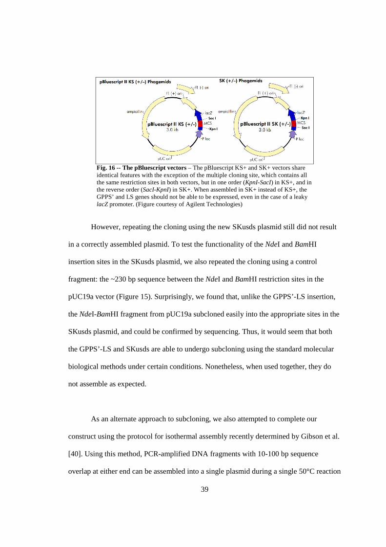
39
Fig. 16 -- The pBluescript vectors – The pBluescript KS+ and SK+ vectors share identical features with the exception of the multiple cloning site, which contains all the same restriction sites in both vectors, but in one order (KpnI-SacI) in KS+, and in the reverse order (SacI-KpnI) in SK+. When assembled in SK+ instead of KS+, the GPPS’ and LS genes should not be able to be expressed, even in the case of a leaky lacZ promoter. (Figure courtesy of Agilent Technologies)
However, repeating the cloning using the new SKusds plasmid still did not result
in a correctly assembled plasmid. To test the functionality of the NdeI and BamHI
insertion sites in the SKusds plasmid, we also repeated the cloning using a control
fragment: the ~230 bp sequence between the NdeI and BamHI restriction sites in the
pUC19a vector (Figure 15). Surprisingly, we found that, unlike the GPPS’-LS insertion,
the NdeI-BamHI fragment from pUC19a subcloned easily into the appropriate sites in the
SKusds plasmid, and could be confirmed by sequencing. Thus, it would seem that both
the GPPS’-LS and SKusds are able to undergo subcloning using the standard molecular
biological methods under certain conditions. Nonetheless, when used together, they do
not assemble as expected.
As an alternate approach to subcloning, we also attempted to complete our
construct using the protocol for isothermal assembly recently determined by Gibson et al.
[40]. Using this method, PCR-amplified DNA fragments with 10-100 bp sequence
overlap at either end can be assembled into a single plasmid during a single 50°C reaction

40
in which a heat-sensitive 5’ exonuclease degrades the 5’ ends of both sequences, allowing
the complementary ends to anneal, while thermophilic polymerase and ligase complete
the construct (Figure 13). In our work, it so happened that the PCR-amplified segment of
the SKusds vector was virtually the same length as the vector itself. It was also found that
the pUC19a vector carrying the GPPS’ and LS genes, in its super-coiled form migrated
the same distance on an agarose gel as the linear GPPS’-LS fragment (Figure 17).
1 2
3 4
FIG. 17 Relative plasmid and fragment lengths for isothermal assembly – In our isothermal assembly reaction, the length of the SKusds plasmid and the BamHI-NdeI fragment are virtually the same (lanes 1 & 2). In addition, the super-coiled form of the pUC19a plasmid with the GPPS’-LS insert migrates the same distance on an agarose gel as does the linear GPPS’-LS fragment itself (lanes 3 & 4). Thus, separating these to two fragments from the template DNA used in the cloning could not be done by simple gel purification, necessitating the additional of a DpnI digest post-PCR, to digest any template plasmid contaminant.
Thus, these fragments could not be separated from the template DNA in each
PCR reaction simply by gel purification. To minimize contamination of both DNA
fragments by the template plasmids, we added an additional post-PCR digest with DpnI,
which digests methylated DNA, to the protocol published by Gibson et al.
However, despite this extra precaution taken to reduce plasmid contamination, our
isothermal construction did not yield any correctly-assembled plasmids. Instead,
sequencing of several clones transformed with the isothermally assembly reaction
product repeatedly showed that the only plasmid being propagated was, surprisingly, the
original SKusds plasmid. Thus, this method has so far been similarly unsuccessful in
constructing plasmid capable of transforming Synechocystis with the GPPS’-LS tandem
gene pair.

41
DISCUSSION
Kinetic Characterization
Our analysis of (+)-4R-limonene synthase cloned from C. sinensis (orange) found that
this enzyme has a Km of 130µM. This is consistent with the preliminary kinetic analysis
of the same clone preformed in the lab previously [16]. A previous characterization done
by Lücker et al. [11] had found that the Km of (+)-limonene synthase cloned from C.
limon (lemon) was only 0.70 µM, less than 1/100 of the value we determined for our
clone. The value published by Lücker et al. appears to agree with additional reports
which have determined Km values in the low micromolar range for a wide variety of other
monoterpene synthases [8, 11].
It is not clear why our (+)-limonene synthase appears to behave so differently
from the one cloned by Lücker et al. from C. limon. However, it is important to note that
the study performed by Lücker et al. also observed evidence of substrate inhibition
kinetics, with activity for (+)-limonene synthase and two additional monoterpene
cyclases decreasing at substrate concentrations greater than 10 µM, including substrate
concentrations as high as 100- and 180 µM. Thus, the reported Km of 0.70 µM for (+)-
limonene synthase from C. limon was subsequently determined by ignoring all substrate
inhibition effects (i.e., excluding data from reactions run at geranyl pyrophosphate
concentrations greater than 10 µM). Contrary to the findings of Lücker et al., the data
from the reactions observed in our study do not show any indication of substrate
inhibition, even at substrate concentrations as high as 600 µM. Rather, the reaction rate
42
continues to increase with substrate concentration as the reaction approaches its
maximum velocity. Furthermore, the maximum reaction velocity per enzyme molecule
reported by Lücker et al., even when extrapolating to ignore substrate inhibition, is
several orders of magnitude slower than the maximum velocity observed per molecule of
our enzyme. Thus, we suspect that there is some element of the assay protocol other than
the enzyme mechanism itself that is causing Lücker et al. to report evidence of a substrate
inhibition behavior which is not actually characteristic of the enzyme, thereby lowering
the observed Km significantly from its true value.
No kcat value for (+)-limonene synthase has yet been published. However, a
previous study using (-)-limonene sythase extracted from Mentha x piperita (peppermint)
and Mentha spicata (spearmint) determined a kcat of 0.3 s-1 for native (-)-limonene
sythase [15]. This value is comparable to the observed kcat of 0.11 s-1 for our recombinant
(+)-limonene synthase, especially given that previous studies have noted that
monoterpene synthase activity can be slightly compromised by recombinant, rather than
native, expression [8].
Crystal Structure Determination
The fold of our (+)-limonene synthase apoprotein matches that described previously by
Hyatt et al. [14] for (-)-limonene synthase cocrystallized with substrate analog, indicating
that our enzyme is most likely crystallizing in its native, or at least in its active,
conformation, despite the absence of a ligand. In addition, the positions of all three

43
manganese ions observed in the t = 4 structure agree well with the manganese observed
coordinating the substrate analog in the (-)-limonene structure.
In completing the crystal soaking experiments described in Chapter 3, we had
hoped to trap reaction intermediates in the enzyme active site, allowing us to better
understand the mechanism by which geranyl pyrophosphate substrate is converted to (+)-
limonene. Previous trials had suggested that soaking with substrate and Mn2+ results in
the appearance of pyrophosphate in the active site (P. Ramasamy, data not yet published),
a phenomenon which was similarly observed in this set of soaking experiments.
However, the lack of electron density corresponding to a geranyl pyrophosphate molecule
in the active site suggests that substrate is not properly bound in any of the crystals
observed. This supposition is further supported by the observation that the N-terminal tail
is highly disordered all three structures analyzed – had substrate bound, we would expect
to see an ordered, “closed” conformation of the tail as it moved to cap the active site in
preparation for hydrolysis of the pyrophosphate group. In addition, the two loop regions
which cap the active site from above (497-504) and below (574-584) in the substrate-
bound structure by Hyatt et al. are found in our structure to be either similarly disordered,
as in the case of the 574-584 loop, or else significantly shifted away from the active site,
as in the case of the 407-504 helical loop. These key differences support the absence of
properly bound substrate in the active site of our substrate-soaked crystals.
It is important to note that all three crystals were soaked in MnCl2 solution for a
full hour before the introduction of geranyl pyrophosphate for substrate soaking. In light

44
of this fact, it is curious to observe that the number of bound manganese atoms increases
from one bound manganese with no substrate soaking, to two bound after one minute of
substrate soaking, to all three bound with four or more minutes of substrate soaking,
despite the fact that the presence of the substrate itself in the active site is not observed in
any of these three structures. One possible explanation for this phenomenon is that
geranyl pyrophosphate is recruiting Mn2+ ions to their appropriate positions in the active
site, as if to initiate substrate binding, but is sterically hindered from actually entering the
active site while the enzyme is in its rigid, crystallized formation. In a crystal formation,
the active site of our soaked crystals may be constrained to a single particular
conformation, preventing it from accommodating the introduction of a molecule as bulky
as geranyl pyrophosphate, while still allowing it to take up the Mn2+ ions that would be
necessary for substrate binding. However, this explanation would not explain the
suspected appearance of pyrophosphate in the active site.
Alternatively, it could be that the substrate is, in fact, able to enter the active site,
but that inability of the N-terminal tail and the two active site loops to move to their
capping positions while crystallized leaves the substrate in the active site too flexible to
generate a significant electron density, giving the impression that it has not bound.
Similarly, it is possible that geranyl pyrophosphate is able to enter the active site and
even to undergo the hydrolysis of the pyrophosphate, but that the inability of the N-
terminal tail and the two active site loops to shield the active site from the surrounding
solvent results in the reaction of the remaining cation with water to form an alcohol. Both
species can then diffuse from the active site, explaining the time-dependent appearance of

45
the suspected pyrophosphate density between 1 minute and 4 minutes (P. Ramasamy,
data not yet published). Finally, it is possible that geranyl pyrophosphate enters the active
site but is simply converted to (+)-limonene too quickly to be observed bound to the
active site, after which the active site reverts back to its “open” conformation. In all three
of these cases, the three Mn2+ observed in the t = 4 would be required to coordinate the
hydrolyzed pyrophosphate.
Regardless of the precise nature of the substrate in the crystal active site, our
results show that (+)-limonene synthase is able to crystallize in its active conformation in
the absence of Mn2+, and that soaking with geranyl pyrophosphate can subsequently
place all three Mn2+ ions into their correct positions in the active site of a folded enzyme.
These observations further support a reaction mechanism by which Mn2+ is not required
for correct enzyme folding, but is recruited to the active site by geranyl pyrophosphate,
and must be correctly positioned in the active site prior to substrate binding and
hydrolysis.
Limonene Production in Synechocystis
The difficulties we experienced in constructing a plasmid with which to transform the
geranyl pyrophosphate synthase and (+)-limonene synthase genes into the Synechocystis
genome remain unresolved. It has been shown that the two genes can be combined into a
single fragment flanked by NdeI and BamHI sites in an alternate plasmid, pUC19a,
indicating that the nucleotide sequences themselves are not toxic to the E. coli cells. It
has similarly been shown that a ~230 basepair control fragment excised from the pUC19a

46
vector can be successfully subcloned into the correct NdeI and BamHI sites in the SKusds
plasmid, suggesting that these restriction sites are accessible and fully functional. These
results indicate that the restriction sites themselves are not the cause of the problems
experienced with subcloning the GPPS’ and LS genes into the SKusds vector. The similar
failure of the Gibson isothermal assembly method, which does not rely on the use of
restriction enzymes, to generate the correct construct further indicates that the restriction
sites in question are not the source of the problem.
It has recently come to our attention that, in addition to a translation start site, the
region directly upstream of the psbA2 gene in Synechocystis contains a promoter region
for the psbA2 gene. In Synechocystis, this promoter is, not surprisingly given the gene’s
function, light-inducible, causing the cell to produce higher levels of photosystem II in
high-light conditions. The sequence is of E. coli consensus type, and it has been shown
that when found in E. coli, this promoter is constitutively active [41]. In light of this
finding, it now seems very likely that our GPPS’-LS fragment, when inserted properly
after the upstream fragment, is being constitutively transcribed and expressed by the E.
coli cell, producing a build-up of a toxic compound that quickly kills the cell. This would
explain why we are clearly able to subclone the upstream fragment and the GPPS’-LS
fragment independent of one another, but are never able to see them correctly assembled.
It has been shown that E. coli can tolerate limonene up to a concentration 0.25%
(v/v) [27], or 1.5 mM. It is not clear at this point whether our E. coli, when transformed
with the GPPS’-LS insertion correctly placed behind the psbA2 promoter, could produce

47
limonene at a sufficiently high concentration so as to cause E. coli cell death. A previous
study by Carter et al. [28] reports the successful production of small amounts of geranyl
pyrophosphate and (-)-limonene as intermediates in the construction of (-)-carvone and
(-)-carveol pathways in E. coli, but notes that this comes at the cost of a high metabolic
burden to the cells. Thus, it remains likely that either the production of limonene, or the
diversion of its precursors from key metabolic pathways, in our subcloning experiments
is significantly hindering the survival of cells transformed with the desired plasmid,
effectively selecting against our goal construct.
Further Directions
Further studies of (+)-4R-limonene synthase are needed to continue all three initiatives
described in Chapters 2-4 of this thesis. The significant disparity observed between the
kinetics of our (+)-limonene synthase from C. sinensis and the kinetics of the same
enzyme from C. limon described by Lücker et al. [11] has yet to be resolved. It seems
improbable that such a large inconsistency could simply be the result of speciation, given
that the two enzymes demonstrate 95% sequence identity [16]. The (+)-limonene
synthase gene from C. limon has previously been cloned, but never expressed or
kinetically characterized, in our lab by S. N. Olsen [16]. It would be necessary to perform
a Michaelis-Menton analysis of this C. limon clone in order to more fully understand the
true nature of the (+)-limonene synthase reaction kinetics. Such an analysis would further
allow us to determine whether the difference observed between the behavior of our
enzyme from C. sinensis and the one from C. limon studied by Lücker et al. is rooted in
48
the enzymes’ dissimilar species of origin, or in some overlooked detail of the assay
methodologies used in their respective characterizations.
Similarly, much work remains to be done to further elucidate the reaction
mechanism of (+)-limonene synthase by crystal structure determination. The three
structures analyzed in this study require additional refinement prior to submission to the
PDB. However, these three existing structures could only be determined to a resolution of
~3.0Å; crystallization conditions need to be further optimized in order to produce more
stable crystals capable of diffracting to a higher resolution and allowing us to observe
more clearly the changes occurring in the active site. In addition, our study shows that
soaking in substrate solution, even for as long as 10 minutes, does not yield a crystal
structure which includes a properly bound geranyl pyrophosphate molecule, although the
presence of a pyrophosphate at certain time points in the soaking is suspected. The exact
reason for this observation is not yet understood, but it is possible that the constraints
placed on the enzyme by crystal formation are inhibiting proper substrate binding or
reaction. Thus, in order to observe the enzyme reaction mechanism further, it may be
necessary to employ cocrystallization methods with a substrate analog, like the 2-
flourogeranayl pyrophosphate used by Hyatt et al. [14], rather than soaking with
substrate. The addition of such analogs for cocrystallization may also serve to stabilize
the enzyme structure further, allowing the structure to be determined at higher
resolutions.
49
Finally, the construction of a plasmid with which to transform Synechocystis with
the geranyl pyrophosphate synthase and (+)-limonene synthase genes remains to be
completed. In light of new findings that the upstream fragment contains a promoter that is
constitutively active in E. coli, it seems likely the GPPS’ and LS genes, when inserted at
their proper location in the SKusds plasmid, are simply toxic to the cell. This new
hypothesis will now need to be tested by adding deactivating mutations to both genes,
and repeating the subcloning into the appropriate sites in SKusds. If the deactivated genes
can be successfully used to generate our desired construct, this will be strong evidence
that the subcloning technique is sound, but that the product of the active GPPS’ and LS
genes is too toxic for the plasmid to be carried in E. coli. If we find this to be the case,
further work will include putting the two genes under an inducible promoter, instead of
the constitutively active one found in the upstream fragment, allowing us to suppress
gene expression until the entire construct has been moved into Synechocystis.
Once the insertion of the geranyl pyrophosphate synthase and (+)-limonene
synthase genes has been completed, the only remaining step in the plasmid construction
will be the addition of an antibiotic-resistance gene after the GPPS’-LS gene fragment,
which will allow for selection of correctly transformed Synechocystis clones. In our
original experimental design, we had planned to use the kanamycin-resistance gene,
KanR, amplified from the pET28a vector, for this purpose. The region directly upstream
of KanR gene includes a Shine-Delgarno sequence that we hope will facilitate the
polycistronic expression of this gene immediately following the expression of (+)-
limonene synthase. Upon completion of the plasmid, Synechocystis can successfully be

50
transformed according to previously determined protocols [22, 23], allowing us to
produce (+)-limonene photosynthetically. A method for the extraction and analysis of this
potential biofuel from the cell culture can then be optimized.
51
APPENDIX: SUPPLEMENTARY FIGURES
BG11 Media Composition
To 700 mL dH2O add:
10 mL 150.0 g/L NaNO3
10 mL 7.8 g/L K2HPO4
10 mL 2.0 g/L Na2CO3
10 mL 5.8 g/L Na2SiO3 · 9H2O
10 mL 7.5 g/L MgSO4 · 7H2O
10 mL 3.5 g/L CaCl2 · 2H2O
10 mL 0.1 g/L diNa EDTA
10 mL 0.6 g/L each Fe citrate-citric acid
1 mL Trace Elements:
H3BO3 2.86 g/L
MnCl2 · 4H2O 1.80 g/L
ZnSO4 · 7H2O 0.22 g/L
NaMoO4 · 2H2O 0.39 g/L
CuSO4 · 5H2O 0.08 g/L
Co(NO3)2 · 6H2O 0.05 g/L
Add salts to distilled water in order given. Bring to 1 liter with dH2O.
FIG . A1 BG11 mineral media composition – The media described above was used for Synechocystis liquid culture and for the production of BG11 agar plates. Protocol courtesy of Professor Mary Allen, Wellesley College, Wellesley, MA.

52
Synechocystis psbA2 Flanking Sequences
5’CTTGCCCAGATGCAGGCCTTCTGGCGATCGCCATGGTGAGCAACGATTGCGGCTTTAGCGTTCCAGTGGATATTTGCTGGGGGTTAA
TGAAACATTGTGGCGGAACCCAGGGACAATGTGACCAAAAAATTCAGGGATATCAATAAGTATTAGGTATATGGATCATAATTGTATGC
CCGACTATTGCTTAAACTGACTGACCACTGACCTTAAGAGTAATGGCGTGCAAGGCCCAGTGATCAATTTCATTATTTTTCATTATTTC
ATCTCCATTGTCCCTGAAAATCAGTTGTGTCGCCCCTCTACACAGCCCAGAACTATGGTAAAGGCGCACGAAAAACCGCCAGGTAAACT
CTTCTCAACCCCCAAAACGCCCTCTGTTTACCCATGGAAAAAACGACAATTACAAGAAAGTAAAACTTATGTCATCTATAAGCTTCGTG
TATATTAACTTCCTGTTACAAAGCTTTACAAAACTCTCATTAATCCTTTAGACTAAGTTTAGTCAGTTCCAATCTGAACATCGACAAAT
ACATAAGGAATTATAACCAAATGACAACGACTCTCCAACAGCGCGAAAGCGCTTCCTTGTGGGAACAGTTTTGTCAGTGGGTGACCTCT
ACCAACAACCGGATTTATGTCGGTTGGTTCGGTACCTTGATGATCCCCACCCTCTTAACTGCCACCACTTGCTTCATCATTGCCTTCAT
CGCCGCTCCCCCCGTTGACATCGACGGTATCCGTGAGCCCGTTGCTGGTTCTTTGCTTTACGGTAACAACATCATCTCTGGTGCTGTTG
TACCTTCTTCCAACGCTATCGGTTTGCACTTCTACCCCATCTGGGAAGCCGCTTCCTTAGATGAGTGGTTGTACAACGGTGGTCCTTAC
CAGTTGGTAGTATTCCACTTCCTCATCGGCATTTTCTGCTACATGGGTCGTCAGTGGGAACTTTCCTACCGCTTAGGTATGCGTCCTTG
GATTTGTGTGGCTTACTCTGCCCCCGTATCCGCTGCCACCGCCGTATTCTTGATCTACCCCATTGGTCAAGGCTCCTTCTCTGATGGTA
TGCCCTTGGGTATTTCTGGTACCTTCAACTTCATGATCGTGTTCCAAGCTGAGCACAACATCCTGATGCACCCCTTCCACATGTTAGGT
GTGGCTGGTGTATTCGGTGGTAGCTTGTTCTCCGCCATGCACGGTTCCTTGGTAACCTCCTCCTTGGTGCGTGAAACCACCGAAGTTGA
ATCCCAGAACTACGGTTACAAATTCGGTCAAGAAGAAGAAACCTACAACATCGTTGCCGCCCACGGCTACTTTGGTCGGTTGATCTTCC
AATATGCTTCTTTCAACAACAGCCGTTCCTTGCACTTCTTCTTGGGTGCTTGGCCTGTAATCGGCATCTGGTTCACTGCTATGGGTGTA
AGCACCATGGCGTTCAACCTGAACGGTTTCAACTTCAACCAGTCCATCTTGGATAGCCAAGGCCGGGTAATCGGCACCTGGGCTGATGT
ATTGAACCGAGCCAACATCGGTTTTGAAGTAATGCACGAACGCAATGCCCACAACTTCCCCCTCGACTTAGCGTCTGGGGAGCAAGCTC
CTGTGGCTTTGACCGCTCCTGCTGTCAACGGTTAATTCCTTGGTGTAATGCCAACTGAATAATCTGCAAATTGCACTCTCCTTCAATGG
GGGGTGCTTTTTGCTTGACTGAGTAATCTTCTGATTGCTGATCTTGATTGCCATCGATCGCCGGGGAGTCCGGGGCAGTTACCATTAGA
GAGTCTAGAGAATTAATCCATCTTCGATAGAGGAATTATGGGGGAAGAACCTGTGCCGGCGGATAAAGCATTAGGCAAGAAATTCAAGA
AAAAAAATGCCTCCTGGAGCATTGAAGAAAGCGAAGCTCTGTACCGGGTTGAGGCCTGGGGGGCACCTTATTTTGCCATTAATGCCGCT
GGTAACATAACCGTCTCTCCCAACGGCGATCGGGGCGGTTCGTTAGATTTGTTGGAACTGGTGGAAGCCCTGCGGCAAAGAAAGCTCGG
CTTACCCCTATTAATTCGTTTTTCCGATATTTTGGCCGATCGCCTAGAGCGATTGAATAGTTGTTTTGCCAAGGCGATCGCCCGTTACA
FIG . A2 Upstream and downstream psbA2 flanking sequences – A portion of the Synechocystis genome is shown, indicating the psbA2 gene (in italics) and the 0.5 kb fragments directly up- and downstream (in blue). These fragments were amplified by PCR and inserted into the pBluescript KS+ vector to make the plasmid KSusds.
GPPS Mutagenesis
5’TTTGATTTCAAGGAGTACTTGCATTCCAAAGCAATATCAGTGAATGAGGCACTGGAGAGGGCTGTCCCACTTCGCTATCCTGAAAAA
ATACATAAGCTATGAGGTATTCTCTTCTAGCAGGAGGGAAGCGGATTCGTCCTATTCTGACTATTGCAGCATGTGAGCTTGTGGGAGGG
AGTGAGGAGCTCGCCATGCCAACTGCCTGTGCAATGGAGATGATCCACACAATGTCTTTGATTCATGATGATTTGCCTTCCATGGACAA
TGATGATTTACGCCGAGGTAAGCTTACAAACCACAAGGTCTTTGGCGAAGGCACTGCTGTTCTTGCAGGGGATGCACTTCTTTCATTTG
CATTTGAGCACATTGCAGTGTCCACAAGGAAAACTGTGGCGAGTCATAGGGTTTTGAGGGTGGTATCTGAATTGGGTAAGGCAATAGGC
TCTCAAGGGGTTGCTGGTGGGCAGGTTGCTGATATTACCAGTGAAGGGGATCCATCTGTTGGGCTTGAGACTCTCGAATGGATTCACAT
TCACAAGACTGCAGTGCTCTTGGAGTGCGCCGTTGTGAGTGGGGCGATCATCGGTGGCGCTTCAGAGAATGAAATTGAGAGAACTGGAA
GGTATGCCCGTTGCGTTGGGCTTCTGTTTCAGGTTGTGGATGACATACTCGATGTCACCAGATCATCAGAAGAATTGGGAAAGACTGCA
GGAAAGGATTTGGTTAGTGATAAGGCCACTTATCCCAAGCTGATGGGTTTGGAGAAAGCGAAGGAATTTGCTGATGAATTGTTGGACAG
AGCTAAGGAGGAGTTATCTTGCTTCAACCCAGCTAAGGCTGCACCTCTGTTGGGTCTTGCAGATTACATTGCATTAAGACAGAACTGA
FIG . A3 GPPS silent mutation – The GPPS originally synthesized (Genewiz) was found to contain a BamHI restriction site, 5’-GGATCC-3’ (underlined). The silent mutation GGG � GGA was introduced, changing the former restriction site to 5’-AGATCC-3’. GPPS gene, GenBank Accession #AF513111 [39].
53
REFERENCES
1. de Cassia da Silveira e Sa, R., L.N. Andrade, and D.P. de Sousa, A review on anti-inflammatory activity of monoterpenes. Molecules, 2013. 18(1): p. 1227-54.
2. Chidambara Murthy, K.N., G.K. Jayaprakasha, and B.S. Patil, D-limonene rich volatile oil from blood oranges inhibits angiogenesis, metastasis and cell death in human colon cancer cells. Life Sci, 2012. 91(11-12): p. 429-39.
3. Lange, B.M. and A. Ahkami, Metabolic engineering of plant monoterpenes, sesquiterpenes and diterpenes--current status and future opportunities. Plant Biotechnol J, 2013. 11(2): p. 169-96.
4. Ohara, K., et al., Monoterpene engineering in a woody plant Eucalyptus camaldulensis using a limonene synthase cDNA. Plant Biotechnol J, 2010. 8(1): p. 28-37.
5. Dewick, P.M., The biosynthesis of C5-C25 terpenoid compounds. Nat Prod Rep, 2002. 19(2): p. 181-222.
6. Bohlmann, J., G. Meyer-Gauen, and R. Croteau, Plant terpenoid synthases: molecular biology and phylogenetic analysis. Proc Natl Acad Sci U S A, 1998. 95(8): p. 4126-33.
7. Maruyama, T., et al., Molecular cloning, functional expression and characterization of d-limonene synthase from Schizonepeta tenuifolia. Biol Pharm Bull, 2001. 24(4): p. 373-7.
8. Wise, M.L., et al., Monoterpene synthases from common sage (Salvia officinalis). cDNA isolation, characterization, and functional expression of (+)-sabinene synthase, 1,8-cineole synthase, and (+)-bornyl diphosphate synthase. J Biol Chem, 1998. 273(24): p. 14891-9.
9. Kampranis, S.C., et al., Rational conversion of substrate and product specificity in a Salvia monoterpene synthase: structural insights into the evolution of terpene synthase function. Plant Cell, 2007. 19(6): p. 1994-2005.
10. Williams, D.C., et al., Truncation of limonene synthase preprotein provides a fully active 'pseudomature' form of this monoterpene cyclase and reveals the function of the amino-terminal arginine pair. Biochemistry, 1998. 37(35): p. 12213-20.
11. Lucker, J., et al., Monoterpene biosynthesis in lemon (Citrus limon). cDNA isolation and functional analysis of four monoterpene synthases. Eur J Biochem, 2002. 269(13): p. 3160-71.
12. Shaw, P.E., Review of Quantitative-Analyses of Citrus Essential Oils. Journal of Agricultural and Food Chemistry, 1979. 27(2): p. 246-257.
13. Rajaonarivony, J.I., J. Gershenzon, and R. Croteau, Characterization and mechanism of (4S)-limonene synthase, a monoterpene cyclase from the glandular trichomes of peppermint (Mentha x piperita). Arch Biochem Biophys, 1992. 296(1): p. 49-57.
14. Hyatt, D.C., et al., Structure of limonene synthase, a simple model for terpenoid cyclase catalysis. Proc Natl Acad Sci U S A, 2007. 104(13): p. 5360-5.

54
15. Alonso, W.R., et al., Purification of 4S-limonene synthase, a monoterpene cyclase from the glandular trichomes of peppermint (Mentha x piperita) and spearmint (Mentha spicata). J Biol Chem, 1992. 267(11): p. 7582-7.
16. Olsen, S.N., Isolation, Purification, and Characterization of (+)-4R-limonene synthase: The first step in exploring enzyme stereospecificity in terpenoid biosynthesis, in Department of Biochemistry2011, Brandeis University.
17. Whittington, D.A., et al., Bornyl diphosphate synthase: structure and strategy for carbocation manipulation by a terpenoid cyclase. Proc Natl Acad Sci U S A, 2002. 99(24): p. 15375-80.
18. Starks, C.M., et al., Structural basis for cyclic terpene biosynthesis by tobacco 5-epi-aristolochene synthase. Science, 1997. 277(5333): p. 1815-20.
19. Christianson, D.W., Structural biology and chemistry of the terpenoid cyclases. Chem Rev, 2006. 106(8): p. 3412-42.
20. Aguirre von Wobeser, E., et al., Concerted changes in gene expression and cell physiology of the cyanobacterium Synechocystis sp. strain PCC 6803 during transitions between nitrogen and light-limited growth. Plant Physiol, 2011. 155(3): p. 1445-57.
21. Kaneko, T., et al., Structural analysis of four large plasmids harboring in a unicellular cyanobacterium, Synechocystis sp. PCC 6803. DNA Res, 2003. 10(5): p. 221-8.
22. Kufryk, G.I., et al., Transformation of the cyanobacterium Synechocystis sp. PCC 6803 as a tool for genetic mapping: optimization of efficiency. FEMS Microbiol Lett, 2002. 206(2): p. 215-9.
23. Zang, X., et al., Optimum conditions for transformation of Synechocystis sp. PCC 6803. J Microbiol, 2007. 45(3): p. 241-5.
24. Mohamed, A. and C. Jansson, Influence of light on accumulation of photosynthesis-specific transcripts in the cyanobacterium Synechocystis 6803. Plant Mol Biol, 1989. 13(6): p. 693-700.
25. Sharkey, T.D., et al., Evolution of the isoprene biosynthetic pathway in kudzu. Plant Physiol, 2005. 137(2): p. 700-12.
26. Lindberg, P., S. Park, and A. Melis, Engineering a platform for photosynthetic isoprene production in cyanobacteria, using Synechocystis as the model organism. Metab Eng, 2010. 12(1): p. 70-9.
27. Dunlop, M.J., Engineering microbes for tolerance to next-generation biofuels. Biotechnol Biofuels, 2011. 4: p. 32.
28. Carter, O.A., R.J. Peters, and R. Croteau, Monoterpene biosynthesis pathway construction in Escherichia coli. Phytochemistry, 2003. 64(2): p. 425-33.
29. Weyman, P.D., et al., Heterologous expression of Alteromonas macleodii and Thiocapsa roseopersicina [NiFe] hydrogenases in Synechococcus elongatus. PLoS One, 2011. 6(5): p. e20126.
30. O'Maille, P.E., J. Chappell, and J.P. Noel, A single-vial analytical and quantitative gas chromatography-mass spectrometry assay for terpene synthases. Anal Biochem, 2004. 335(2): p. 210-7.
31. Leslie, A.G.W. and H.R. Powell, Processing Diffraction Data with Mosflm. Evolving Methods for Macromolecular Crystallography, 245, 41-51 ISBN 978-1-4020-6314-5, 2007.
55
32. Evans, P.R., Scaling and assessment of data quality. Acta Cryst, 2006. D62: p. 72-78.
33. Collaborative Computational Project and N. 4, The CCP4 Suite: Programs for Protein Crystallography. Acta Cryst, 1994. D50(760-763).
34. Potterton, E., et al., A graphical user interface to the CCP4 program suite. Acta Cryst., 2003. D59: p. 1131-1137.
35. McCoy, A.J., et al., Phaser crystallographic software. J. Appl. Cryst., 207. 40: p. 658-674.
36. Afonine, P.V., et al., Towards automated crystallographic structure refinement with phenix.refine. Acta Cryst., 2012. D68: p. 352-367.
37. Adams, P.D., et al., PHENIX: a comprehensive Python-based system for macromolecular structure solution. Acta Cryst., 2010. D66: p. 213-221.
38. Emsley, P., et al., Features and Development of COOT. Acta Cryst., 2010. D66: p. 486-501.
39. Burke, C. and R. Croteau, Geranyl diphosphate synthase from Abies grandis: cDNA isolation, functional expression, and characterization. Arch Biochem Biophys, 2002. 405(1): p. 130-6.
40. Gibson, D.G., et al., Enzymatic assembly of DNA molecules up to several hundred kilobases. Nat Methods, 2009. 6(5): p. 343-5.
41. Shibato, J., M. Asayama, and M. Shirai, Specific recognition of the cyanobacterial psbA promoter by RNA polymerases containing principal sigma factors. Biochim Biophys Acta, 1998. 1442(2-3): p. 296-303.