386091/FULLTEXT01.pdf
58
In vitro & in vivo approaches in the characterization of XTH gene products Nomchit Kaewthai Doctoral Thesis in Biotechnology Stockholm, Sweden 2011
Transcript of 386091/FULLTEXT01.pdf

In vitro & in vivo approaches in the characterization of
XTH gene products
Nomchit Kaewthai
Doctoral Thesis in Biotechnology
Stockholm, Sweden 2011
ii
© Nomchit Kaewthai
TRITA-BIO Report 2011: 1 ISSN 1654-2312 ISBN 978-91-7415-848-9 School of Biotechnology Royal Institute of Technology AlbaNova University Centre SE-10691 Stockholm Sweden Printed at US-AB Universitetsservice Box 700 14 10044 Stockholm Sweden
Cover illustration: Confocal microscopy showing Arabidopsis thaliana root labeled with different probes. Green: Xyloglucan probed with CCRC-M1 antibody. Yellow: in situ xyloglucan endo-hydrolase activity probed with XXXG-Res substrate. Red: in situ xyloglucan endo-transglycosylase activity probed with XXXG-SR substrate.

iii
ABSTRACT
The xyloglucan endo-transglycosylase/hydrolase (XTH) genes are found in all vascular and some nonvascular plants. The XTH genes encode proteins which comprise a subfamily of glycoside hydrolase (GH) family 16 in the Carbohydrate-Active enZYmes (CAZY) classification. The XTH gene products are believed to play intrinsic role in cell wall modification during growth and development throughout the lifetime of the plant. In the present investigation, biochemical and reverse genetic approaches were used to better understand the functions of individual members of the XTH gene family of two important plants: the model organism Arabidopsis thaliana and the grain crop barley (Hordeum vulgare). A phylogenetic tree of the xyloglucan-active enzymes of GH16 has previously been constructed, where enzymes with similar activities have been shown to cluster together. Several members of phylogenetic Group I/II and III-B, predicted to exhibit xyloglucan endo-transglycosylase activity (EC 2.4.1.207) and members of Group III-A, predicted to exhibit xyloglucan endo-hydrolase activity (EC 3.2.1.151), were included to analyze the functional diversity of XTH gene products. A heterologous expression system using the yeast Pichia pastoris was found to be effective for recombinant protein production with a success rate of ca. 50%. XTH gene products were obtained in soluble and active forms for subsequent biochemical characterization.
In order to be able to screen larger numbers of protein producing clones, a fast and easy method is required to identify clones expressing active protein in high enough amounts. Thus, a miniaturized XET/XEH assay for high-throughput analysis was developed, which was able to identify activities with good precision and with a reduced time and materials consumption and a reduced work load.
Enzyme kinetic analysis indicated that the XET or XEH activity of all XTH gene products characterized in the present study corresponded to predictions based on the previously revised phylogenetic clustering. To gain insight into the biological function of the predominant XEHs AtXTH31 and AtXTH32, which are highly expressed in rapidly developing tissues, a reverse genetic approach was employed using T-DNA insertion lines of the A. thaliana Columbia ecotype. Genotypic and phenotypic characterization, together with in situ assays of XET and XEH activities, in single- and double-knock-out mutants indicated that these Group III-A enzymes are active in expanding tissues of the A. thaliana roots and hypocotyl. Although suppression of in muro XEH activity was clearly observed in the double-knock-out, no significant growth phenotype was observed, with the exception that radicle emergence appeared to be faster than in the wild type plants.
Keywords: Arabidopis thaliana, Hordeum vulgare, plant cell wall, xyloglucan, glycoside hydrolase family 16, xyloglucan endo-transglycosylase/hydrolase gene family, xyloglucan endo-transglycosylase, xyloglucan endo-hydrolase, heterologous protein expression, Pichia pastoris, T-DNA insertion, in situ XET/XEH assay, high-throughput screening

iv
SAMMANFATTNING
Växtgenfamiljen xyloglukan-endo-transglykosylaser/hydrolaser (XTHer) återfinns i alla landlevande
växter. XTH-generna innehåller den genetiska koden för xyloglukan-endo-transglykosylas/hydrolas-
enzymerna hemmahörande i en underfamilj till glykosidhydrolasfamilj 16 (GH16) enligt Carbohydrate-
Active enZYmes (CAZY) klassificationen. XTH-genprodukterna tros vara en integrerad del av
cellväggsmodifieringen under en växts utveckling. I denna avhandling har biokemiska- och genetiska
metoder använts för att utröna funktionen hos enskilda XTH-gener i två olika växtmodeller: backtrav
(Arabidopsis thaliana) och korn (Hordeum vulgare). Ett fylogenetiskt träd för de xyloglukan-aktiva
enzymerna i GH16 har tidigare konstruerats, och enzymer med lika aktiviteter har visat sig gruppera sig
tillsammans i kluster. Genom att inkludera medlemmar från både Grupp I/II och III/B som har
xyloglukan-endo-transglykosylasaktivitet (E.C.2.4.1.207) och medlemmar från Grupp III-A som har
xyloglukan-endo-hydrolasaktivitet (E.C.3.2.1.151) kunde de olika gruppernas inverkan på växtcellväggen
studeras i de två arterna. Jästen Pichia pastoris visade sig vara en effektiv värd för heterologt uttryck där
ungefär varannan XTH-gen gav ett fungerande enzym. De heterologt producerade XTH-genprodukterna
utrycktes i både löslig- och aktiv form vilket möjliggjorde biokemisk karaktärisering och funktionsstudier
i backtrav.
För att kunna analysera ett stort antal mikrobiella kloner krävs en snabb och enkel metod för
identifiering av positiva kloner som producerar tillräckliga mängder protein. Därför utvecklades en
miniatyriserad XET/XEH metod för high-throughput screening som visade sig kunna identifiera aktivitet
med god säkerhet och med minskning av tidsåtgång, material och arbetsbelastning.
Resultaten från den biokemiska analysen visade att aktiviteten hos alla karaktäriserade XTH-
genprodukter i denna avhandling följde den aktivitet som tidigare fylogenetisk gruppering antytt. För att
förstå den biologiska funktionen hos två övervägande hydrolytiska XEHer, AtXTH31 och AtXTH32,
uttrycka i vävnader som genomgår snabb utveckling användes T-DNA insertionslinjer för att skapa
knockout-växter av AtXTH31 och AtXTH32 i A. thaliana ekotyp Columbia. Genom att helt och hållet slå ut
dessa två gener kunde funktionen och effekten av den motsvarande enzymaktiviteten studeras i
cellväggen. Genotypisk och fenotypisk karaktärisering tillsammans med mätning av XEH/XET aktivitet in
situ av xth31-1, xth32-1 och xth31-1/xth32-1 indikerade att dessa två gener från Grupp III-A utrycks i
expanderande vävnader i roten och i hypokotylen hos A. thaliana. Endast en mild fenotyp observerades
där grodden var snabbare ur fröet hos vildtypen än hos dubbelmutanten.

v

vi
LIST OF PUBLICATIONS
I. Nomchit Kaewthai, Andrew J. Harvey, Maria Hrmova, Harry Brumer, Ines Ezcurra, Tuula T. Teeri,
Geoffrey B. Fincher. 2010. Heterologous expression of diverse barley XTH genes in the yeast
Pichia pastoris. Plant Biotechnology, 27, 251-258.
II. Maria Hrmova, Vladimir Farkas, Andrew J. Harvey, Jelle Lahnstein, Bente Wischmann, Nomchit
Kaewthai, Ines Ezcurra, Tuula T. Teeri, Geoffrey B Fincher. 2009. Substrate specificity and
catalytic mechanism of a xyloglucan xyloglucosyl transferase HvXET6 from barley (Hordeum
vulgare L.). FEBS J., 276, 437-56.
III. An Maris, *Nomchit Kaewthai, *Jens M. Eklöf, Janice G. Miller, Harry Brumer, Stephen C. Fry, Jean-
Pierre Verbelen and Kris Vissenberg. 2011. Differences in enzymic properties of five
recombinant xyloglucan endotransglucosylase/hydrolase (XTH) proteins of Arabidopsis thaliana.
J. Exp. Bot., 62, 261-271. * These authors contributed equally to the work
IV. *Nomchit Kaewthai, *Delphine Gendre, *Jens M. Eklöf, Farid M.Ibatullin, Ines Ezcurra, Rhishikesh
P. Bhalerao and Harry Brumer. 2010. Group III-A XTH genes encode predominant xyloglucan
endo hydrolase active in expanding tissues of Arabidopsis thaliana. Manuscript. * These authors
contributed equally to the work
V. Nomchit Kaewthai, Harry Brumer and Ines Ezcurra. 2010. Development of a high-throughput assay
for screening xyloglucan endo-transglycosylase and endo-xyloglucanase expression in crude
microbial supernatants. Manuscript.

vii
The author’s contribution:
Publication I: Experimental design for protein expression, troubleshooting expression, screening
for positive clones, glycan analysis, drafting of the manuscript.
Publication II: Clone screening and protein expression.
Publication III: Expression, purification and kinetic analysis of AtXTH13. Kinetic analysis was
performed together with Jens M. Eklöf. Wrote respective parts of the manuscript.
Publication IV: Experimental design, cloning and expression of AtXTH31 and AtXTH32,
identification of T-DNA insertion lines, generation of double mutants, back crossing of mutant
plants, phenotypic characterization and data analysis, Semi-q-RT-PCR, RT-PCR of mutant, in situ
XEH and XET assays were performed together with Delphine Gendre, UPSC. Drafted and improved
the manuscript.
Publication V: Experimental design, method verification, high-throughput experiments, drafted and
revised the manuscript including figures.

viii
LIST OF ABBREVIATIONS
XET Xyloglucan endo-transglycosylase
XEH Xyloglucan endo-hydrolase
XTH Xyloglucan endo-transglycosylase/hydrolase
XGO Xyloglucan oligosaccharide
XXXG-SR Sulforhodamine-conjugated xyloglucan oligosaccharide
XXXG-Res Resorufin-conjugated oligosaccharide
Xyl Xylose
TXG Tamarind xyloglucan
HEC Hydroxyethyl cellulose
WSCA Water soluble cellulose acetate
MLG Mixed-linkage β-glucan
CMC Carboxymethylcellulose
HRGP Hydroxyproline-rich glycoprotein
GRP Glycine-rich protein
PRP Proline-rich protein
BCA Bicinchoninic Acid reducing sugar assay HPLC High Performance Anion-Exchange Chromatography
HPAEC-PAD High Performance Anion-Exchange Chromatography with Pulsed Amperometric Detection
IEF Isoelectric focusing
PCR Polymerase chain reaction
RT-PCR Reverse transcriptase polymerase chain reaction
ORFs open reading frames
SDS-PAGE Sodium dodecyl sulfate polyacrylamide gel electrophoresis
G-layers Gelatinous layers
LM5 Monoclonal antibody against rhamnogalacturonan-I-associated (1→4) -linked β-D-galactan
CCRC-M1 Monoclonal antibody against α-L-fucosylated xyloglucan
GH5 Glycoside hydrolase family 5
GH16 Glycoside hydrolase family 16
CAZY Carbohydrate Active enZYme

ix
TABLE OF CONTENT
1. Introduction ................................................................................................................................. 1
1.1 Plant growth and development .................................................................................................... 2
1.2 Plant cell walls ............................................................................................................................... 5
1.3 Plant cell wall components ........................................................................................................... 6
1.4 Plant cell wall synthesis and expansion ........................................................................................ 9
1.5 The XTH gene family ................................................................................................................... 11
1.5.1 Diversity of the family ....................................................................................................... 11
1.5.2 Phylogenetic tree for biochemical function annotation of XTH gene products ............... 11
1.6 Structure and function of XTH gene products ............................................................................ 12
1.6.1 Overall structure of XTH gene products ........................................................................... 12
1.6.2 Catalytic motif of XTH proteins ......................................................................................... 13
1.6.3 Structure determines functions ........................................................................................ 13
1.6.4 Substrate recognition and binding of XETs and XEHs ....................................................... 14
1.7 Mechanism and substrate........................................................................................................... 15
1.8 Approaches for characterizing XTH genes .................................................................................. 16
2. Heterologous expression of XTH genes ....................................................................................... 18
3. Assays for high-throughput screening of heterologous XTH gene expression .............................. 21
4. In vitro characterization of XTH gene products ........................................................................... 24
4.1 Assays used for in vitro characterization of XTH gene products ................................................. 24
4.2 In vitro characterization of XTH gene products .......................................................................... 25
4.2.1 Substrate specificity of XTH gene products ...................................................................... 26
4.2.2 pH dependence ................................................................................................................. 26
4.2.3 Detailed kinetic properties of XETs and XEHs ................................................................... 27
5. In vivo characterization of XTH gene products in the plant cell wall ............................................ 29
5.1 Biological roles of members of XTH genes .................................................................................. 29
5.2 Expression profiles of AtXTH genes ............................................................................................ 30
5.3 Probing roles of XTH gene products by reverse genetics ........................................................... 32
5.4 Immunolocalization of substrates for XTH gene products ......................................................... 33
5.5 Assays used for in vivo characterization ..................................................................................... 33
6. Concluding remarks and future perspectives .............................................................................. 37

x
7. Acknowledgements .................................................................................................................... 38
8. References ................................................................................................................................. 40

In vitro & in vivo approaches in the characterization of XTH gene products
1
1. Introduction
Plants are sustainable natural resources because they naturally regenerate, and harbor chlorophyll,
enabling them to use sunlight to convert water and carbon dioxide to sugars, which are used as
fundamental building blocks in the plethora of biochemical pathways required to drive growth and
development throughout the plant lifecycle. Hence, plants play many key roles in natural ecosystems,
such as sources of nutrients for diverse organisms, and hosts for various symbionts. In addition to their
ecological importance, plants (and various parts of plants) have enormous economic significance.
Notably, as the overwhelmingly predominant components of wood and fibrous plant materials, plant
cell walls have been used for millennia to make fire, paper and a huge array of construction materials
and textiles. More recently, plant cell walls have drawn further attention since they can be converted
into biofuels, which can be used as substitutes for dwindling supplies of fossil fuels (Sticklen, 2008; Minic
et al., 2009). Efforts have been made, through various approaches, to optimize the production rates and
composition of plant cell walls for various purposes, among other things, biofuel production. For
instance, intense efforts have focused recently on using genetic engineering to increase the biomass or
reduce proportions of specific components of plant cell walls for specific purposes, e.g. to reduce lignin
contents to make the material more suitable for biofuel production (Sticklen, 2006). In addition, of
course, plant cell walls are major sources of food and feeds, since they are chiefly composed of
polysaccharides (Chapple and Carpita, 1998), and extracts from them are extensively used in the food
industry, for instance pectin is widely used as a food additive and gelling agent.
Furthermore, better understanding of the mechanisms involved in cell wall construction and remodeling
has led to technological innovations, e.g. ‘biomimetic’ techniques to modify cellulose surfaces using
xyloglucan endo-transglycosylase (XET) enzymes to introduce desired functional groups anchored to
xyloglucan, which has naturally high affinity to cellulose (Teeri and Brumer, 2003; Zhou et al., 2006;
Teeri et al., 2007; Zhou et al., 2007; Zhou et al., 2009). Similar techniques have been used to produce
artificial, biocompatible blood vessels from bacterial cellulose (Bodin et al., 2007). Thus, novel cellulose-
based materials with desired properties can be obtained. Such techniques require more cooperative
development before they can be applied at industrial scale, but they show great potential for numerous
applications.
Plant cell walls have complex structures, and both their formation and modification are complicated
processes. Hence, huge amounts of information are required for their rigorous analysis. Thus, the

In vitro & in vivo approaches in the characterization of XTH gene products
2
compilation of comprehensive genomic databases of model plants, such as the dicot Arabidopsis,
Arabidopsis thaliana (Kaul et al., 2000) and the monocot Rice, Oryza sativa (Goff et al., 2002; Yu et al.,
2002), together with draft genomes of the black cottonwood tree, Populus trichocarpa (Tuskan et al.,
2006) and various other species (http://www.phytozome.org), has greatly facilitated analyses of plant
cell wall modeling processes and the functions of the genes involved. In addition, this information has
opened avenues (using diverse techniques) to explore the structure and function of cell wall
components and mechanisms of wall assembly, modification and degradation during the course of
growth and development of numerous plant species, such as Arabidopsis, rice, poplar, mosses and
tomato.
Knowledge gained from all of these efforts has greatly improved our understanding of the plant cell wall
and greatly assisted attempts to modify plant cell walls to meet requirements of various applications.
However, further information is required in order to develop innovative, environmentally friendly,
sustainable, industrial-scale uses of this attractive, renewable raw material. Thus, the overall aim of the
studies in this thesis was to acquire further valuable knowledge of the processes involved in the plant
cell wall formation and modification.
1.1 Plant growth and development
Plants are unique among the eukaryotes because they are autotrophs, i.e. they can manufacture
substances that meet their needs for both energy and organic building blocks for survival, growth and
reproduction. Further, since terrestrial plants are generally rooted in a specific location they have
evolved highly specialized tissues and organs that allow them to harvest carbon dioxide and sunlight
(usually leaves in higher plants) and mineral nutrients and water (roots) without moving. They have also
evolved supporting structures (branches and stems). Key components of these organs are the cell walls
that provide both rigidity and flexibility, allowing growth and development. Further fundamental
features of higher plants are meristems, consisting of one or more cells that are able to generate the
specialized tissues, organs and reproductive systems throughout the life of the plant (Graham et al.,
2000).

In vitro & in vivo approaches in the characterization of XTH gene products
3
Figure 1. Diagram of the evolutionary tree of the plant kingdom, which includes the red and green algae and terrestrial plants. The seed plants consist of two categories: gymnosperms (which produce naked seeds, such as conifer) and angiosperms or flowering plants (which produce seeds contained in vessels such as fruits). The angiosperms are divided into two classes: monocots (rice, wheat, bamboo etc.) and dicots, such as potato and Arabidopsis (Taiz and Zeiger, 2010).
Generally, growth in all plants is defined as an irreversible increase in volume and biomass resulting
from cell growth. It involves three basic developmental processes: cell division, cell expansion, and cell
differentiation (which gives rise to specialized cells and organs). The growth is localized in the regions of
cell division called meristems. In young plants, the most active meristems are apical meristems, so
called because they are located at the tips of the stem, root and axillary buds in stem nodes that give
rise to branch shoots (Taiz and Zeiger, 2010).
The life cycles and developmental stages of primitive plants, such as bryophytes (mosses, liverworts and
hornworts), and higher plants differ, due to differences in their body complexity (Figure 2A & B). The
former may have simple root and leaf-like structures, but they lack specialized vascular tissues (i.e.
tissues that transport water and nutrients within the plant), and produce spores rather than seeds for
their reproduction (Graham et al., 2000).
The development of seed plants can be divided into three major stages: embryogenesis, vegetative
development and reproductive development. Embryogenesis describes the process in which a zygote
(the initial cell formed when a new organism is produced via sexual reproduction) begins to divide by
mitosis to produce a multicellular organism. This process occurs in the ovule and results in an embryo,
which is often capable of withstanding long periods of dormancy and harsh environmental conditions.
Vegetative development begins with breaking of the dormant state (if such a state has been entered)

In vitro & in vivo approaches in the characterization of XTH gene products
4
and germination of the embryo. Depending on the species, germination generally occurs in response to
signals indicating that environmental factors, such as moisture levels, temperatures and the light regime
are appropriate. Upon germination, the storage materials in seeds are mobilized, providing nutrients for
the first period of vegetative growth, in which a primordial root (the radicle) emerges from the seed,
then a young plant (seedling) continues to develop through the activity of root and shoot apical
meristems. The seedling then undergoes photomorphogenesis and further development of the shoot,
enabling further vegetative development to maturity. After the period of vegetative growth, plants shift
to reproductive development. In flowering plants, this transition involves the formation of specialized
floral meristems, from which flowers and finally seeds develop.
Among the terrestrial plants, most attention has centered on seed plants, due to their high economic
importance. Furthermore, to facilitate attempts of plant physiologists, geneticists and biochemists to
understand the processes involved in plant growth and development, certain “model plants” have been
identified on which to focus particular attention. In this context, the dicot model plant Arabidopsis
(Figure 2C) has been especially important (Boyes et al., 2001). The enormous amounts of information
that have been acquired regarding developmental stages and processes in this and other model plants
have greatly assisted elucidation of corresponding stages and processes (and the roles of specific genes
and gene products) in plants generally.

In vitro & in vivo approaches in the characterization of XTH gene products
5
Figure 2. Life cycles and development process of bryophytes and terrestrial plants. A, bryophyte (moss); B, terrestrial plant (angiosperm) C, the nine growth and developmental stages of the model plant, A. thaliana for phenotypic analysis. Diagrams from http://www.teara.govt.nz/, Wikipedia based on (Judd et al., 1999) and (adapted) from Genevestigator, respectively.
1.2 Plant cell walls
Plant cell walls are dynamic compartments that change throughout the life of the cell (Carpita and
McCann, 2000) and have diverse biological functions, including regulation of cell volume, determination
of cell shape and protection of the protoplast. Although plant cell walls are relatively thin they are
mechanically strong (Taiz and Zeiger, 2010), and highly organized composites of diverse polysaccharides,
proteins and various aromatic substances (Figure 3). The structure and composition of the walls have
changed, and diversified amongst plant taxa, throughout the course of evolution via adjustments to
both the components that are present and their relative proportions (Popper and Fry, 2003; Sarkar et

In vitro & in vivo approaches in the characterization of XTH gene products
6
al., 2009). However, based on structural and biochemical differences, together with the developmental
stage of the cells that generate them, plant cell walls can be generally classified into two types: primary
and secondary (Taiz and Zeiger, 2010). Generally, a gelatinous layer, rich in pectic polysaccharides, called
the middle lamella (Taiz and Zeiger, 2010) is formed initially between plant cells (gluing them together),
then the primary wall is generated, inside the middle lamella, during cell division and its surface area
rapidly increases during cell expansion (Carpita and McCann, 2000; Taiz and Zeiger, 2010). Usually,
primary cell walls are architecturally simple and thin with an exception of those found in collenchyma or
in the epidermis which may be thicker and multilayered. These walls provide mechanical strength and
plasticity, allowing cell expansion and cell division during cell growth. The cell wall is then thickened and
strengthened by formation of the secondary cell wall between the plasma membrane and the primary
cell wall, once the cell has ceased to grow, in conjunction with a distinctive fortification process
(lignification), usually resulting in three layers, known as S1, S2 and S3 (Marjamaa et al., 2007). These
elements of a typical plant cell wall are illustrated in Figure 3, and their components are described in the
following sections.
Figure 3. Plant cell wall structures. A, the primary cell wall; B, the secondary cell wall (modified from Sticklen
2008).
1.3 Plant cell wall components
Plant cell walls consist of complex mixtures of polysaccharides, enzymes, phenolic polymers and several
classes of structural proteins, such as hydroxyproline-rich glycoproteins (HRGPs) localized in the
cambium and vascular parenchyma, glycine-rich proteins (GRPs) in primary xylem and phloem, and
proline-rich proteins (PRPs) in xylem, fibers and cortical tissues. The cell components determine the
wall’s physical and chemical characteristics. The proportions of polysaccharides present in the primary

In vitro & in vivo approaches in the characterization of XTH gene products
7
cell wall differ from those in the secondary cell wall. In addition, there are differences in the
components of the primary wall both between primitive and higher plants (Popper and Fry, 2003), and
amongst flowering plants, notably between monocots and dicots (Figure 4). Nevertheless, a major
component of both types of wall is cellulose (see below), which may account for >90% of the dry mass in
cotton fibers, for instance (Taiz and Zeiger, 2010).
Figure 4. The composition of typical primary cell walls by dry mass. A, the primary cell wall of grass coleoptiles consists of ca. 70% hemicelluloses, 20-25% cellulose and 10% pectin; B, the primary cell wall of land plants generally consists of ca. 25% cellulose, 30% hemicelluloses, 40% pectin and 2-5% structural proteins (dry mass) (Taiz and Zeiger, 2010).
Cellulose
Cellulose is the most abundant plant polysaccharide since it is a major component of both primary cell
walls and secondary walls, accounting for ca. 25% and up to 90% of their dry mass, respectively (Taiz and
Zeiger, 2010). Cellulose is a linear polymer of (1→ 4)-linked units of the (1→ 4)-β-D-glucose disaccharide
cellobiose (Figure 5). In plant cell walls, individual cellulose chains are hydrogen-bonded to each other,
resulting in highly ordered microfibrils that vary in width and length, depending on the biological source.
For example, microfibrils in land plants are ca. 5-12 nm wide, and are assembled in aggregates
(macrofibrils) of ca. 30-50 chains (Taiz and Zeiger, 2010). The length of the microfibrils also vary among
plant species, since the length of the individual glucan chains varies from ca. 2000 to more than 25,000
glucose residues (Brown et al., 1996). The microfibrils are stiff and embedded in a strongly hydrogen-
bonded cellulose-xyloglucan network that profoundly contributes to the strength of the plant cell wall.

In vitro & in vivo approaches in the characterization of XTH gene products
8
OO
HOHO
HO
OOHO
OH
OH
HO
H
n
Figure 5. Structural diagram of cellobiose, the repeating unit of linear (1→4)-linked β-D-glucopyranose chains of cellulose (Taiz and Zeiger, 2010).
Hemicelluloses
Hemicelluloses are matrix polysaccharides that can tightly bind to the surface of cellulose. They include
xyloglucan (XG), xylan, glucomannan and arabinoxylan. In the primary cell wall of land plants (excluding
grasses and closely related taxa) the most abundant hemicellulose is xyloglucan, which accounts for up
to 20% of its dry mass.
The occurrence of xyloglucan in plant species seems to be consistent across the plant kingdom, including
Charophycean green algae (Domozych et al., 2009; Domozych et al., 2010), bryophytes (Popper and Fry,
2003) and all other land plants. Xyloglucan is a major structural polysaccharide of the expanding cell
wall of flowering plants (Fry, 1989; Brett and Waldron, 1996), and the gelatinous layers of tension wood
(Mellerowicz et al., 2008). Particularly in seed plants, xyloglucan is present as a predominant storage
polysaccharide in seeds of many dicotyledonous species (Reid, 1985), such as tamarind, nasturtium and
in the fruits of persimmon (Cutillas-Iturralde et al., 1998). The fundamental structure of xyloglucan
consists of a backbone of ca. 300 to 3000 (1→4)-linked β-D-glucose residues (Fry, 1989). The repeating
unit is generally a xyloglucan oligosaccharide (XGO), either nanosaccharide or heptasaccharide (Brett
and Waldron, 1996), containing four glucose units carrying three (1→6)-linked α-D-xylose substituents,
which may also carry (1→2)-linked β-D-galactose substituents (Figure 6). Arabinose or fucose may be
additional substituents in some cases (Brett and Waldron, 1996; Vincken et al., 1997). Two main classes
of xyloglucan (XXXG and XXGG) are recognized, according to their branching pattern (Vincken et al.,
1997). Xyloglucan has been shown to play a key physical role in the cell wall (in addition to hydrogen
bonding), since the fucosylated side chains bind to cellulose microfilbrils in the formation of the nascent
cellulose-xyloglucan network (Nishitani, 1997 and references therein) that contributes to the strength of
the plant cell wall. Furthermore, xyloglucan oligosaccharides such as XXFG have been shown to
antagonize induction of growth by auxin in pea stem segments at about 10-9 M (Fry et al., 1993).

In vitro & in vivo approaches in the characterization of XTH gene products
9
OO
HOHO
O
OOHO
OHO
O
HOOH
OH
OO
HOOH
O
OH
O
OH
O
OHOH
O
HO
O
HO
O
OHO
HOOH
OH
O
OH
O
OH
OH
H
OH
n
x
y
RO
HO
4Glcpβ1
Xylpα1
4Glcpβ1 4Glcpβ1 4Glcpβ166 6
Xylpα1 Xylpα1
(Galpβ1)x (Galpβ1)y
2 2
L-Fucpα1
2
n
Figure 6. Typical repeating units of xyloglucan, the most abundant hemicellulose in the cell walls of higher plants (but small amounts are found in the walls of grasses), shown in both structural and abbreviated representations. The substituents at x and y positions differ in various types of xyloglucan oligosaccharides: (1) XXXG, x=0, y=0, R=H; (2) XXLG, x=0, y=1, R=H; (3) XLXG, x=1, y=0, R=H; (4) XLLG, x=1, y=1, R=H; (5) XXFG, x=0, y=1, R=α-L- Fucose-(1-2); (6) XLFG, x=1, y=1, R= α-L- Fucose-(1-2) (Fry et al., 1993).
Pectins
Pectins are the most soluble of the primary cell wall polysaccharides. Some pectins, such as
rhamnogalacturonan, can be large and complex with highly branched regions, while others (e.g.
homogalacturonan) do not contain branched regions (Taiz and Zeiger, 2010). Pectins are considered to
have many functions, for example they are key determinants of wall porosity and provide charged
surfaces that modulate wall pH and ion balance. By limiting the wall porosity, pectin may also regulate
the wall-loosening enzymes and affect cell growth. In addition, pectins serve as recognition molecules in
plant defense and symbiotic systems (Buchanan et al., 2000).
1.4 Plant cell wall synthesis and expansion
Cell wall biosynthesis involves both self-assembly and enzyme-mediated processes. The details of wall
assembly are not well understood, but some of the key components involved (mentioned above) have
been intensively studied. Cellulose microfibrils are assembled at the plasma membrane surface while
other individual polymers, are synthesized in the Golgi apparatus, secreted into the extracellular space
via vesicles (Figure 7A). These polymers are then assembled by a combination of uncatalyzed bonding
and, in the case of xyloglucans, reactions mediated by the key enzyme xyloglucan endo-transglycosylase
(XET), can cut the xyloglucan backbone and rejoin the cut xyloglucan with another “acceptor” xyloglucan
(Figure 7C). This activity of XET is important in the formation of the cellulose-xyloglucan network
because it facilitates re-arrangement of the network (Figure 7B) and simultaneously integrates newly
synthesized xyloglucan into the wall (Nishitani, 1997; Rose and Bennett, 1999; Rose et al., 2002). The

In vitro & in vivo approaches in the characterization of XTH gene products
10
cellulose-xyloglucan network is embedded in the pectin gel. In addition to XET, other enzymes assist in
the assembly process by preparing polymers in forms that are ready to be integrated into the wall. For
example, pectin methyl esterases catalyze hydrolysis of the methyl esters of pectin molecules, thereby
freeing the carboxyl group and enhancing gel formation in the cellulose-xyloglucan network.
During cell growth in response to mechanical stress, turgor pressure and environmental signals, cell
walls need to be loosened to allow integration of new cell wall polymers in both expansion and
elongation. XET is believed to be a key regulator of these processes, as it is in cell wall assembly. The
expansion and elongation may be closely coordinated and dynamically balanced in rapidly growing
tissues or organs, such as growing root hairs (Taiz and Zeiger, 2010).
In addition to modification during cell growth, the plant cell wall may also be modified after growth is
complete. For example, softening as fruits ripen results from cell wall loosening due to the activities of
hydrolytic and transglycosylating enzymes (Rose and Bennett, 1999). Additionally, cell wall loosening
may be caused by other proteins, e.g. expansins, transcripts of which are present in ripening fruits (Rose
et al., 1997), and enzymes, e.g. pectin methyl esterases, which are highly expressed in softening fruits
(Harriman et al., 1991).
Figure 7. The action of XET in cell wall synthesis and expansion. A, Electron micrograph showing Golgi stacks and vesicles containing xyloglucan (large arrows) and glycosylated proteins (small arrows) (Taiz and Zeiger, 2010); B, Schematic diagram of the cellulose-xyloglucan network in a plant primary cell wall (Rose and Bennett, 1999); C, illustration of XET activity in cutting and rejoining xyloglucan chains (Cosgrove, 2005).

In vitro & in vivo approaches in the characterization of XTH gene products
11
1.5 The XTH gene family
1.5.1 Diversity of the family
The plant xyloglucan endo-transglycosylase/hydrolase (XTH) genes encode proteins with either or both
xyloglucan endo-transglycosylase (EC 2.4.1.207) and xyloglucan endo-hydrolase (EC 3.2.1.151) activities.
XTH gene products comprise a subfamily of glycoside hydrolase (GH) family 16 of Carbohydrate-Active
enZYmes (CAZY) (Cantarel et al., 2009). XTH genes are present in all vascular plants investigated to date,
and some nonvascular plants such as mosses. Each plant genome carries ca. 30 XTH genes. For
example, in A. thaliana there are 33 open reading frames (ORFs) potentially encoding XTH gene products
(Yokoyama and Nishitani, 2001a); tomato (Lycopersicon esculentum cv T5) has 25 XTH genes (Saladie et
al., 2006), and genomes of the monocots rice (Oryza sativa) and barley (Hordeum vulgare) possess 29
(Yokoyama et al., 2004) and at least 22 XTH genes (Strohmeier et al., 2004), respectively. Further, 32
members of XTH gene family have been recently identified in a non-seed plant; the moss Physcomitrella
patens (Yokoyama et al., 2010). Thus monocotyledons (e.g. rice), dicotyledons (e.g. A. thaliana), and
bryophytes (mosses) appear to possess similar numbers of XTH genes. However, they differ in cell
architecture, and the architectural differences are reflected in variations in the genes involved in their
cell wall dynamics (Rose et al., 2002).
1.5.2 Phylogenetic tree for biochemical function annotation of XTH gene products
The Arabidopsis XTH gene family was initially divided into three major phylogenetic groups based on
their structure and organization: group 1 genes contained four exons, while group 2 had two or three
exons (with the exception of AtXTH26, which has four), and group 3 genes had four or five exons
(Yokoyama and Nishitani, 2001a; Rose et al., 2002). However, the first phylogenetic tree could not be
used to distinguish the biochemical functions of the individual XTH gene products, due to the small size
(~33kDa) and high sequence similarities among the XTH gene products. Hence, a new phylogenetic tree,
based on the sequences of proteins encoded by XTHs of various plants, including Arabidopsis, rice, black
cottonwood, tomato and hybrid aspen (Baumann et al., 2007), has been recently proposed and is
currently used. In this tree the former groups I and II are merged (Yokoyama and Nishitani, 2001a) and
group III has been subdivided into two major clades, designated groups III-A and III-B (Figure 8). The
current phylogenetic tree can guide biochemical function annotations in investigations of the precise
functions of XTH gene products.

In vitro & in vivo approaches in the characterization of XTH gene products
12
Figure 8. The current phylogenetic tree of XTHs, based on amino acid sequences of 130 full-length XTH gene products and Bacillus licheniformis lichenase (1GBG) (Baumann et al., 2007).
1.6 Structure and function of XTH gene products
1.6.1 Overall structure of XTH gene products
The first XTH gene products were discovered in extracts of various plants in the early 1990s (Farkas et
al., 1992; Fry et al., 1992; Nishitani and Tominaga, 1992), and the first structures of XTHs (PttXET16-34,
PDB ID 1un1, true XET and TmNXG1, PDB ID 2uwa, predominant XEH) were published in 2004
(Johansson et al., 2004), and 2007 (Baumann et al., 2007), respectively. The elucidated structures
revealed important features of XTH gene products, and both the structures and identified motifs have
been highly useful for predicting XTH functions in attempts to elucidate the roles of XTH genes in plants.

In vitro & in vivo approaches in the characterization of XTH gene products
13
Moreover, these two structures have served as comparative models for structure/function studies in
following years. The overall structures of TmNXG1 and PttXET16-34 reveal that XTH gene products
exhibit the typical β-jellyroll type structure of GH16 family enzymes (Johansson et al., 2004; Baumann et
al., 2007). Conserved cysteine residues help to stabilize a C-terminal extension by forming disulfide
bonds (Johansson et al., 2004; Baumann et al., 2007). In addition, an N-glycosylation site is required to
stabilize some XET proteins, e.g. AtXTH22 (TCH4) and AtXTH24 (Meri-5) (Campbell and Braam, 1999b),
PttXET16-34 (Kallas et al., 2005) and AtXTH14 and AtXTH26 (Maris et al., 2009). However, a
glycosylation site does not seem to be required by group III-A XTHs, since no investigated members of
the group carry such a site, e.g. TmNXG1 (Baumann et al., 2007), AtXTH31 and AtXTH32 proteins (Paper
IV).
1.6.2 Catalytic motif of XTH proteins
The conserved sequence that functions as a catalytic motif in all XTH gene products is 83(H/W/R)-(D/N)-E-(I/L/F/V)-D-(F/I/L/M)-E-(F/L)-(L/M)-G92 (catalytic residues in boldface, most frequent
amino acid underlined, PttXET16-34 numbering). The catalytic residues in this motif are identical to that
of endoglucanase from Bacillus species (Eklöf and Brumer, 2010). Glu-85 acts as a “catalytic
nucleophile” that attacks the anomeric carbon of the xyloglucan glucose backbone in subsite -1 to
obtain the glycosyl-enzyme intermediate in the first step of the catalytic reactions. Glu-89 functions
alternately as a general acid that protonates the departing xyloglucan chain and as a general base that
activates the incoming acceptor substrate. The precise functions of the Asp-87 residue have not been
yet identified. However, it might modulate the ionization state of the catalytic nucleophile, as in other
GH16 enzymes (Planas, 2000; Eklöf and Brumer, 2010).
1.6.3 Structure determines functions
XTH gene products catalyze hydrolytic (XEH, E.C.3.2.1.151) and/or transglycosylation (XET, E.C.2.4.1.207)
reactions, with varying specificity, and intensive structure-function studies have revealed some
determinants of these activities in recent years (Baumann et al., 2007). These studies, based on
PttXET16-34 and TmNXG1, have shown that the length of loop 2 (a key motif) differs among group I/II
and group III members; being two and five amino acids longer in members of group III-B and group III-A,
respectively. Furthermore, kinetic data have clearly shown that the TmNXG1 mutant (TmNXG1-∆YNIIG),
which lacks loop 2, has a greatly enhanced transglycosylation:hydrolysis activity ratio (Baumann et al.

In vitro & in vivo approaches in the characterization of XTH gene products
14
2007), indicating that loop 2 is a major determinant of XET/XEH functionality. However, the identity of
other determinants remains unclear.
1.6.4 Substrate recognition and binding of XETs and XEHs
In order to catalyze any reaction, an enzyme must be able to recognize its substrate, thus substrate
recognition is one of the most important aspects of catalysis. In case of GH16 xylogucan active enzymes,
the active site cleft is comprised of 4 negative and 3 positive subsites in which the catalytic residues are
located between subsites -1 and +1 (Saura-Valls et al., 2008; Mark et al., 2009). These seven subsites
bind anhydroglucose backbone of xyloglucan oligosaccharides or polysaccharide donor substrates. The
glucose unit of substrate molecules at subsite -1 must be undecorated and unmodified at C-6 (Mark et
al., 2009). Furthermore, the branching substitution on the xyloglucan backbone has been shown to be
important for substrate recognition. Particularly, xylosyl substitution at the +2’ subsite appears to be
essential for activity (Saura-Valls et al., 2008). Xyl (+2’) recognition is a major specificity determinant
which is common to both XETs and XEHs of GH16 (Eklöf and Brumer, 2010 and references therein). In
addition to xylosylation, other substitutions, such as galactosylation and (fuco) galactosylation also
influence XET activity to some degree (Eklöf and Brumer, 2010 and references therein), which might
reflect the preferences of the enzymes toward different substrates.
Once the enzymes recognize moieties in their substrates, they form an enzyme-substrate intermediate
complex. During this process, the glucose backbone binds tightly in the enzyme’s cleft through
hydrophobic stacking interaction at the -3 to +2 subsites. The enzyme-substrate interactions of XET and
XEH enzymes has been nicely demonstrated by molecular dynamics simulations and experimental
studies based on a true XET (PttXET16-34) and a predominantly XEH enzyme (TmNXG1). These studies
indicated that PttXET16-34 may bind donor substrate with higher affinity at the positive subsites than at
the negative subsites (Figure 9) (Saura-Valls et al., 2008), whereas TmNXG1 tightly binds at the negative
subsites (Mark et al., 2009). Moreover, the XTH enzymes require sugar residues at both acceptor and
donor sites to properly orient the glycosidic bond relative to the catalytic residues (Johansson et al.,
2004).

In vitro & in vivo approaches in the characterization of XTH gene products
15
Figure 9. Structure of XTH gene products and a closely related GH16 β-1,3;1,4-glucanase. A, Overlay of PttXET16-34 (gold with red loops) and TmNXG1 (silver with blue loops) demonstrating the extension of loop 2. B, Surface representative of PttXET16-34 illustrating its binding to octadecasaccharide (XLLGXLLG) at the positive subsites. C, Surface representative of a β-1,3;1,4-glucanase with the loop narrowing the negative subsites in copper. Adapted from Eklöf and Brumer (2010).
1.7 Mechanism and substrate
In addition to the features of the substrate that aid recognition by the enzymes, other structural
elements of the substrates may also affect the enzymes activities. Experimental data have shown that a
minimal donor substrate for XETs and XEHs is XXXGXXXG (Baumann et al., 2007; Ibatullin et al., 2008;
Saura-Valls et al., 2008). However, there are no strong indications that XTH gene products require
longer substrates (Eklöf and Brumer 2010). Additionally, Xyl substitution of donor substrates at the Glc
at subsite +2’ has been shown to contribute strongly to transition state stabilization (Saura-Valls et al.,
2008) and thus influence XET activity. On the other hand, the acceptor substrate in transglycosylation
reactions may be either a polymer molecule or a suitable xyloglucan oligosaccharide (XGO) (Fry, 1997).
However, in either case, the xyloxyl residue side chain at the reducing end is important for acceptor
activity (Nishitani, 1997).
XETs and XEHs catalyze their reactions via a double displacement/retaining mechanism, which results in
a net retention of anomeric configuration, as for GH16 family members generally and illustrated in
Figure 10 (Henrissat and Davies, 1997). The mechanism involves two catalytic carboxylic amino acid side
chains, one acting in nucleophile attack and the second in a general acid base mechanism. The first step
occurs when the C-1 carbon at an unsubstituted backbone glucose, G according to xyloglucan
oligosaccharide nomenclature (Figure 6), is attacked to yield a covalent glycosyl-enzyme intermediate.

In vitro & in vivo approaches in the characterization of XTH gene products
16
Once this intermediate has formed it further undergoes one of two reactions, depending on the
acceptor. In the hydrolytic pathway, in which water is an acceptor, relatively short xyloglucans or XGOs
are obtained as hydrolytic products, while in transglycosylation an XGO or a polysaccharide is the
acceptor substrate, yielding the acceptor-integrated product. In these second steps, another carboxyl
amino acid side chain is required to activate the glycosyl acceptor substrate through general acid base
catalysis (Baumann et al., 2007 and references therein). Both kinetic data obtained from experiments
with purified XET from nasturtium seed (Baran et al., 2000) and other data provided by Saura-Valls et al.
(2006) have clearly shown that PttXET16-34 catalyzes the reaction via a ping-pong mechanism involving
competitive inhibition by the acceptor substrate on binding to the free enzyme and the donor substrate
on binding to the acceptor subsites of the enzyme.
Figure 10. Schematic representation of the enzymatic mechanism of XTH gene products with xyloglucan substrates.
1.8 Approaches for characterizing XTH genes
Several approaches have been used to investigate the proteins involved in plant cell wall formation and
modification, including proteomic (Irshad et al., 2008), genetic (Ito and Nishitani, 1999; Park et al., 2004;
Matsui et al., 2005; Osato et al., 2006; Lee et al., 2010; Miedes et al., 2010), biological (Vissenberg et al.,
2000; Vissenberg et al., 2001; Bourquin et al., 2002; Vissenberg et al., 2005; Nishikubo et al., 2007) and

In vitro & in vivo approaches in the characterization of XTH gene products
17
biochemical approaches (Steele et al., 2001; Kallas et al., 2005; Baumann et al., 2007). In the studies in
this thesis, my coworkers and I (hereafter we) employed biochemical approaches for in vitro
characterization of the functions of XTH gene products (Papers II, III and IV), and genetic approaches for
in vivo characterization of XTH genes functions. The overall aim was to improve understanding of their
functions, especially the predominantly hydrolytic XTHs derived from Arabidopsis, which potentially play
important roles during plant growth (Paper IV).

In vitro & in vivo approaches in the characterization of XTH gene products
18
2. Heterologous expression of XTH genes
Attempts to express diverse XTH genes from various plant models in various expression systems have
had varying degrees of success. For example, XTH genes from fruits have been heterologously
expressed in Escherichia coli, but subsequent protein solubilisation and refolding steps were required to
yield low amounts of active enzymes (Arrowsmith and Desilva, 1995; Schröder et al., 1998). This is
because XTH gene products contain a structurally-important N-glycosylation site (Johansson et al., 2004;
Kallas et al., 2005), which precludes the use of bacterial systems, such as E. coli, as general expression
hosts. Among eukaryotic hosts capable of post-translational glycosylation of proteins, the Spodoptera
frugiperda SF9 insect cell system was the first to be successfully used for expressing XTH genes,
including BRU1 from Glycine max (Oh et al., 1998) and AtXTH14, AtXTH22, AtXTH24 and AtXTH27 from
A. thaliana (Campbell and Braam, 1999b). In these cases the enzymes were obtained in soluble, active
form, but the levels of expression were still low. Furthermore, protein production is significantly more
technically demanding in animal cells than in microbial hosts. Lately, Md-XTH2 derived from ripe apple
has been successfully expressed under GDP constitutive promoter and terminator in Saccharomyces
cerevisiae (Goulao et al., 2008).
Currently, Pichia pastoris is being used as a host for expressing XTH genes, partly because of the yeast’s
capacity to perform post-translational modifications, and partly because the system is generally easy to
use compared with animal cells and other potential systems. Moreover, levels of endogenous, non-
recombinant proteins secreted from P. pastoris are very low, which facilitates downstream purification
(Daly and Hearn, 2005). This system requires linearized foreign DNA that can be inserted with high
efficiency via homologous recombination to generate stable cell lines. The expression of several XTH
genes from various plants in P. pastoris has now been reported, including individual members from
tomato (Catalá et al., 2001; Saladie et al., 2006), nasturtium (Baumann et al., 2007), hybrid aspen
(Johansson et al., 2004; Kallas et al., 2005), silver birch, Gerbera hybrida (Toikkanen et al., 2007),
Selaginella kraussiana (Van Sandt et al., 2006) and Arabidopsis (Maris et al., 2009).
In our studies, P. pastoris was chosen as the host for recombinant expression of XTH gene targets from
barley for structure/function studies (Papers I and II) and from Arabidopsis for biochemical
characterization (Papers III and IV). The barley targets (nine HvXTH genes) were each cloned into pPICZ
alpha-C vector and subsequently expressed in P. pastoris strain SMDH1168H. In the following expression
trials, the Bradford assay (Bradford, 1976) was used for rapid identification of positive clones (Paper I).

In vitro & in vivo approaches in the characterization of XTH gene products
19
Roughly 50% of the tested HvXTH genes were successfully expressed using this system. Importantly, the
HvXTH gene products were secreted in soluble, active form except for HvXTH2, which was inactive. A
similar expression system was also used to express seven XTH genes from A. thaliana (AtXTH12,
AtXTH13, AtXTH17, AtXTH18, ATXTH19 in the study reported in Paper III, and both AtXTH31 and
AtXTH32 in the study reported in Paper IV). Notably, of these, only the AtXTH32 gene was not
expressed.
Successful expression of these genes was followed by mass spectrometric analysis, to confirm that the
expected proteins had been produced by P. pastoris. The acquired data showed that full length XTH
proteins were obtained, fused with engineered c-Myc epitope and hexahistidine tags (Papers I-IV).
These tags were added to facilitate XTH proteins verification using corresponding antibodies (Papers I
and IV). However, some of the AtXTH proteins were obtained in mixtures of full length and truncated
proteins lacking the hexahistidine or partial c-Myc epitope (Papers III and IV). Nonetheless, the function
of the truncated proteins was retained.
A possible reason for the failure to express some putative XTH genes in P. pastoris was that nucleotide
sequence-based factors may have been responsible, due to incompatible codon usage in Pichia and A.
thaliana. To address this possibility, we analyzed the nucleotide sequence of AtXTH32, and found that
ca. 20% of its codons are rarely used in P. pastoris. Therefore, a variant of the AtXTH32 gene with
codons optimized towards preferences of P. pastoris was expressed in the same system as previously
used. However, the expression again failed, suggesting that codon usage was not responsible for poor
expression of this gene. Further analysis focused on the transcription level, by examining the stability of
the full length and truncated transcripts. The findings showed that P. pastoris produced sufficient
transcripts of the gene, but not detectable soluble protein, suggesting that poor translation and folding
were responsible for the failure to express some recombinant XTH genes.
Another, potentially more appropriate host for Arabidopsis XTH gene expression, is Nicotiana
benthamiana (tobacco), since (being a plant) it is more closely genetically related to A. thaliana.
Therefore, genes of interest were introduced into N. benthamiana via Agrobacterium-mediated
transformation and Agroinfiltration. This system has been used to express various heterologous
proteins efficiently, for instance GFP (Earley et al., 2006; Sainsbury and Lomonossoff, 2008; Sainsbury et
al., 2009; Joensuu et al., 2010). In the study reported in Paper IV, AtXTH32 was inserted into the pEAQ-
HT vector for expression in N. benthamiana. The results showed that the system did not successfully
express the gene. However, a similar system using pCambia vectors for expression in tobacco was

In vitro & in vivo approaches in the characterization of XTH gene products
20
successful for two barley XTHs (HvXTH4 and HvXTH5), although the yields of their products appeared to
be very low (data not shown, discussed in Paper I). Thus, plant-based heterologous expression of XTH
genes has not proved suitable for obtaining products for biochemical characterization.
The successful expression of diverse XTHs in these investigations provided proteins in active form for
further biochemical characterization. HvXET6 protein from barley (Paper I) was selected and
subsequently biochemically characterized in great detail in Prof. Fincher’s lab (Paper II). In parallel,
AtXTH12, AtXTH13, AtXTH17, AtXTH18 ATXTH19 were characterized in Prof. Vissenberg’s lab (Paper III).
AtXTH13 and AtXTH31 were mainly characterized in our laboratory (Papers III and IV).

In vitro & in vivo approaches in the characterization of XTH gene products
21
3. Assays for high-throughput screening of heterologous XTH gene expression
Screening methods are often essential tools for identifying systems that are expressing target enzymes,
the optimal choice obviously depending on the properties of the enzymes. In our efforts to identify
clones expressing the target XTH proteins, we used the Bradford assay and SDS-PAGE (Paper I), followed
by enzyme activity assays. Another sensitive and specific screening method is based on the use of a
fluorescently labeled oligosaccharide substrate (XGO-SR) as a glycosyl acceptor (Fry, 1997; Mohand and
Farkas, 2006). This method has proved to be effective in screening for hetero-transglycosylating
activities in extracts from nasturtium (Tropaeolum majus). A combination of fluorescent enzyme assays
and IEF has also been used to identify plant extracts containing putative XET isoforms (Farkas et al.,
2005).
Ongoing efforts in our laboratory to clone, heterologously express, and characterize diverse enzymes
with activities towards xyloglucan (Baumann et al., 2007; Gloster et al., 2007) and attempts to design
GH16 XET/XEH chimeric enzymes with SCHEMA (Gullfot, 2010) have necessitated the development of a
high-throughput assay for both XET and XEH activities. Therefore, in the study described in Paper V we
developed a high-throughput screening method for detecting heterologous expression of XTH genes in
the yeast P. pastoris, with potential applicability to other cases. Another, spectrophotometric assay for
XET activity has been developed by Soluvá et al. (1995), based on the ability of xyloglucan-derived
subunit oligosaccharides (XGOs) to stimulate selective breakdown of xyloglucan by
endotransglycosylation while serving as additional glycosyl acceptors. High Mr xyloglucan (> 10 kDa) can
bind tri-iodide anions to yield a blue-green color (λmax 620 nm) (Kooiman, 1960), but the binding
diminishes as the length of the polysaccharide chain is decreased by enzymatic activity. We selected
this assay for miniaturization (and compatibility with a 96-well plate format) due to its low cost,
simplicity, speed, and the possibility it provides for analyzing multiple samples simultaneously, by
reducing the assay volume and replacing the centrifugation step with filtration using a MultiScreen
Vacuum Manifold. Among other optimization steps, the assay volume was reduced ca. 5-fold to
accommodate constraints of the 96-well format, which both reduced reagent consumption and
parallelized the assay procedure. In addition, the incorporation of a parallel filtration step dramatically
improved measurements of culture supernatants through the removal of interfering particulate
substances.

In vitro & in vivo approaches in the characterization of XTH gene products
22
In the high-throughput, 96-well plate expression system, large numbers of clones can be assayed
simultaneously (Figure 11). The convenience of the procedure reduces time consumption for all of the
solution handling and clone/reaction labeling steps. In expression trials 83% of positive clones were
successfully identified as producing either the strict XET PttXET16-34 or the XEH TmNXG1. Thus, this 96-
well plate screening method is a powerful tool for identifying clones expressing enzymes with target
properties in directed evolution and protein engineering efforts.

In vitro & in vivo approaches in the characterization of XTH gene products
23
Figure 11. Schematic representation of the high-throughput screening of XET/XEH-expressing clones, involving cultivation (1-3) followed by assays in microplate format (4-11).

In vitro & in vivo approaches in the characterization of XTH gene products
24
4. In vitro characterization of XTH gene products
4.1 Assays used for in vitro characterization of XTH gene products
Currently, there are at least five in situ XET assays that are capable of detecting XET activity at various
levels. The most sensitive XET assay involves the reaction of high molecular mass xyloglucan with a
radio-labeled oligosaccharide (Fry et al., 1992). We used this assay to determine pH and temperature
optima for five AtXTH isoforms (Paper III). In another, non-isotopic labeling assay, developed by
Nishitani (1992), a conjugated fluorescent pyridylamino-xyloglucan is used as acceptor substrate and
XET activity is monitored by HPLC (Nishitani, 1992). This assay was applied to study kinetics of HvXET6 in
detail (Paper II). Another fluorescence, dot blotting XET assay, exploiting the affinity between xylogucan
and cellulose, developed by Fry (1997) can be performed by spotting the enzyme under investigation
onto substrate-impregnated paper then incubating the paper under humid conditions, using fluorescent
oligosaccharides as acceptor substrates. The un-reacted oligosaccharides are washed off, while
fluorescent products remain hydrogen-bonded to the cellulose of the paper. The paper is then
inspected for fluorescence. This dot blot assay was used to preliminarily characterize five AtXTH
isoforms in precipitates from Pichia culture supernatant, as described in Paper III. This assay could
clearly distinguish between supernatants containing XET protein and those containing no detectable XET
protein.
In addition, several assays have been developed to assess the potential xyloglucanase activity (XEH) of
XTH isoforms. One such assay, based on the viscosity of the solution after treatment with the
investigated enzyme (Lorences and Fry, 1993), was applied in the study described in Paper III. The
results showed that AtXTH isoforms that exhibit XET activity were unable to catalyze the hydrolysis of
xyloglucan. In addition, a more accurate assay, the BCA (bicinchoninic acid) and Somogyi-Nelson
reducing sugar assay, were applied to determine XEH activity (Papers I, II, III and IV). In addition, a High
Performance Anion-Exchange Chromatography with Pulsed Amperometric Detection (HPAEC-PAD) assay
devised by Baumann et al. (2007) was used to analyze oligosaccharides produced by the action of XET
and XEH enzymes (Papers III and IV). This assay is convenient and provides high confidence indications
of the activities of enzymes that can potentially catalyze hydrolysis and transglycosylation reactions,
such as XTH enzymes. The assay is suitable for initial rate kinetic determinations that require tests with
a wide range of substrate concentrations.

In vitro & in vivo approaches in the characterization of XTH gene products
25
4.2 In vitro characterization of XTH gene products
In the studies described in Papers II, III and IV, biochemical approaches were adopted for
structure/function studies of XTH gene products. The XTH gene products were each characterized in
vitro using the assays listed in Table 1 (the HvXTH isoforms by Prof. Fincher’s group, the AtXTH isoforms
by Prof. Vissenberg’s group, and AtXTH13 and AtXTH31 in our laboratory). The results revealed
properties of the enzymes that are also listed in Table 1.
Table 1. Gene bank accession numbers of HvXTH and AtXTH genes and properties of proteins expressed in P. pastoris.
XTH isoform
GeneBank accession number
Predicted* number of
N-glycosylation sites
Activity/Assay used in the determination of activities Paper
XET XEH HvXTH1 X91660 2 n.da n.da
I
HvXTH2 X93173 1 n.dab n.dab
I
HvXTH3 X93174 1 Yes Radioactive XET assay
No Somogyi-Nelson reducing sugar
I
HvXTH4 X93175 1 Yes Radioactive XET assay
No Somogyi-Nelson reducing sugar
I
HvXTH5 X91659 1 Yes Radioactive XET assay
No Somogyi-Nelson reducing sugar
I,II
HvXTH6 EU247793 1 Yes Radioactive XET assay
No Somogyi-Nelson reducing sugar
I,II
HvXTH7 FJ917200 0 n.da
n.da I
HvXTH8 FJ917201 1 Yes Radioactive XET assay
No Somogyi-Nelson reducing sugar
I
HvXTH9 FJ917202 1 n.da
n.da I
AtXTH12 NM_125134.1 1 Yes XET Dot-blot activity
No Viscosity
III
AtXTH13 NM_125135.1 1 Yes XET Dot-blot activity
No Somogyi-Nelson reducing sugar/HPLC
III
AtXTH17 BT030008.1 1 Yes XET Dot-blot activity
No Viscosity
III
AtXTH18 NM_119174.1 2 Yes XET Dot-blot activity
No Viscosity
III
AtXTH19 NM_119175.2 1 Yes XET Dot-blot activity
No Viscosity
III
AtXTH31 BT006326.1 0 No HPLC
Yes BCA reducing sugar/ HPLC
IV
AtXTH32 AY133846.1 0 n.da
n.da IV
*N-glycosylation predictions obtained using the NetNGlyc 10 server (http://www.cbs.dtu.dk/services/NetNGlyc/)
an.d., not detected.
bprotein was produced in a soluble, but inactive form.

In vitro & in vivo approaches in the characterization of XTH gene products
26
4.2.1 Substrate specificity of XTH gene products
Various polysaccharides in natural environments could potentially be used by XTH gene products,
depending on their specificity. Thus, the specificity and potential donor and acceptor substrates of each
of the enzymes were examined. The natural substrate for XTH gene products is the abundant xyloglucan
in plant cell walls. Natural substrates, such as tamarind xyloglucan (TXG), are good donor substrates for
all XETs and XEHs characterized to date (Steele et al., 2001; Baumann et al., 2007), including HvXET6
(Paper II) and AtXTH12, AtXTH13, AtXTH17, AtXTH18, AtXTH19 (Paper III) and AtXTH31 (Paper IV). Non-
xyloglucan polymers, such as hydroxyethyl cellulose (HEC), can also act as donors with trace activity
(Papers II and III). Other substrates, such as water-soluble cellulose acetate (WSCA), mixed-linkage β-
glucan (MLG) and carboxymethylcellulose (CMC) exhibit low donor substrate activity (Paper III).
However, TmNXG1 and AtXTH31 are inactive towards HEC and CMC (Baumann et al. 2007, Paper IV). In
addition, experiments with various acceptor substrates have shown that the preferences of XETs for
acceptor substrates vary. For instance, XETs derived from Arabidopsis show highest preference for
fucosylated forms (XXFGol), while G6-ol is the poorest of the tested acceptor substrates (Paper III).
Additionally, due to the limitations of using xyloglucan as a substrate in kinetic determinations of XTH
enzymes (Saura-Valls et al., 2006), we used Glc8-based XGOs as substrates for in vitro kinetic studies
(Papers II and III) and XXXG-Res (Ibatullin et al., 2009) for in situ activity analysis (Paper IV).
4.2.2 pH dependence
XTH enzymes are active over a wide pH range and have bell-shaped pH profiles, corresponding to two
ionizable groups. In experiments reported in Paper III, some isoforms (AtXTH17, AtXTH18 and AtXTH19)
showed optimum activity at pH 7, but were inactive at pH < 5.5. In contrast, the predominant XEH
(AtXTH31) exhibited a pH optimum at 4.75, in accordance with that previously reported for TmNXG1
(Baumann et al., 2007). Nonetheless, the pH optima of XTH gene products are consistently acidic to
neutral, corresponding to the pH in the apoplast environment of Arabidopsis roots (Bibikova et al.,
1998). Despite their tissue specificity, all these AtXTHs are expressed in roots, indicating that the pH
dependence varies within the same tissue and species (Campbell and Braam, 1999b; Steele and Fry,
2000).
In vitro & in vivo approaches in the characterization of XTH gene products
27
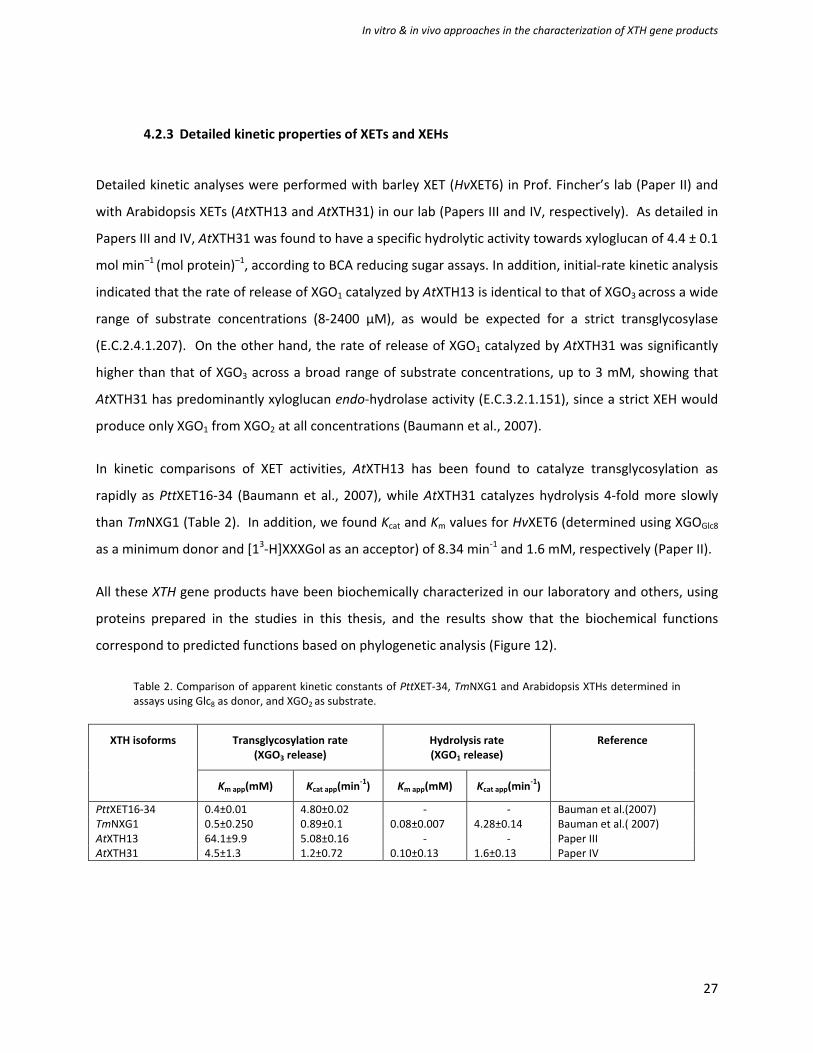
4.2.3 Detailed kinetic properties of XETs and XEHs
Detailed kinetic analyses were performed with barley XET (HvXET6) in Prof. Fincher’s lab (Paper II) and
with Arabidopsis XETs (AtXTH13 and AtXTH31) in our lab (Papers III and IV, respectively). As detailed in
Papers III and IV, AtXTH31 was found to have a specific hydrolytic activity towards xyloglucan of 4.4 ± 0.1
mol min–1 (mol protein)–1, according to BCA reducing sugar assays. In addition, initial-rate kinetic analysis
indicated that the rate of release of XGO1 catalyzed by AtXTH13 is identical to that of XGO3 across a wide
range of substrate concentrations (8-2400 µM), as would be expected for a strict transglycosylase
(E.C.2.4.1.207). On the other hand, the rate of release of XGO1 catalyzed by AtXTH31 was significantly
higher than that of XGO3 across a broad range of substrate concentrations, up to 3 mM, showing that
AtXTH31 has predominantly xyloglucan endo-hydrolase activity (E.C.3.2.1.151), since a strict XEH would
produce only XGO1 from XGO2 at all concentrations (Baumann et al., 2007).
In kinetic comparisons of XET activities, AtXTH13 has been found to catalyze transglycosylation as
rapidly as PttXET16-34 (Baumann et al., 2007), while AtXTH31 catalyzes hydrolysis 4-fold more slowly
than TmNXG1 (Table 2). In addition, we found Kcat and Km values for HvXET6 (determined using XGOGlc8
as a minimum donor and [13-H]XXXGol as an acceptor) of 8.34 min-1 and 1.6 mM, respectively (Paper II).
All these XTH gene products have been biochemically characterized in our laboratory and others, using
proteins prepared in the studies in this thesis, and the results show that the biochemical functions
correspond to predicted functions based on phylogenetic analysis (Figure 12).
Table 2. Comparison of apparent kinetic constants of PttXET-34, TmNXG1 and Arabidopsis XTHs determined in assays using Glc8 as donor, and XGO2 as substrate.
XTH isoforms Transglycosylation rate (XGO3 release)
Hydrolysis rate (XGO1 release)
Reference
Km app(mM) Kcat app(min-1) Km app(mM) Kcat app(min-1)
PttXET16-34 0.4±0.01 4.80±0.02 - - Bauman et al.(2007) TmNXG1 0.5±0.250 0.89±0.1 0.08±0.007 4.28±0.14 Bauman et al.( 2007) AtXTH13 64.1±9.9 5.08±0.16 - - Paper III AtXTH31 4.5±1.3 1.2±0.72 0.10±0.13 1.6±0.13 Paper IV

In vitro & in vivo approaches in the characterization of XTH gene products
28
Figure 12. Phylogenetic tree of the XTH gene family (Baumann et al., 2007), based on amino acid sequences of rice, Arabidopsis, black cottonwood and some barley XTHs. The XTH gene products characterized in this thesis are highlighted in red (Xyloglucan endo-transglycosylases) and blue (xyloglucan-endo-hydrolases). PttXET16-34 and TmNXG1 are highlighted accordingly.

In vitro & in vivo approaches in the characterization of XTH gene products
29
5. In vivo characterization of XTH gene products in the plant cell wall
5.1 Biological roles of members of XTH genes
XTHs are long-lived proteins in plants, and are believed to be active in many processes related to cell
differentiation, cell growth and maintenance after cell death (Nishikubo et al., 2007). XETs cleave
xyloglucan chains and rejoin them in the xyloglucan-cellulose network, allowing cells to expand (Fry et
al., 1992; Rose and Bennett, 1999; Cosgrove, 2005). This process occurs during cell expansion and
elongation throughout plant growth. XETs are expressed in response to hormonal and environmental
signals, time, tissue and organ specifically (Yokoyama and Nishitani, 2001a; Becnel et al., 2006). Clear
functions of various XETs in cell wall modification during plant growth have been demonstrated. For
instance, the chickpea isoform CaXTH1 is involved in expansion processes (Romo et al., 2005), AtXTH14
and AtXTH26 in strengthening the side-walls of root hairs and cell walls in the root differentiation zone
following cell expansion (Maris et al., 2009), AtXTH27 functions during the development of tracheary
elements (Matsui et al., 2005), while GhXTH1 plays a role in cotton fiber elongation (Michailidis et al.,
2009; Lee et al., 2010). In woody plants, PttXET16-34 has a demonstrated role in restructuring primary
walls in xylem and phloem fibers during secondary wall formation (Bourquin et al., 2002). Remarkably,
induction of XET activity has been observed in mature G-layers of tension wood in response to
mechanical stress (Nishikubo et al., 2007). This activity was found to persist for several years, thus the
enzyme functions after cell death in repairing cross-links as they are broken during the associated
shrinking (Nishikubo et al., 2007). In addition, XETs are involved in cell wall softening in ripening fruits
such as tomato and kiwi. In this process, XETs are proposed to act as cell wall loosening agents (Fry et
al., 1992; Schröder et al., 1998; Saladie et al., 2006; Van Sandt et al., 2006; Atkinson et al., 2009).
XTHs also participate in cell wall modification in plant interactions with various microbial symbionts,
including those between arbuscular mycorrhiza and Medicago (Maldonado-Mendoza et al., 2005) and
between Frankia and Datisca glomerata in root nodules (N.K. and Katharina Pawloski, unpublished
data). They are also involved in interactions between plants and pathogens. For instance, in tomato
fruits infected with Penicillium expansum the fruits’ cell wall is modified, resulting in softening and wall
disassembly, thereby facilitating fungal colonization and further progress of the infection (Albert et al.,
2004; Miedes and Lorences, 2007).

In vitro & in vivo approaches in the characterization of XTH gene products
30
5.2 Expression profiles of AtXTH genes
Comprehensive expression analysis by quantitative real-time RT-PCR of the AtXTH genes revealed that
most of the members exhibit distinct expression profiles in terms of tissue specificity and responses to
hormonal signals, although some of the members have similar expression profiles (Yokoyama and
Nishitani, 2001a). Additionally, most of the AtXTH genes are distributed in almost every organ, with
varying levels of expression, some predominantly in specific tissues (Becnel et al., 2006; Jamet et al.,
2009). Furthermore, AtXTH genes are most likely expressed in every developmental stage, from seed
germination to silique maturation, as shown in Figure 13 (Zimmermann et al., 2004; Becnel et al., 2006).
AtXTH31 and AtXTH32 genes are highly expressed in stages of rapid development, according to
Genevestigator data, since AtXTH31 appears to be highly up-regulated during germination and in
seedlings, while AtXTH32 is expressed during all developmental stages (Figure 13 and Zimmermann et
al., 2004). In our experiments, semi-quantitative PCR showed that although AtXTH31 transcripts are
more abundant than AtXTH32 transcripts in roots and hypocotyls, AtXTH32 is more abundant in
cotyledons (Paper IV) and highly up-regulated in stems and nodes (Zimmermann et al., 2004).

In vitro & in vivo approaches in the characterization of XTH gene products
31
Figure 13. Expression profiles of Arabidopsis XTHs in indicated developmental stages, based on data obtained from Genevestigator and reported as absolute expression values (Zimmermann et al., 2004) shaded according to the expression level (red>yellow>green).
In vitro & in vivo approaches in the characterization of XTH gene products
32
5.3 Probing roles of XTH gene products by reverse genetics
Reverse genetic approaches have been successfully used to explore the biological roles of various XTH
genes. For instance, AtXtH27, which belongs to group III-B, has been successfully altered by T-DNA
insertion, and the resulting mutant exhibited aberrant tracheary elements in rosette leaves (Matsui et
al., 2005). In addition, the abundance of AtXTH18 (Group II) transcripts has been reduced by RNAi,
yielding a mutant with slight reductions in primary root growth and final cell length (Osato et al., 2006).
We also employed a reverse genetics approach to study the biological functions of AtXTH31 and
AtXTH32 in the cell walls of A. thaliana (Paper IV). These genes belong to Group III-A in the current
phylogenetic tree (Baumann et al., 2007) and we predicted that they would exhibit predominantly XEH
activity. The members of this group have been speculated to play a role in the degradation of storage
xyloglucan in seeds of plants such as nasturtium, thus providing nutrients during germination in
response to auxin and light signals (Hensel et al., 1991; Brandao et al., 2009). In contrast,
Genevestigator data, and our findings (Paper IV), indicated that two predominant XEHs from Arabidopsis
(AtXTH31 and AtXTH32) do not respond significantly to endogenous auxin (Zimmermann et al., 2004).
In our study, we used T-DNA insertion lines with interrupted AtXTH31 or AtXTH32 genes in an A.
thaliana Columbia background. To identify lines harboring T-DNA insertions in these genes, a PCR
screen was employed, using gene-specific and T-DNA left border (LB-b1) primers (Krysan et al., 1999;
Weigel and Glazebrook, 2002), yielding homozygous knock-out single mutants designated xth31-1 and
xth32-1, respectively. In addition, to avoid possible compensation events from closely related genes, a
double mutant (xth31-1/xth32-1) was generated by crossing the single homozygous mutants. The
results showed that these T-DNA insertions efficiently knocked out the activity of AtXTH31 and AtXTH32
genes (Paper IV). T-DNA mutagenesis, with or without subsequent crossing of single mutants, has also
been found to be highly efficient for knocking out many other Arabidopsis genes (Wang, 2008).
Subsequent analyses showed that knocking out the two predominantly hydrolytic XTH genes resulted in
mutants with phenotypic traits that were indistinguishable from those of wild type counterparts in all
growth and developmental stages, including extension of 6 day-old roots, and 5- and 11 day-old
hypocotyls. The only (mild) deviation observed was that the xth31-1/xth32-1 mutant exhibited slightly
more rapid root and hypocotyl extension immediately after germination (Paper IV). A possible
explanation for this observation is that reduction of XEH activity during radicle emergence may relieve
strong inhibition of auxin-stimulated growth (McDougall and Fry, 1989). In accordance with this

In vitro & in vivo approaches in the characterization of XTH gene products
33
hypothesis, AtXTH31 encodes a protein homologous to TmNXG1, which exhibits xyloglucanase activity
(Baumann et al., 2007), and previous evidence of xyloglucanase from azuki bean (Kaku et al., 2002),
suggesting that xyloglucan endo-hydrolysis might also play a role in primary cell wall extension. In
comparison, the observed XET activity in all examined zones of the xth31-1/xth32-1 mutant remained
effectively unchanged versus wild type, indicating that suppression of these XTH genes does not
dramatically alter XET activity.
Turning to XET, a comprehensive expression analysis of the A. thaliana XTH gene family has shown that
AtXTH12, -13, -17, -18 and -19 transcripts are present in Arabidopsis roots (Yokoyama and Nishitani,
2001a). Among these phylogenetically related genes, transcripts of AtXTH18 were the most abundant
(Osato et al., 2006). The previously elucidated function in primary root growth of AtXTH18 (Osato et al.,
2006), together with its biochemical characterization in Paper III, clearly indicate the role of this XET
isoform in Arabidopsis roots.
5.4 Immunolocalization of substrates for XTH gene products
In the study reported in Paper IV, we used the LM5 antibody to probe the rhamnogalacturonan-I-
associated (1→4)-linked β-D-galactan epitope in order to mark the expanding zone in roots (McCartney
et al., 2003). In parallel, the monoclonal antibody CCRC-M1, which recognizes a fucose (Fuc)-containing
epitope of xyloglucan specific to the growing wall (Puhlmann et al., 1994), was used to probe the
localization of substrates for XET and XEH enzymes in roots. The results showed that CCRC-M1 labeled
xyloglucan was present in the root elongation zone and root hairs (Paper IV), in line with previous
reports (Freshour et al., 2003).
5.5 Assays used for in vivo characterization
An assay using a flucorescently labelled substrate has been employed to visualize (and hence localize)
XET activity in vivo in various plant organs, such as roots (Vissenberg et al., 2000; Vissenberg et al., 2001)
and the G-layer in tention wood (Nishikubo et al., 2007). The in situ assay includes sulphorhodamine-
labelled oligosaccharide as the transglycosylation acceptor substrate, which is thus integrated into
xyloglucan chains, thereby marking the location of XET activity. We employed this assay to monitor XET
activity in roots and hypocotyls of A. thaliana ecotype Columbia and the xth31-1/xth32-1 mutant (Paper
IV). XET activity was observed in roots of wild type plants, in accordance with previous observations

In vitro & in vivo approaches in the characterization of XTH gene products
34
(Vissenberg et al., 2000; Vissenberg et al., 2001), and both the expanding and root junction zones of
hypocotyls (Figure 15, Paper IV).
In the study described in Paper IV we also used a new real-time fluorogenic probe (XXXG-Res) for
visualizing glycoside hydrolase activity in planta (Ibatullin et al., 2009), based on the release of a
fluorophore mediated by hydrolytic enzymes (with minor modifications of the previously published
protocol) to visualize XEH activity in roots and hypocotyls of A. thaliana ecotype Columbia and the
xth31-1/xth32-1 mutant. The results clearly showed the presence of XEH activity in the root elongation
zone, lateral roots, root hairs and emerging root hairs in wild type plants (Figure 14), sites at which XET
activity is also present. In the hypocotyl, both XEH and XET activity appeared in the expanding zone and
in the region close to the root-hypocotyl junction as shown in Figure 15 (see also Paper IV). Notably,
significant reductions in XEH activity were observed in elongation zones in both roots (Figure 14) and
hypocotyls (Figure 15) of the xth31-1/xth32-1 double mutant, although the activity was not completely
abolished in some tissues. Although AtXTH31 and AtXTH32 are clearly key players, the observation of
XEH activity in roots and hypocotyls suggests there might be other xyloglucanases capable of
hydrolyzing the fluorogenic XXXG-Res. These could include other GH16 enzymes (i.e. other XTH gene
products) for which xyloglucanase activity has not been identified, GH5 members, and/or a combination
of other exo-acting glycoside hydrolases.

In vitro & in vivo approaches in the characterization of XTH gene products
35
Figure 14. Localization of xyloglucan endo-hydrolase activity in 6 day-old A. thaliana roots using XXXG-Res. A, wild-type Col-O; B, xth31-1/xth32-1 double knock-out; C, Col-O, region corresponding to the top portion of panel A (ca. 2 mm from the root tip); D, xth31-1/xth32-1 double knock-out, region corresponding to the top portion of panel B (ca. 2 mm from the root tip); E, Col-O, emerging lateral root XEH activity detected with XXXG-Res; F, Col-O, lateral root XET activity detected with XXXG-SR. Representative images from analyses of ca. 10 plants from each line are shown.

In vitro & in vivo approaches in the characterization of XTH gene products
36
Figure 15. Localization of xyloglucan endo-hydrolase and xyloglucan endo-transglycosylase activity in 5 day-old A. thaliana etiolated hypocotyls using XXXG-Res and XXXG-SR. XEH activity: A, wild-type Col-0; B, xth31-1/xth32-1 double knock-out. XET activity: C, wild-type Col-0; D xth31-1/xth32-1 double knock-out. Numbered zones show the approximate positions at which images were taken along the length of the hypocotyls. Composite image produced from representative images from analyses of ca. 20 plants from each line (XEH) and ca. four plants from each line (XET).

In vitro & in vivo approaches in the characterization of XTH gene products
37
6. Concluding remarks and future perspectives
Efforts based on both biochemical and reverse genetic approaches have been made to improve
understanding of the functions of XTH genes in the cell walls of various plants, including Arabidopsis
(dicot) and barley (monocot). Members of Group I/II and III-B, which exhibit xyloglucan endo-
transglycosylase activity, and members of Group III-A (xyloglucan endo-hydrolases) were included to
assess their biological roles in the cell walls of species possessing them. The P. pastoris heterologous
expression system was found to be effective for generating some active XTH gene products for
biochemical characterization and structure/function studies, with a ca. 50% success rate. In addition,
the huge number of clones required for the expression trials necessitated a rapid, straightforward
method to identify clones that produced substantial amounts of enzymes of interest. Thus, a high-
throughput assay for xyloglucan endo-transglycosylase and xyloglucanase activities was developed to
facilitate screening in the expression trials, and other applications. All biochemically characterized XTH
gene products exhibited activities corresponding to their predicted functions, based on the current
phylogenetic tree (Baumann et al., 2007). To gain insights into the function of predominantly XEH
enzymes that had not previously been studied, we employed a reverse genetic approach involving T-
DNA mutagenesis to create loss of function lines of the predominant XEH genes in A. thaliana (AtXTH31
and AtXTH32). The results of knocking out the activities of the corresponding gene products indicated
that they might be important players in cell wall modification in rapidly growing cells. However, the
mutants displayed virtually indistinguishable phenotypes from wild type counterparts in all
developmental stages (except that emergence of the radicle appeared to be faster in the double
mutant). Therefore, further study is required to elucidate the precise function of AtXTH31 in radical
emergence, and a more focused study including use of lateral root mutants together with the XXXG-Res
probe to elucidate the importance of XEH activity in this organ. Furthermore, overexpression of
AtXTH31 and AtXTH32 genes would be useful to complement results obtained from T-DNA mutagenesis.
Over expression could possibly result in more significant phenotypic deviations, thereby providing more
profound indications of the biological functions of the genes encoding predominantly XEH enzymes in
the plant cell wall.

In vitro & in vivo approaches in the characterization of XTH gene products
38
7. Acknowledgements
In the extremely cold environment outside, here inside never felt that cold. My time in Stockholm has
been very nice and warm, without you I would not imagine this journey to be possible. I would
especially like to thank you to many people have been involved and supports in different ways:
Prof. Tuula Teeri for accepting me as a student and brought me into the XTH family. Thank you for your
guidance as the first supervisor. Assoc. Prof. Sunee Nithisinprasert for your supports and introducing me
to Sweden. Prof. Vincent Bulone for critical reading and fruitful suggestions of the thesis, and for always
being there out in the lab, supporting and encouraging. Assoc. Prof. Ines Ezcurra for teaching me good
cloning techniques and of course how to grow plants differently. Assoc. Prof. Christina Divne for your
advice regarding health. Asst. Prof. Qi Zhou for many tips of art work and chemistry.
All the former colleagues: Dr. Alex Selvanayagam Gomez, Dr. Ulla Rusander, Dr. Mariane Wittrup Larsen,
Dr. Linda Fransson, Dr. Tien Chye Tan and Dr. Oliver Spadiut are thanked for being excellent senior and
helpful. Dr. Åsa Kallas for teaching me all the molecular techniques in such great details and all helps.
Dr. Martin Baumann for fruitful discussion and smart way of clone screening. Dr. Gea Guerriero for your
kindly help and sharing in the world of cloning, soil and plants, and for your caring and best wishes. Dr.
Gustav Sunquist for all the mass spectrometry data, without you I might lose my way.
All the present colleagues at floor 2, thank you all of you for creating a fantastic atmosphere. Felicia
Leijon, Erik Malm, Cortwa Hooijmaijers, Elzbieta Rzeszutek, Núria Butchosa Robles, Karim Engelmark
Cassimjee and Dr. Macus Ruda thank you for all fika and laugh, they are good enhancer for the great
results. Johanna Fugelstad for all your attempts to make me happy here, I do have a wonderful time
with you. Dr. Aihua Pei for your nice companion and great help. Per-Olof Syrén for your friendship and
badminton! Christain Brown for your help and giving supports. Thank you all members in ‘‘Brumer
Group’’ for creating such a nice working group. Jens Eklöf for being a fantastic lab mate, and optimistic,
yes everything will be OK. Johan Larsbrink for always listening, and teaching me useful Swedish
expression. Thank you both of you for helping with sammanfattning. Dr. Fredrika Gulfot for your help
with translation and your shoulder when I needed. Farid Ibatullin for making such fantastic substrates.
Thank you to Ela Nelson, Lotta Rosenfeldt, Marlene Jonhsson and Kaj Kauko for all administrative and
technical help. Dr. Anna Ohlsson for your tips regarding to RT-PCR. And you, Huiqiong Lin, thank you for
helping with mutants screening.

In vitro & in vivo approaches in the characterization of XTH gene products
39
Thank you all the collaborators for keeping up the good work and being part of making this thesis
happens: Prof. Rishikesh Bhalerao and Delphine Gendre, Prof. Kris Vissenberg, An Maris, Prof. Geoffrey
Fincher, Maria Hrmova and Andrew Harvey. Colleagues at Department of Botany, Stockholm University
are thanked for all help and fruitful discussion about plants, especially, Prof. Katharina Pawloski for
encouraging me and answering all questions which might arise anytime.
Thai friends in Stockholm for sharing, supports, help and all activities, I do have lots of fun. Bergshamra
house mates: P’Tee, Dr. Potjamas Pansri, Dr. Patricia Santos, Anna Amelina, Dr. Agatha Murgoci, Dr.
Maarja Mäe, Dr. Stanislav Tashenov and Preetam for creating a warm house and helping me when I had
such a difficult situation! Other friends in the real world are thanked for never ending our friendship.
Thank you to Singhapan and my family for all supports along the way of my studies.
I would like to thank you The Ministry of Science and Technology, Thailand and Biomime, KTH for
Financial support.
Last but not least, Prof. Harry Brumer for your global thought and attempts of pursuing multidisciplinary
research, every great supports in all situations and many more. No other word better than ‘‘Thank you
for everything you have done for me’’.

In vitro & in vivo approaches in the characterization of XTH gene products
40
8. References
Albert M, Werner M, Proksch P, Fry SC, Kaldenhoff R (2004) The cell wall-modifying xyloglucan endotransglycosylase/hydrolase LeXTH1 is expressed during the defence reaction of tomato against the plant parasite Cuscuta reflexa. Plant Biology 6: 402-407
Arrowsmith DA, Desilva J (1995) Characterization of 2 tomato fruit-expressed cDNAs encoding xyloglucan endo-transglycosylase. Plant Molecular Biology 28: 391-403
Atkinson RG, Johnston SL, Yauk YK, Sharma NN, Schroder R (2009) Analysis of xyloglucan endotransglucosylase/hydrolase (XTH) gene families in kiwifruit and apple. Postharvest Biology and Technology 51: 149-157
Baran R, Sulová Z, Stratilová E, Farkaš V (2000) Ping-pong character of nasturtium-seed xyloglucan endotransglycosylase (XET) reaction. General Physiology and Biophysics 19: 427-440
Baumann MJ, Eklöf JM, Michel G, Kallas ÅM, Teeri TT, Czjzek M, Brumer H (2007) Structural evidence for the evolution of xyloglucanase activity from xyloglucan endo-transglycosylases: Biological implications for cell wall metabolism. Plant Cell 19: 1947-1963
Becnel J, Natarajan M, Kipp A, Braam J (2006) Developmental expression patterns of Arabidopsis XTH genes reported by transgenes and genevestigator. Plant Molecular Biology 61: 451-467
Bibikova TN, Jacob T, Dahse I, Gilroy S (1998) Localized changes in apoplastic and cytoplasmic pH are associated with root hair development in Arabidopsis thaliana. Development 125: 2925-2934
Bodin A, Ahrenstedt L, Fink H, Brumer H, Risberg B, Gatenholm P (2007) Modification of nanocellulose with a xyloglucan-RGD conjugate enhances adhesion and proliferation of endothelial cells: Implications for tissue engineering. Biomacromolecules 8: 3697-3704
Bourquin V, Nishikubo N, Abe H, Brumer H, Denman S, Eklund M, Christiernin M, Teeri TT, Sundberg B, Mellerowicz EJ (2002) Xyloglucan endotransglycosylases have a function during the formation of secondary cell walls of vascular tissues. Plant Cell 14: 3073-3088
Boyes DC, Zayed AM, Ascenzi R, McCaskill AJ, Hoffman NE, Davis KR, Gorlach J (2001) Growth stage-based phenotypic analysis of arabidopsis: A model for high throughput functional genomics in plants. Plant Cell 13: 1499-1510
Bradford MM (1976) A rapid and sensitive method for the quantitation of microgram quantities of protein utilizing the principle of protein-dye binding. Analytical Biochemistry 72: 248-254
Brandao AD, Del Bem LEV, Vincentz M, Buckeridge MS (2009) Expression pattern of four storage xyloglucan mobilization-related genes during seedling development of the rain forest tree Hymenaea courbaril L. Journal of Experimental Botany 60: 1191-1206
Brett CT, Waldron K (1996) Physiology and biochemistry of plant cell walls, Ed 2nd. Chapman & Hall, London
Brown RM, Saxena IM, Kudlicka K (1996) Cellulose biosynthesis in higher plants. Trends in Plant Science 1: 149-156
Buchanan BB, Gruissem W, Jones R (2000) Biochemistry & Molecular Biology of Plants. John Willey & Sons, Rockville
In vitro & in vivo approaches in the characterization of XTH gene products
41
Campbell P, Braam J (1999b) In vitro activities of four xyloglucan endotransglycosylases from Arabidopsis. Plant Journal 18: 371-382
Cantarel B, Coutinho P, Rancurel C, Bernard T, Lombard V, Henrissat B (2009) The Carbohydrate-Active EnZymes database (CAZy): an expert resource for glycogenomics. Nucleic Acids Research 37: D233
Carpita N, McCann M (2000) Biochemisty & Molecular Biology of plants. American society of plant physiology, Rockville
Catalá C, Rose JK, York WS, Albersheim P, Darvill AG, Bennett AB (2001) Characterization of a tomato xyloglucan endotransglycosylase gene that is down-regulated by auxin in etiolated hypocotyls. Plant Physiology 127: 1180-1192
Chapple C, Carpita N (1998) Plant cell walls as targets for biotechnology. Current Opinion in Plant Biology 1: 179-185
Cosgrove D (2005) Growth of the plant cell wall. Nature Reviews Molecular Cell Biology 6: 850 - 861
Cutillas-Iturralde A, Peña MJ, Zarra I, Lorences EP (1998) A xyloglucan from persimmon fruit cell walls. Phytochemistry 48: 607-610
Daly R, Hearn MTW (2005) Expression of heterologous proteins in Pichia pastoris: a useful experimental tool in protein engineering and production. Journal of Molecular Recognition 18: 119-138
Domozych DS, Sorensen I, Pettolino FA, Bacic A, Willats WGT (2010) The cell wall polymers of the charophycean green alga Chara corrallina: Immunobinding and biochemical screening. International Journal of Plant Sciences 171: 345-361
Domozych DS, Sorensen I, Willats WGT (2009) The distribution of cell wall polymers during antheridium development and spermatogenesis in the Charophycean green alga, Chara corallina. Annals of Botany 104: 1045-1056
Earley KW, Haag JR, Pontes O, Opper K, Juehne T, Song KM, Pikaard CS (2006) Gateway-compatible vectors for plant functional genomics and proteomics. Plant Journal 45: 616-629
Eklöf JM, Brumer H (2010) The XTH gene family: An update on enzyme structure, function, and phylogeny in xyloglucan remodeling. Plant Physiology 153: 456-466
Farkas V, Ait-Mohand F, Stratilova E (2005) Sensitive detection of transglycosylating activity of xyloglucan endotransglycosylase/hydrolase (XTH) after isoelectric focusing in polyacrylarnide gels. Plant Physiology and Biochemistry 43: 431-435
Farkas V, Sulova Z, Stratilova E, Hanna R, Maclachlan G (1992) Cleavage of xyloglucan by nasturtium seed xyloglucanase and transglycosylation to xyloglucan subunit oligosaccharides. Archives of Biochemistry and Biophysics 298: 365-370
Freshour G, Bonin CP, Reiter WD, Albersheim P, Darvill AG, Hahn MG (2003) Distribution of fucose-containing xyloglucans in cell walls of the mur1 mutant of Arabidopsis. Plant Physiology 131: 1602-1612
Fry SC (1989) The structure and functions of xyloglucan. Journal of Experimental Botany 40: 1-11
Fry SC (1997) Novel 'dot-blot' assays for glycosyltransferases and glycosylhydrolases: Optimization for xyloglucan endotransglycosylase (XET) activity. Plant Journal 11: 1141-1150
Fry SC, Aldington S, Hetherington PR, Aitken J (1993) Oligosaccharides as signals and substrates in the plant cell wall. Plant Physiology 103: 1-5

In vitro & in vivo approaches in the characterization of XTH gene products
42
Fry SC, Smith RC, Renwick KF, Martin DJ, Hodge SK, Matthews KJ (1992) Xyloglucan endotransglycosylase, a new wall-loosening enzyme activity from plants. Biochemical Journal 282: 821-828
Fry SC, York WS, Albersheim P, Darvill A, Hayashi T, Joseleau JP, Kato Y, Lorences EP, Maclachlan GA, McNeil M, Mort AJ, Reid JSG, Seitz HU, Selvendran RR, Voragen AGJ, White AR (1993) An unambiguous nomenclature for xyloglucan-derived oligosaccharides. Physiological Plantarum 89: 1-3
Gloster TM, Ibatullin FM, Macauley K, Eklöf JM, Roberts S, Turkenburg JP, Bjørnvad ME, Jørgensen PL, Danielsen S, Johansen KS, Borchert TV, Wilson KS, Brumer H, Davies GJ (2007) Characterization and three-dimensional structures of two distinct bacterial xyloglucanases from families GH5 and GH12. Journal of Biological Chemistry 282: 19177-19189
Goff SA, Ricke D, Lan TH, Presting G, Wang RL, Dunn M, Glazebrook J, Sessions A, Oeller P, Varma H, Hadley D, Hutchinson D, Martin C, Katagiri F, Lange BM, Moughamer T, Xia Y, Budworth P, Zhong JP, Miguel T, Paszkowski U, Zhang SP, Colbert M, Sun WL, Chen LL, Cooper B, Park S, Wood TC, Mao L, Quail P, Wing R, Dean R, Yu YS, Zharkikh A, Shen R, Sahasrabudhe S, Thomas A, Cannings R, Gutin A, Pruss D, Reid J, Tavtigian S, Mitchell J, Eldredge G, Scholl T, Miller RM, Bhatnagar S, Adey N, Rubano T, Tusneem N, Robinson R, Feldhaus J, Macalma T, Oliphant A, Briggs S (2002) A draft sequence of the rice genome (Oryza sativa L. ssp japonica). Science 296: 92-100
Goulao LF, Cosgrove DJ, Oliveira CM (2008) Cloning, characterisation and expression analyses of cDNA clones encoding cell wall-modifying enzymes isolated from ripe apples. Postharvest Biology and Technology 48: 37-51
Graham LE, Cook ME, Busse JS (2000) The origin of plants: Body plan changes contributing to a major evolutionary radiation. Proceedings of the National Academy of Sciences of the United States of America 97: 4535-4540
Gullfot F (2010) On the engineering of proteins: methods and applications for carbohydrate-active enzymes. Doctoral Thesis in Biotechnology. Royal Institute of Technology, Stockholm
Harriman RW, Tieman DM, Handa AK (1991) Molecular-cloning of tomato pectin methyl esterase gene and its expression in rutgers, ripening inhibitor, nonripening, and never ripe tomato fruits. Plant Physiology 97: 80-87
Henrissat B, Davies G (1997) Structural and sequence-based classification of glycoside hydrolases. Current Opinion Structural Biology 7: 637-644
Hensel A, Brummell DA, Hanna R, Maclachlan G (1991) Auxin-dependent breakdown of xyloglucan in cotyledons of germinating Nasturtium seeds. Planta 183: 321-326
Ibatullin FM, Banasiak A, Baumann MJ, Greffe L, Takahashi J, Mellerowicz EJ, Brumer H (2009) A real-time fluorogenic assay for the visualization of glycoside hydrolase activity in Planta. Plant Physiology 151: 1741-1750
Ibatullin FM, Baumann MJ, Greffe L, Brumer H (2008) Kinetic analyses of retaining endo-(xylo)glucanases from plant and microbial sources using new chromogenic xylogluco-oligosaccharide aryl glycosides. Biochemistry 47: 7762-7769
Irshad M, Canut H, Borderies G, Pont-Lezica R, Jamet E (2008) A new picture of cell wall protein dynamics in elongating cells of Arabidopsis thaliana: Confirmed actors and newcomers. Bmc Plant Biology 8: 94

In vitro & in vivo approaches in the characterization of XTH gene products
43
Ito H, Nishitani K (1999) Visualization of EXGT-mediated molecular grafting activity by means of a fluorescent-labeled xyloglucan oligomer. Plant and Cell Physiology 40: 1172-1176
Jamet E, Roujol D, San-Clemente H, Irshad M, Soubigou-Taconnat L, Renou J-P, Pont-Lezica R (2009) Cell wall biogenesis of Arabidopsis thaliana elongating cells: transcriptomics complements proteomics. BMC Genomics 10: 505
Joensuu JJ, Conley AJ, Lienemann M, Brandle JE, Linder MB, Menassa R (2010) Hydrophobin fusions for high-level transient protein expression and purification in Nicotiana benthamiana. Plant Physiology 152: 622-633
Johansson P, Brumer III H, Baumann MJ, Kallas ÅM, Henriksson H, Denman SE, Teeri TT, Jones TA (2004) Crystal structures of a xyloglucan endotransglycosylase reveal details of the transglycosylation acceptor binding. Plant Cell 16: 874-886
Judd WS, Campbell CS, Kellogg EA, Stevens PF, Judd WS, Campbell CC, Kellogg EA, Stevens PF (1999) Plant systematics: A phylogenetic approach. Sinauer Associates, Inc.
Kaku T, Tabuchi A, Wakabayashi K, Kamisaka S, Hoson T (2002) Action of xyloglucan hydrolase within the native cell wall architecture and its effect on cell wall extensibility in azuki bean epicotyls. Plant Cell Physiol. 43: 21-26
Kallas ÅM, Piens K, Denman SE, Henriksson H, Faldt J, Johansson P, Brumer H, Teeri TT (2005) Enzymatic properties of native and deglycosylated hybrid aspen (Populus tremula x tremuloides) xyloglucan endotransglycosylase 16A expressed in Pichia pastoris. Biochemical Journal 390: 105-113
Kaul S, Koo HL, Jenkins J, Rizzo M, Rooney T, Tallon LJ, Feldblyum T, Nierman W, Benito MI, Lin XY, Town CD, Venter JC, Fraser CM, Tabata S, Nakamura Y, Kaneko T, Sato S, Asamizu E, Kato T, Kotani H, Sasamoto S, Ecker JR, Theologis A, Federspiel NA, Palm CJ, Osborne BI, Shinn P, Conway AB, Vysotskaia VS, Dewar K, Conn L, Lenz CA, Kim CJ, Hansen NF, Liu SX, Buehler E, Altafi H, Sakano H, Dunn P, Lam B, Pham PK, Chao Q, Nguyen M, Yu GX, Chen HM, Southwick A, Lee JM, Miranda M, Toriumi MJ, Davis RW, Wambutt R, Murphy G, Dusterhoft A, Stiekema W, Pohl T, Entian KD, Terryn N, Volckaert G, Salanoubat M, Choisne N, Rieger M, Ansorge W, Unseld M, Fartmann B, Valle G, Artiguenave F, Weissenbach J, Quetier F, Wilson RK, de la Bastide M, Sekhon M, Huang E, Spiegel L, Gnoj L, Pepin K, Murray J, Johnson D, Habermann K, Dedhia N, Parnell L, Preston R, Hillier L, Chen E, Marra M, Martienssen R, McCombie WR, Mayer K, White O, Bevan M, Lemcke K, Creasy TH, Bielke C, Haas B, Haase D, Maiti R, Rudd S, Peterson J, Schoof H, Frishman D, Morgenstern B, Zaccaria P, Ermolaeva M, Pertea M, Quackenbush J, Volfovsky N, Wu DY, Lowe TM, Salzberg SL, Mewes HW, Rounsley S, Bush D, Subramaniam S, Levin I, Norris S, Schmidt R, Acarkan A, Bancroft I, Brennicke A, Eisen JA, Bureau T, Legault BA, Le QH, Agrawal N, Yu Z, Copenhaver GP, Luo S, Pikaard CS, Preuss D, Paulsen IT, Sussman M, Britt AB, Selinger DA, Pandey R, Mount DW, Chandler VL, Jorgensen RA, Pikaard C, Juergens G, Meyerowitz EM, Dangl J, Jones JDG, Chen M, Chory J, Somerville MC, Ar Gen I (2000) Analysis of the genome sequence of the flowering plant Arabidopsis thaliana. Nature 408: 796-815
Kooiman P (1960) A method for the determination of amyloid in plant seeds. Reclueil Travaux Chimiques des Pays-Bas 79: 675-678
Krysan PJ, Young JC, Sussman MR (1999) T-DNA as an insertional mutagen in Arabidopsis. Plant Cell 11: 2283-2290

In vitro & in vivo approaches in the characterization of XTH gene products
44
Lee J, Burns TH, Light G, Sun Y, Fokar M, Kasukabe Y, Fujisawa K, Maekawa Y, Allen RD (2010) Xyloglucan endotransglycosylase/hydrolase genes in cotton and their role in fiber elongation. Planta 232: 1191-1205
Lorences EP, Fry SC (1993) Xyloglucan oligosaccharides with at least two α-D-xylose residues act as acceptor substrates for xyloglucan endotransglycosylase and promote the depolymerisation of xyloglucan. Physiologia Plantarum 88: 105-112
Maldonado-Mendoza IE, Dewbre GR, Blaylock L, Harrison MJ (2005) Expression of a xyloglucan endotransglucosylase/hydrolase gene, Mt-XTH1, from Medicago truncatula is induced systemically in mycorrhizal roots. Gene 345: 191-197
Maris A, Suslov D, Fry SC, Verbelen JP, Vissenberg K (2009) Enzymic characterization of two recombinant xyloglucan endotransglucosylase/hydrolase (XTH) proteins of Arabidopsis and their effect on root growth and cell wall extension. Journal of Experimental Botany 60: 3959-3972
Marjamaa K, Kukkola E, Fagerstedt K (2007) Lignification in development. International Journal of Plant Development Biology 1: 160–169
Mark P, Baumann MJ, Eklöf JM, Gullfot F, Michel G, Kallas ÅM, Teeri TT, Brumer H, Czjzek M (2009) Analysis of nasturtium TmNXG1 complexes by crystallography and molecular dynamics provides detailed insight into substrate recognition by family GH16 xyloglucan endo-transglycosylases and endo-hydrolases. Proteins-Structure Function and Bioinformatics 75: 820-836
Matsui A, Yokoyama R, Seki M, Ito T, Shinozaki K, Takahashi T, Komeda Y, Nishitani K (2005) AtXTH27 plays an essential role in cell wall modification during the development of tracheary elements. Plant Journal 42: 525-534
McCartney L, Steele-King CG, Jordan E, Knox JP (2003) Cell wall pectic (1 -> 4)-beta-D-galactan marks the acceleration of cell elongation in the Arabidopsis seedling root meristem. Plant Journal 33: 447-454
McDougall G, Fry S (1989) Structure-activity relationships for xyloglucan oligosaccharides with antiauxin activity. Plant Physiology 89: 883
Mellerowicz EJ, Immerzeel P, Hayashi T (2008) Xyloglucan: The molecular muscle of trees. Annals of Botany 102: 659-665
Michailidis G, Argiriou A, Darzentas N, Tsaftaris A (2009) Analysis of xyloglucan endotransglycosylase/hydrolase (XTH) genes from allotetraploid (Gossypium hirsutum) cotton and its diploid progenitors expressed during fiber elongation. Journal of Plant Physiology 166: 403-416
Miedes E, Herbers K, Sonnewald U, Lorences EP (2010) Over expression of cell wall enzyme reduces xyloglucan depolymerization and softening of transgenic tomato fruits. Journal of Agricultural and Food Chemistry 58: 5708-5713
Miedes E, Lorences EP (2007) The implication of xyloglucan endotransglucosylase/hydrolase (XTHs) in tomato fruit infection by Penicillium expansum link. A. Journal of Agricultural and Food Chemistry 55: 9021-9026
Minic Z, Jamet E, San-Clemente H, Pelletier S, Renou JP, Rihouey C, Okinyo DPO, Proux C, Lerouge P, Jouanin L (2009) Transcriptomic analysis of Arabidopsis developing stems: a close-up on cell wall genes. BMC Plant Biology 9

In vitro & in vivo approaches in the characterization of XTH gene products
45
Mohand FA, Farkas V (2006) Screening for hetero-transglycosylating activities in extracts from nasturtium (Tropaeolum majus)l. Carbohydrate Research 341: 577-581
Nishikubo N, Awano T, Banasiak A, Bourquin V, Ibatullin F, Funada R, Brumer H, Teeri TT, Hayashi T, Sundberg B, Mellerowicz EJ (2007) Xyloglucan endo-transglycosylase (XET) functions in gelatinous layers of tension wood fibers in poplar - A glimpse into the mechanism of the balancing act of trees. Plant and Cell Physiology 48: 843-855
Nishitani K (1992) A novel method for detection of endo-xyloglucan transferase. Plant Cell Physiol. 33: 1159-1164
Nishitani K (1997) The role of endoxyloglucan transferase in the organization of plant cell walls. International Review of Cytology 173: 157-206
Nishitani K, Tominaga R (1992) Endoxyloglucan transferase, A novel class of glycosyltransferase that catalyzes transfer of a segment of xyloglucan molecule to another xyloglucan molecule. Journal of Biological Chemistry 267: 21058-21064
Oh M-H, Romanow WG, Smith RC, Zamski E, Sasse J, Clouse SD (1998) Soybean Bru1 encodes a functional xyloglucan endotransglycosylase that is highly expressed in inner epicotyl tissues during brassinosteroid-promoted elongation. Plant Cell Physiol. 39: 124-130
Osato Y, Yokoyama R, Nishitani K (2006) A principal role for AtXTH18 in Arabidopsis thaliana root growth: a functional analysis using RNAi plants. Journal of Plant Research 119: 153-162
Park YW, Baba K, Furuta Y, Iida I, Sameshima K, Arai M, Hayashi T (2004) Enhancement of growth and cellulose accumulation by overexpression of xyloglucanase in poplar. Febs Letters 564: 183-187
Planas N (2000) Bacterial 1,3-1,4-beta-glucanases: structure, function and protein engineering. Biochimica Et Biophysica Acta-Protein Structure and Molecular Enzymology 1543: 361-382
Popper ZA, Fry SC (2003) Primary cell wall composition of bryophytes and charophytes. Annals of Botany 91: 1-12
Puhlmann J, Bucheli E, Swain MJ, Dunning N, Albersheim P, Darvill AG, Hahn MG (1994) Generation of monoclonal-antibodies against plant cell-wall polysaccharide. I. Characterization of a monoclonal-antibody to a terminal-antibody to a terminal alpha-(1-2)-linked fucosyl-containing epitope. Plant Physiology 104: 699-710
Reid JSG (1985) Cell-wall storage carbohydrate in seeds-Biochemistry of the seed gums and hemicellulose. Advance in Bototany Research 11: 125-155
Romo S, Jimenez T, Labrador E, Dopico B (2005) The gene for a xyloglucan endotransglucosylase/hydrolase from Cicer arietinum is strongly expressed in elongating tissues. Plant Physiology and Biochemistry 43: 169-176
Rose JKC, Bennett AB (1999) Cooperative disassembly of the cellulose-xyloglucan network of plant cell walls: parallels between cell expansion and fruit ripening. Trends in Plant Science 4: 176-183
Rose JKC, Braam J, Fry SC, Nishitani K (2002) The XTH family of enzymes involved in xyloglucan endotransglucosylation and endohydrolysis: Current perspectives and a new unifying nomenclature. Plant and Cell Physiology 43: 1421-1435
Rose JKC, Lee HH, Bennett AB (1997) Expression of a divergent expansin gene is fruit-specific and ripening-regulated. Proceedings of the National Academy of Sciences of the United States of America 94: 5955-5960
In vitro & in vivo approaches in the characterization of XTH gene products
46
Sainsbury F, Lomonossoff GP (2008) Extremely high-level and rapid transient protein production in plants without the use of viral replication. Plant Physiology 148: 1212-1218
Sainsbury F, Thuenemann EC, Lomonossoff GP (2009) pEAQ: versatile expression vectors for easy and quick transient expression of heterologous proteins in plants. Plant Biotechnology Journal 7: 682-693
Saladie M, Rose JKC, Cosgrove DJ, Catala C (2006) Characterization of a new xyloglucan endotransglucosylase/hydrolase (XTH) from ripening tomato fruit and implications for the diverse modes of enzymic action. Plant Journal 47: 282-295
Sarkar P, Bosneaga E, Auer M (2009) Plant cell walls throughout evolution: towards a molecular understanding of their design principles. Journal of Experimental Botany 60: 3615-3635
Saura-Valls M, Faure R, Brumer H, Teeri TT, Cottaz S, Driguez H, Planas A (2008) Active-site mapping of a Populus xyloglucan endo-transglycosylase with a library of xylogluco-oligosaccharides. Journal of Biological Chemistry 283: 21853-21863
Saura-Valls M, Faure R, Ragas S, Piens K, Brumer H, Teeri TT, Cottaz S, Driguez H, Planas A (2006) Kinetic analysis using low-molecular mass xyloglucan oligosaccharides defines the catalytic mechanism of a Populus xyloglucan endotransglycosylase. Biochemical Journal 395: 99-106
Schröder R, Atkinson RG, Langenkamper G, Redgwell RJ (1998) Biochemical and molecular characterisation of xyloglucan endotransglycosylase from ripe kiwifruit. Planta 204: 242-251
Steele NM, Fry SC (2000) Differences in catalytic properties between native isoenzymes of xyloglucan endotransglycosylase (XET). Phytochemistry 54: 667-680
Steele NM, Sulova Z, Campbell P, Braam J, Farkas V, Fry SC (2001) Ten isoenzymes of xyloglucan endotransglycosylase from plant cell walls select and cleave the donor substrate stochastically. Biochemical Journal 355: 671-679
Sticklen M (2006) Plant genetic engineering to improve biomass characteristics for biofuels. Current Opinion in Biotechnology 17: 315-319
Sticklen MB (2008) Plant genetic engineering for biofuel production: towards affordable cellulosic ethanol. Nature Reviews Genetics 9: 433-443
Strohmeier M, Hrmova M, Fischer M, Harvey AJ, Fincher GB, Pleiss J (2004) Molecular modeling of family GH16 glycoside hydrolases: Potential roles for xyloglucan transglucosylases/hydrolases in cell wall modification in the poaceae. Protein Science 13: 3200-3213
Taiz L, Zeiger E (2010) Plant physiology. Sinauer Associates,Inc., Publishers, Sunderland
Teeri TT, Brumer H (2003) Discovery, characterisation and applications of enzymes from the wood-forming tissues of poplar: Glycosyl transferases and xyloglucan endotransglycosylases. . Biocatalysis and Biotransformation 21: 173-179
Teeri TT, Brumer H, Daniel G, Gatenholm P (2007) Biomimetic engineering of cellulose-based materials. Trends in Biotechnology 25: 299-306
Toikkanen JH, Niku-Paavola ML, Bailey M, Immanen J, Rintala E, Elomaa P, Helariutta Y, Teeri TH, Fagerstrom R (2007) Expression of xyloglucan endotransglycosylases of Gerbera hybrida and Betula pendula in Pichia pastoris. Journal of Biotechnology 130: 161-170
Tuskan GA, DiFazio S, Jansson S, Bohlmann J, Grigoriev I, Hellsten U, Putnam N, Ralph S, Rombauts S, Salamov A, Schein J, Sterck L, Aerts A, Bhalerao RR, Bhalerao RP, Blaudez D, Boerjan W, Brun

In vitro & in vivo approaches in the characterization of XTH gene products
47
A, Brunner A, Busov V, Campbell M, Carlson J, Chalot M, Chapman J, Chen GL, Cooper D, Coutinho PM, Couturier J, Covert S, Cronk Q, Cunningham R, Davis J, Degroeve S, Dejardin A, Depamphilis C, Detter J, Dirks B, Dubchak I, Duplessis S, Ehlting J, Ellis B, Gendler K, Goodstein D, Gribskov M, Grimwood J, Groover A, Gunter L, Hamberger B, Heinze B, Helariutta Y, Henrissat B, Holligan D, Holt R, Huang W, Islam-Faridi N, Jones S, Jones-Rhoades M, Jorgensen R, Joshi C, Kangasjarvi J, Karlsson J, Kelleher C, Kirkpatrick R, Kirst M, Kohler A, Kalluri U, Larimer F, Leebens-Mack J, Leple JC, Locascio P, Lou Y, Lucas S, Martin F, Montanini B, Napoli C, Nelson DR, Nelson C, Nieminen K, Nilsson O, Pereda V, Peter G, Philippe R, Pilate G, Poliakov A, Razumovskaya J, Richardson P, Rinaldi C, Ritland K, Rouze P, Ryaboy D, Schmutz J, Schrader J, Segerman B, Shin H, Siddiqui A, Sterky F, Terry A, Tsai CJ, Uberbacher E, Unneberg P, Vahala J, Wall K, Wessler S, Yang G, Yin T, Douglas C, Marra M, Sandberg G, Van de Peer Y, Rokhsar D (2006) The genome of black cottonwood, Populus trichocarpa (Torr. & Gray). Science 313: 1596-1604
Van Sandt VST, Guisez Y, Verbelen JP, Vissenberg K (2006) Analysis of a xyloglucan endotransglycosylase/hydrolase (XTH) from the lycopodiophyte Selaginella kraussiana suggests that XTH sequence characteristics and function are highly conserved during the evolution of vascular plants. Journal of Experimental Botany 57: 2909-2922
Wang Y (2008) How effective is T-DNA insertional mutagenesis in Arabidopsis? Journal of Biochemical Technology 1: 11
Weigel D, Glazebrook J (2002) Arabidopsis: a laboratory manual. Cold Spring Harbor Laboratory Press, New York
Vincken JP, York WS, Beldman G, Voragen AGJ (1997) Two general branching patterns of xyloglucan, XXXG and XXGG. Plant Physiology 114: 9-13
Vissenberg K, Fry SC, Pauly M, Hofte H, Verbelen JP (2005) XTH acts at the microfibril-matrix interface during cell elongation. Journal of Experimental Botany 56: 673-683
Vissenberg K, Fry SC, Verbelen JP (2001) Root hair initiation is coupled to a highly localized increase of xyloglucan endotransglycosylase action in arabidopsis roots. Plant Physiology 127: 1125-1135
Vissenberg K, Martinez-Vilchez IM, Verbelen JP, Miller JG, Fry SC (2000) In vivo colocalization of xyloglucan endotransglycosylase activity and its donor substrate in the elongation zone of arabidopsis roots. Plant Cell 12: 1229-1237
Yokoyama R, Nishitani K (2001a) A comprehensive expression analysis of all members of a gene family encoding cell-wall enzymes allowed us to predict cis-regulatory regions involved in cell-wall construction in specific organs of Arabidopsis. Plant Cell Physiology 42: 1025-1033
Yokoyama R, Rose JK, Nishitani K (2004) A surprising diversity and abundance of xyloglucan endotransglucosylase/hydrolases in rice. Classification and expression analysis. Plant Physiology 134: 1088-1099
Yokoyama R, Uwagaki Y, Sasaki H, Harada T, Hiwatashi Y, Hasebe M, Nishitani K (2010) Biological implications of the occurrence of 32 members of the XTH (xyloglucan endotransglucosylase/hydrolase) family of proteins in the bryophyte Physcomitrella patens. The Plant Journal 64: 645-656
Yu J, Hu SN, Wang J, Wong GKS, Li SG, Liu B, Deng YJ, Dai L, Zhou Y, Zhang XQ, Cao ML, Liu J, Sun JD, Tang JB, Chen YJ, Huang XB, Lin W, Ye C, Tong W, Cong LJ, Geng JN, Han YJ, Li L, Li W, Hu GQ, Huang XG, Li WJ, Li J, Liu ZW, Liu JP, Qi QH, Liu JS, Li T, Wang XG, Lu H, Wu TT, Zhu M, Ni PX, Han H, Dong W, Ren XY, Feng XL, Cui P, Li XR, Wang H, Xu X, Zhai WX, Xu Z, Zhang JS, He SJ,

In vitro & in vivo approaches in the characterization of XTH gene products
48
Zhang JG, Xu JC, Zhang KL, Zheng XW, Dong JH, Zeng WY, Tao L, Ye J, Tan J, Ren XD, Chen XW, He J, Liu DF, Tian W, Tian CG, Xia HG, Bao QY, Li G, Gao H, Cao T, Zhao WM, Li P, Chen W, Wang XD, Zhang Y, Hu JF, Liu S, Yang J, Zhang GY, Xiong YQ, Li ZJ, Mao L, Zhou CS, Zhu Z, Chen RS, Hao BL, Zheng WM, Chen SY, Guo W, Li GJ, Liu SQ, Tao M, Zhu LH, Yuan LP, Yang HM (2002) A draft sequence of the rice genome (Oryza sativa L. ssp indica). Science 296: 79-92
Zhou Q, Baumann MJ, Brumer H, Teeri TT (2006) The influence of surface chemical composition on the adsorption of xyloglucan to chemical and mechanical pulps. Carbohydrate Polymers 63: 449-458
Zhou Q, Brumer H, Teeri TT (2009) Self-Organization of Cellulose Nanocrystals Adsorbed with Xyloglucan Oligosaccharide−Poly(ethylene glycol)−Polystyrene Triblock Copolymer. Macromolecules 42: 5430-5432
Zhou Q, Rutland MW, Teeri TT, Brumer H (2007) Xyloglucan in cellulose modification. Cellulose 14: 625-641
Zimmermann P, Hirsch-Hoffmann M, Hennig L, Gruissem W (2004) GENEVESTIGATOR. Arabidopsis microarray database and analysis toolbox. Plant Physiology 136: 2621-2632